. 2015 Oct;22(10):1654-64.
doi: 10.1038/cdd.2015.14. Epub 2015 Mar 6.

Osteoblast-induced osteoclast apoptosis by fas ligand/FAS pathway is required for maintenance of bone mass

Affiliations

Osteoblast-induced osteoclast apoptosis by fas ligand/FAS pathway is required for maintenance of bone mass

L Wang et al. Cell Death Differ. 2015 Oct.

Abstract

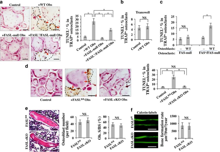
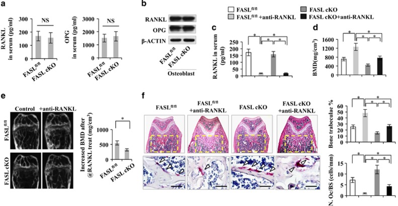
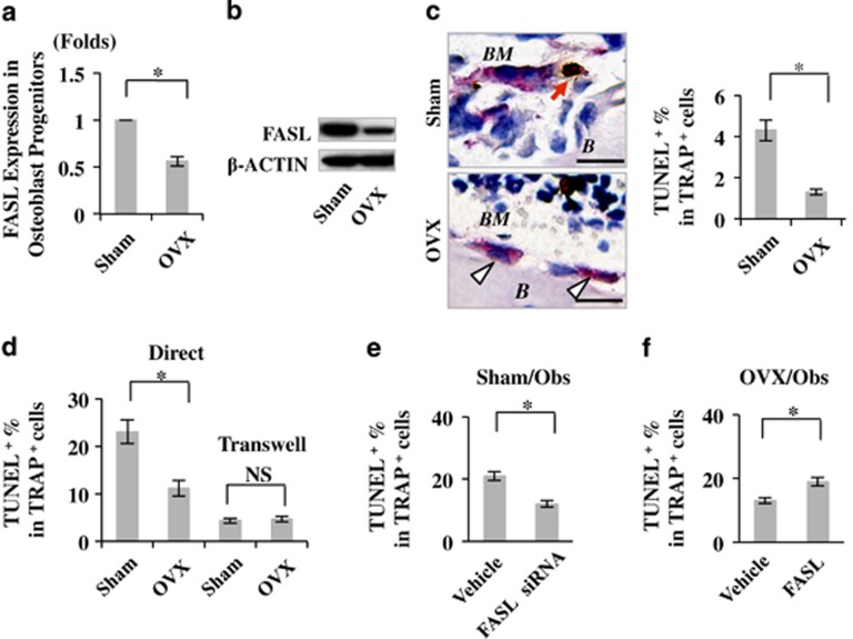
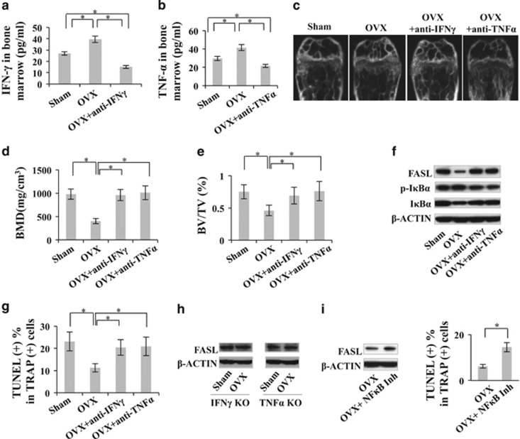
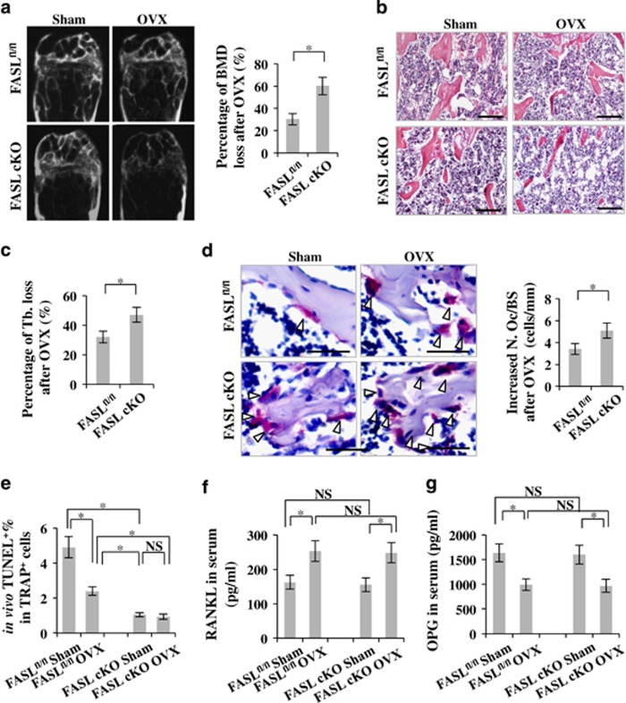
The interplay between osteoblasts and osteoclasts has a crucial role in maintaining bone homeostasis. In this study, we reveal that osteoblasts are capable of inducing osteoclast apoptosis by FAS ligand (FASL)/FAS signaling. Conditional knockout of FASL in osteoblasts results in elevated osteoclast numbers and activity, along with reduced bone mass, suggesting that osteoblast-produced FASL is required to maintain physiological bone mass. More interestingly, we show that osteoblasts from ovariectomized (OVX) osteoporotic mice exhibit decreased FASL expression that results from the IFN-γ- and TNF-α-activated NF-κB pathway, leading to reduced osteoclast apoptosis and increased bone resorption. Systemic administration of either IFN-γ or TNF-α ameliorates the osteoporotic phenotype in OVX mice and rescues FASL expression in osteoblasts. In addition, ovariectomy induces more significant bone loss in FASL conditional knockout mice than in control group with increased osteoclast activity in which the levels of RANKL and OPG remain unchanged. Taken together, this study suggests that osteoblast-induced osteoclast apoptosis via FASL/FAS signaling is a previously unrecognized mechanism that has an important role in the maintenance of bone mass in both physiological conditions and OVX osteoporosis.


Figures

Figure 1
Figure 1
FASL cKO mice show osteopenic phenotype and decreased osteoclast apoptosis. (a) μCT analysis showed markedly reduced BMD and BV/TV in femurs of adult FASL cKO mice when compared with the control littermates (FASLfl/fl). (b) H&E and von Kossa staining showed reduced bone trabeculae in the femurs of FASL cKO mice, and histomorphometric analyses revealed that bone trabeculae percentage in FASL cKO mice were markedly lower than those metrics as measured in control littermates. (c) TRAP staining showed that FASL cKO mice exhibited an increased number of osteoclasts/bone surface (N. Oc/BS) and osteoclast surface/bone surface ratio (Oc. S/BS). (d) TUNEL-TRAP double staining showed that FASL cKO mice presented a significantly reduced number of TUNEL+TRAP+ apoptotic osteoclasts compared with the control littermates. (e) Using an in vivo titanium particles implantation assay, we showed that titanium particles induced more bone resorption in calvarial bones of FASL cKO mice than observed in control littermates. B, bone; Red arrows, apoptotic nuclear; white triangles, TRAP+ cells in c and d and bone resorption pits in e; Error bars represent mean±S.E.M., n=8 animals per group, and all the experiments were performed in triplicate. *P<0.05. Scale bar, 100 μm
Figure 2
Figure 2
Osteoblasts are capable of inducing osteoclast apoptosis via FASL/FAS pathway. (a) After coculture of osteoblasts from C57BL6 mice (WT Obs), FASL-mutated B6Smn.C3- FASLgld/J mice (FASL-null Obs) and FASL-transfected FASL-null osteoblasts (FASL+/FASL-null Obs) with matured osteoclasts, TUNEL-TRAP double staining showed a marked osteoclast apoptosis induced by WT Obs and FASL+/FASL-null Obs, but not FASL-null Obs. (b) When osteoblasts and osteoclasts were cocultured in a transwell system, no marked osteoclast apoptosis was observed. (c) When cocultured with osteoclasts from FAS-mutated C3MRLFAS lpr/J mice (FAS-null osteoclasts), WT osteoblasts failed to induce matured osteoclast apoptosis, whereas WT osteoblasts were able to induce FAS-transfected FAS-null osteoclast (FAS+/FAS-null osteoclasts) apoptosis in the coculture system. (d) TUNEL-TRAP double staining showed that osteoblasts derived from FASL cKO mice exhibited decreased capacity to induce osteoclast apoptosis compared with those from the control littermates when cocultured with matured WT osteoclasts. (e) H&E staining showed no significant difference in osteoblast number per field and osteoblast surface/bone surface ratio (Ob. S/BS) in the femurs between FASL cKO mice and the control littermates. (f) Calcein labeling showed no significant difference in the amount of newly formed bone between FASL cKO mice and the control littermates. Dashed lines, TUNELTRAP+ cells; red arrows, apoptotic nuclears; error bars represent mean±S.E.M., and all the experiments were performed in triplicate, n=8 animals per group. *P<0.05. Scale bar, 100 μm
Figure 3
Figure 3
Blockage of RANKL shows limited ability to rescue the osteopenic phenotype in FASL cKO mice. (a and b) ELISA and western blot showed that there was no significant difference in the RANKL and OPG expression in both serum and osteoblast progenitors/osteoblast cell culture lysis of the FASL cKO mice and control littermates. (c) ELISA showed that RANKL neutralizing antibody was able to substantially block RANKL level in serum. (d and e) μCT showed that blockage of RANKL using its neutralizing antibody in control mice resulted in a marked increase in the BMD in femurs, whereas it showed compromised ability to increase the BMD in FASL cKO mice. (f) HE staining showed that blockage of RANKL in control mice resulted in a stronger ability to increase bone trabeculae percentage when compared with FASL cKO mice; TRAP staining showed that blockage of RANKL in control mice resulted in a stronger ability to reduce the osteoclast activity when compared with FASL cKO mice. White triangles, TRAP+ cells; Error bars represent mean±S.E.M., n=6 animals per group. *P<0.05. Scale bar, 100 μm
Figure 4
Figure 4
Reduced expression of FASL in OVX osteoblast progenitors. (a and b) PCR array and western blot showed that FASL was downregulated in OVX-derived osteoblast progenitors. (c) TUNEL-TRAP double staining showed that OVX mice presented a reduced number of apoptotic osteoclasts in femurs compared with the control group. (d) TUNEL-TRAP double staining showed that OVX-derived osteoblasts induced fewer apoptotic osteoclasts when compared with the control sham osteoblasts in the coculture system. (e) Knockdown of FASL expression in osteoblasts by siRNA resulted in reduced capacity to induce osteoclast apoptosis in the coculture system. (f) Overexpression of FASL in OVX-derived osteoblasts by lentivirus transfection rescued the capacity to induce osteoclast apoptosis in the coculture system. B, bone; BM, bone marrow; white triangles, TRAP+ cells; red arrows, TUNEL+TRAP+ cells; error bars represent mean±S.E.M., and all the experiments were performed in triplicate, n=8 animals per group. *P<0.05. Scale bar, 100 μm
Figure 5
Figure 5
FASL deficiency in OVX-derived osteoblast progenitors is mediated by IFN-γ/TNF-α-induced NFκB activation. (a and b) ELISA showed that systemic injection of either IFN-γ or TNF-α neutralizing antibody into OVX mice was able to markedly decrease the levels of IFN-γ and TNF-α, respectively, in the bone marrow. (ce) μCT showed that either IFN-γ or TNF-α neutralizing antibody could increase BMD and BV/TV of femurs. (f) Western blot showed that OVX-derived osteoblasts showed downregulation of FASL and upregulation of p-NFκB and p-IκB compared with those from sham control group, and either IFN-γ or TNF-α neutralizing antibody administration rescued the FASL expression with downregulation of p-IκB in the osteoblasts derived from OVX mice. (g) TUNEL-TRAP double staining showed that either IFN-γ or TNF-α neutralizing antibody administration restored their capacity to induce osteoclast apoptosis in vitro. (h) Western blot showed that OVX failed to induce FASL downregulation in osteoblast progenitors of either IFN-γ knockout or TNF-α knockout mice. (i) Western blot and TUNEL-TRAP double staining showed that NFκB inhibitor IKK Inhibitor VII could rescue FASL expression in OVX osteoblasts, as well as restore their ability to induce osteoclast apoptosis in vitro. White triangles, TRAP+ cells; red arrows, TUNEL+TRAP+ cells; error bars represent mean±S.E.M., n=6 animals per group. *P<0.05. Scale bar, 100 μm
Figure 6
Figure 6
OVX induces more dramatic bone loss in FASL cKO mice. (ac) μCT and HE staining showed that FASL cKO mice presented more dramatic decreases in bone mineral density and bone trabeculae percentage in the femurs when compared with their wild-type littermates. (d) TRAP staining showed that FASL cKO mice presented more dramatically elevated osteoclast activity when compared with their control littermates. (e) TUNEL-TRAP double staining showed that although OVX was able to induce decreased ratio of apoptotic osteoclasts in wild-type mice, FASL cKO mice presented the lowest ratio of apoptotic osteoclasts regardless of sham or OVX procedures. (f and g) ELISA showed that the OVX-induced increase of RANKL/OPG levels were comparable between FASL cKO mice and their wild-type littermates. Error bars represent mean±S.E.M., n=6 animals per group. *P<0.05. Scale bar, 100 μm

Similar articles

Cited by

References

    1. Rankin EB, Wu C, Khatri R, Wilson TL, Andersen R, Araldi E, et al. The HIF signaling pathway in osteoblasts directly modulates erythropoiesis through the production of EPO. Cell. 2012;149:63–74. - PMC - PubMed
    1. Calvi LM, Adams GB, Weibrecht KW, Weber JM, Olson DP, Knight MC, et al. Osteoblastic cells regulate the haematopoietic stem cell niche. Nature. 2003;425:841–846. - PubMed
    1. Greenbaum A, Hsu YM, Day RB, Schuettpelz LG, Christopher MJ, Borgerding JN, et al. CXCL12 in early mesenchymal progenitors is required for haematopoietic stem-cell maintenance. Nature. 2013;495:227–230. - PMC - PubMed
    1. Zhang J, Niu C, Ye L, Huang H, He X, Tong WG, et al. Identification of the haematopoietic stem cell niche and control of the niche size. Nature. 2003;425:836–841. - PubMed
    1. Liu Y, Wang L, Kikuiri T, Akiyama K, Chen C, Xu X, et al. Mesenchymal stem cell-based tissue regeneration is governed by recipient T lymphocytes via IFN-gamma and TNF-alpha. Nat Med. 2011;17:1594–1601. - PMC - PubMed

Publication types