Review
. 2015;55(1):50-9.
doi: 10.2176/nmc.ra.2014-0188. Epub 2014 Dec 20.

Pathophysiology, diagnosis, and treatment of radiation necrosis in the brain

Affiliations
Review

Pathophysiology, diagnosis, and treatment of radiation necrosis in the brain

Shin-Ichi Miyatake et al. Neurol Med Chir (Tokyo). 2015.

Abstract

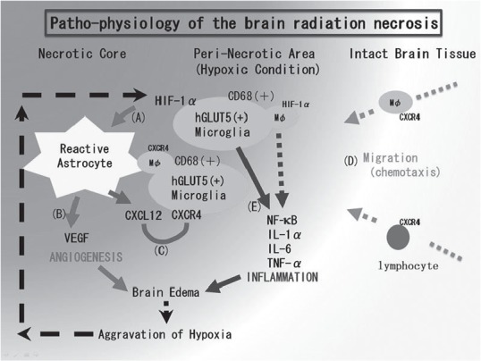
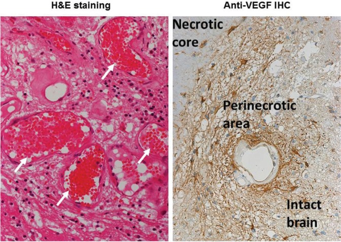
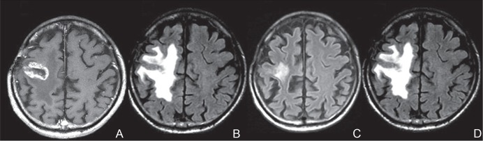
New radiation modalities have made it possible to prolong the survival of individuals with malignant brain tumors, but symptomatic radiation necrosis becomes a serious problem that can negatively affect a patient's quality of life through severe and lifelong effects. Here we review the relevant literature and introduce our original concept of the pathophysiology of brain radiation necrosis following the treatment of brain, head, and neck tumors. Regarding the pathophysiology of radiation necrosis, we introduce two major hypotheses: glial cell damage or vascular damage. For the differential diagnosis of radiation necrosis and tumor recurrence, we focus on the role of positron emission tomography. Finally, in accord with our hypothesis regarding the pathophysiology, we describe the promising effects of the anti-vascular endothelial growth factor antibody bevacizumab on symptomatic radiation necrosis in the brain.


Conflict of interest statement

Conflicts of Interest Disclosure

There is no conflict of interest to disclose for any of the authors.

Figures

Fig. 1.
Fig. 1.
The pathophysiology of brain radiation necrosis: our hypothesis. A: Vascular damage around the irradiated tumor tissue causes tissue ischemia. This hypoxia induces hGLUT5-positive microglia to express hypoxia inducible factor-1 alpha (HIF-1α) around the necrotic core. B: Under HIF-1α regulation, vascular endothelial growth factor (VEGF) is expressed in reactive astrocytes, causing leaky and fragile angiogenesis. C: CXCL12/CXCR4 signaling is also regulated by HIF-1α. D: CXCL12-expressing reactive astrocytes might draw CXCR4-expressing macrophages and lymphocytes by chemotaxis into the perinecrotic area. E: These accumulated hGLUT5-positive microglia producing NF-κB and pro-inflammatory cytokines seem to aggravate radiation necrosis. This figure was taken from our recent publication (Reference 42) with the permission of the publisher. CXCL12: C-X-C motif chemokine 12, CXCR4: C-X-C chemokine receptor type 4, hGLUT5: human glucose transporter 5, IL: interleukin, NF-κB: nuclear factor-kappa B, TNF: tumor necrosis factor.
Fig. 2.
Fig. 2.
Hypoxia inducible factor-1 alpha (HIF-1α) immunohistochemistry of radiation necrosis. A, B: The results of HIF-1α immunohistochemistry on the radiation necrosis in a patient with recurrent glioblastoma multiforme (GBM) who was treated by re-irradiation with boron neutron capture therapy (BNCT). The (A) intact brain area and (B) peri-necrotic area are shown. C, D: HIF-1α immunohistochemistry in patients with radiation necrosis from GBM and metastatic brain tumors, respectively. The former was treated with proton beam radiation and X-ray treatment as an initial treatment, while the latter was treated with repetitive BNCT at the recurrence. Int: intact brain, Ne: necrotic center, Pe: peri-necrotic area. The original objective magnification is ×40.
Fig. 3.
Fig. 3.
Surgical specimen of radiation necrosis derived from a metastatic brain tumor caused by stereotactic radiosurgery (SRS). Hematoxylin and Eosin staining shows marked angiogenesis (indicated by white arrows) with perilesional edema. Anti-vascular endothelial growth factor (VEGF) immunohistochemistry shows the abundant expression of VEGF in the perinecrotic area. The VEGF-producing cells seemed to be reactive astrocytes.
Fig. 4.
Fig. 4.
A representative case of radiation necrosis treated with bevacizumab. The original disease was a metastatic brain tumor from lung cancer. The metastasis was treated with SRS. One year after the SRS, marked enhancement (A) and perilesional edema (B) were recognized on magnetic resonance imaging (MRI). At the time of the MRI, the patient could not walk by himself. After three cycles of bevacizumab treatment 5 mg/kg biweekly, an MRI showed a marked decrease of the edema (C) and he could walk again. Unfortunately, 3 months after the bevacizumab treatment, MRI showed aggravation of the edema (D) with clinical symptom deterioration. Due to financial problems, the patient could not undergo a re-challenge of bevacizumab treatment. A: Gd-enhanced T1-weighted image. B–D: Fluid-attenuated inversion recovery images. SRS: stereotactic radiosurgery.

Republished in

References

    1. Blonigen BJ, Steinmetz RD, Levin L, Lamba MA, Warnick RE, Breneman JC: Irradiated volume as a predictor of brain radionecrosis after linear accelerator stereotactic radiosurgery. Int J Radiat Oncol Biol Phys 77: 996– 1001, 2010. - PubMed
    1. Minniti G, Clarke E, Lanzetta G, Osti MF, Trasimeni G, Bozzao A, Romano A, Enrici RM: Stereotactic radiosurgery for brain metastases: analysis of outcome and risk of brain radionecrosis. Radiat Oncol 6: 48, 2011. - PMC - PubMed
    1. Minniti G, D'Angelillo RM, Scaringi C, Trodella LE, Clarke E, Matteucci P, Osti MF, Ramella S, Enrici RM, Trodella L: Fractionated stereotactic radiosurgery for patients with brain metastases. J Neurooncol 117: 295– 301, 2014. - PubMed
    1. Telera S, Fabi A, Pace A, Vidiri A, Anelli V, Carapella CM, Marucci L, Crispo F, Sperduti I, Pompili A: Radionecrosis induced by stereotactic radiosurgery of brain metastases: results of surgery and outcome of disease. J Neurooncol 113: 313– 325, 2013. - PubMed
    1. Brandsma D, Stalpers L, Taal W, Sminia P, van den Bent MJ: Clinical features, mechanisms, and management of pseudoprogression in malignant gliomas. Lancet Oncol 9: 453– 461, 2008. - PubMed