

Myopic foveoschisis: a clinical review
- PMID: 25744445
- PMCID: PMC4429265
- DOI: 10.1038/eye.2014.311
Myopic foveoschisis: a clinical review
Abstract
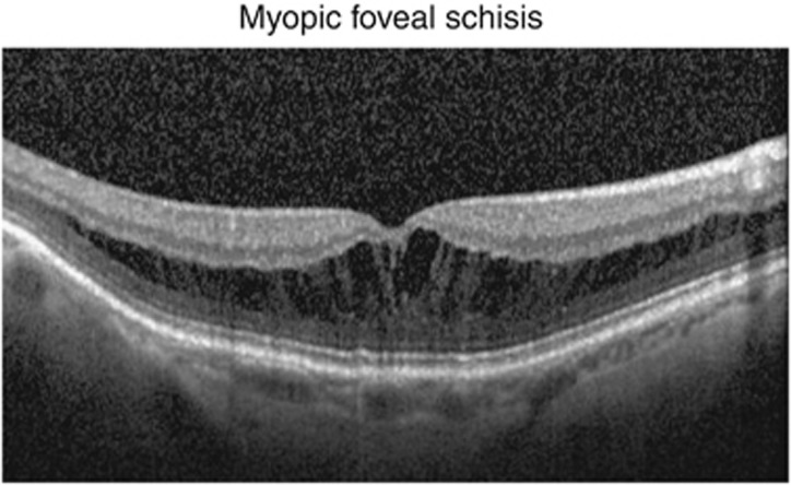
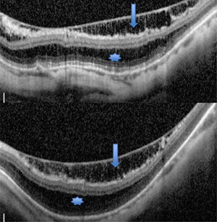
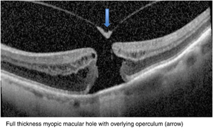
To review the literature on epidemiology, clinical features, diagnostic imaging, natural history, management, therapeutic approaches, and prognosis of myopic foveoschisis. A systematic Pubmed search was conducted using search terms: myopia, myopic, staphyloma, foveoschisis, and myopic foveoschisis. The evidence base for each section was organised and reviewed. Where possible an authors' interpretation or conclusion is provided for each section. The term myopic foveoschisis was first coined in 1999. It is associated with posterior staphyloma in high myopia, and is often asymptomatic initially but progresses slowly, leading to loss of central vision from foveal detachment or macular hole formation. Optical coherence tomography is used to diagnose the splitting of the neural retina into a thicker inner layer and a thinner outer layer, but compound variants of the splits have been identified. Vitrectomy with an internal limiting membrane peel and gas tamponade is the preferred approach for eyes with vision decline. There has been a surge of new information on myopic foveoschisis. Advances in optical coherence tomography will continually improve our understanding of the pathogenesis of retinal splitting, and the mechanisms that lead to macular damage and visual loss. Currently, there is a good level of consensus that surgical intervention should be considered when there is progressive visual decline from myopic foveoschisis.
Figures
Comment in
-
Reply: 'Myopic foveoschisis: an ectatic retinopathy, not a schisis'.Eye (Lond). 2016 Feb;30(2):329-30. doi: 10.1038/eye.2015.234. Epub 2015 Nov 20. Eye (Lond). 2016. PMID: 26584793 Free PMC article. No abstract available.
-
Myopic foveoschisis: an ectatic retinopathy, not a schisis.Eye (Lond). 2016 Feb;30(2):328-9. doi: 10.1038/eye.2015.233. Epub 2015 Nov 20. Eye (Lond). 2016. PMID: 26584797 Free PMC article. No abstract available.
-
Myopic traction maculopathy.Eye (Lond). 2016 Jul;30(7):1025. doi: 10.1038/eye.2016.38. Epub 2016 Mar 11. Eye (Lond). 2016. PMID: 26965013 Free PMC article. No abstract available.
References
-
- Benhamou N, Massin P, Haouchine B, Erginay A, Gaudric A. Macular retinoschisis in highly myopic eyes. Am J Ophthalmol. 2002;133 (6:794–800. - PubMed
-
- Ikuno Y, Gomi F, Tano Y. Potent retinal arteriolar traction as a possible cause of myopic foveoschisis. Am J Ophthalmol. 2005;139 (3:462–467. - PubMed
-
- Ikuno Y, Sayanagi K, Soga K, Oshima Y, Ohji M, Tano Y. Foveal anatomical status and surgical results in vitrectomy for myopic foveoschisis. Jpn J Ophthalmol. 2008;52 (4:269–276. - PubMed
Publication types
MeSH terms
LinkOut - more resources
Full Text Sources
Other Literature Sources
