. 2015 Feb 6;14(1):33.
doi: 10.1186/s12943-015-0311-7.

Structure-function analysis of USP1: insights into the role of Ser313 phosphorylation site and the effect of cancer-associated mutations on autocleavage

Affiliations

Structure-function analysis of USP1: insights into the role of Ser313 phosphorylation site and the effect of cancer-associated mutations on autocleavage

Anne Olazabal-Herrero et al. Mol Cancer. .

Abstract

Background: In complex with its cofactor UAF1, the USP1 deubiquitinase plays an important role in cellular processes related to cancer, including the response to DNA damage. The USP1/UAF1 complex is emerging as a novel target in cancer therapy, but several aspects of its function and regulation remain to be further clarified. These include the role of the serine 313 phosphorylation site, the relative contribution of different USP1 sequence motifs to UAF1 binding, and the potential effect of cancer-associated mutations on USP1 regulation by autocleavage.

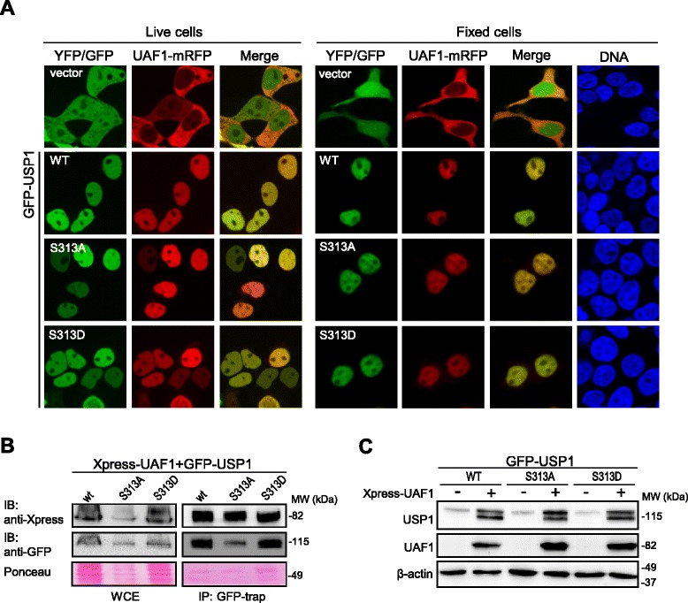
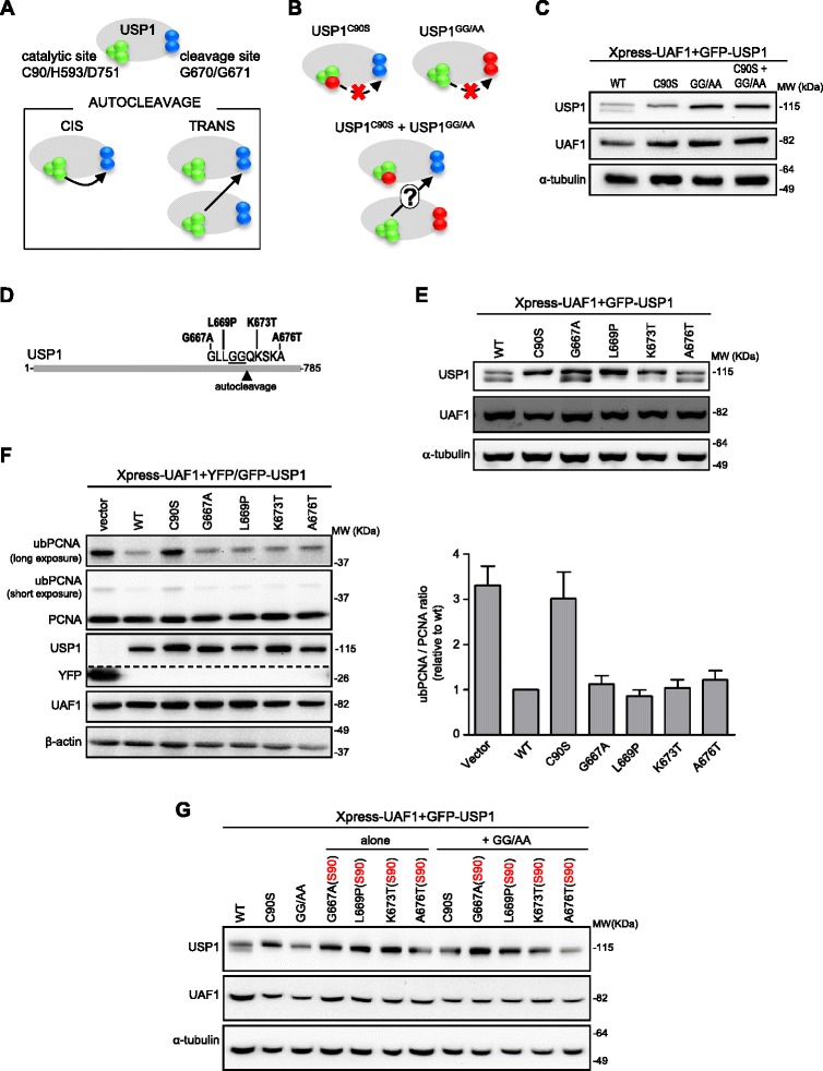
Methods: We have generated a large set of USP1 structural variants, including a catalytically inactive form (C90S), non-phosphorylatable (S313A) and phosphomimetic (S313D) mutants, deletion mutants lacking potential UAF1 binding sites, a mutant (GG/AA) unable to undergo autocleavage at the well-characterized G670/G671 diglycine motif, and four USP1 mutants identified in tumor samples that cluster around this cleavage site (G667A, L669P, K673T and A676T). Using cell-based assays, we have determined the ability of these mutants to bind UAF1, to reverse DNA damage-induced monoubiquitination of PCNA, and to undergo autocleavage.

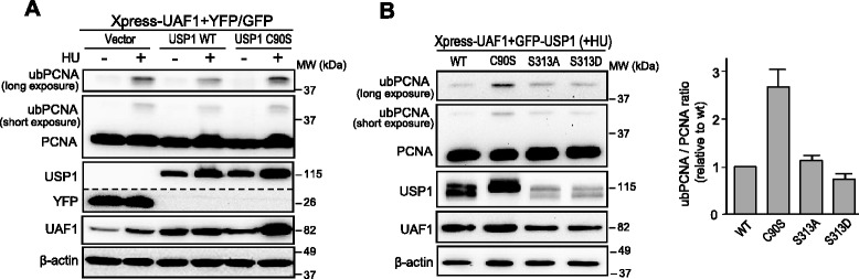
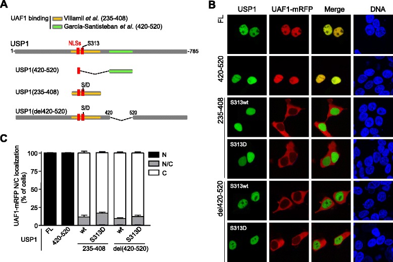
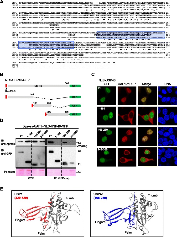
Results: A non-phosphorylatable S313A mutant of USP1 retained the ability to bind UAF1 and to reverse PCNA ubiquitination in cell-based assays. Regardless of the presence of a phosphomimetic S313D mutation, deletion of USP1 fragment 420-520 disrupted UAF1 binding, as determined using a nuclear relocation assay. The UAF1 binding site in a second UAF1-interacting DUB, USP46, was mapped to a region homologous to USP1(420-520). Regarding USP1 autocleavage, co-expression of the C90S and GG/AA mutants did not result in cleavage, while the cancer-associated mutation L669P was found to reduce cleavage efficiency.

Conclusions: USP1 phosphorylation at S313 is not critical for PCNA deubiquitination, neither for binding to UAF1 in a cellular environment. In this context, USP1 amino acid motif 420-520 is necessary and sufficient for UAF1 binding. This motif, and a homologous amino acid segment that mediates USP46 binding to UAF1, map to the Fingers sub-domain of these DUBs. On the other hand, our results support the view that USP1 autocleavage may occur in cis, and can be altered by a cancer-associated mutation.


Figures

Figure 1
Figure 1
UAF1 binds and stabilizes a non-phosphorylatable S313A mutant version of USP1 in cell-based assays. A. Confocal images show representative examples of 293T cells co-expressing UAF1-mRFP with YFP (vector), GFP-USP1 wild type (WT), GFP-USP1S313A or GFP-USP1S313D. Left panels show live cell images, whereas right panels show images of fixed cells. Fixed cells were counterstained with Hoechst to show the nuclei (DNA panels). UAF1-mRFP is cytoplasmic when co-expressed with YFP, but relocates to the nucleus when co-expressed with GFP-USP1 wild type, GFP-USP1S313A and GFP-USP1S313D. B. Co-IP analysis of co-transfected 293T cells, showing that Xpress-UAF1 readily co-immunoprecipitates with GFP-USP1 wild type, GFP-USP1S313A and GFP-USP1S313D. A section of the membrane stained with Ponceau is shown to gauge protein loading. WCE, whole cell extract. C. Immunoblot analysis of 293T cells transfected with GFP-USP1 wild type, GFP-USP1S313A and GFP-USP1S313D, alone (−) or in combination with Xpress-UAF1 (+). Anti-GFP antibody was used to detect GFP-USP1 proteins and anti-Xpress antibody was used to detect Xpress-UAF1. β-actin was used as a loading control. UAF1 co-expression markedly increases the levels of GFP-USP1 wild type, GFP-USP1S313A and GFP-USP1S313D to a similar extent. The lower molecular weight band in (+) samples corresponds to the well-characterized N-terminal fragment that results from USP1 autocleavage at the G670/G671 diglycine motif (see below).
Figure 2
Figure 2
USP1-mediated PCNA deubiquitination is not abrogated by the S313A mutation. A. Immunoblot analysis of 293T cells co-transfected with Xpress-UAF1 and YFP-vector, GFP-USP1 wild type (WT) or the catalytically inactive GFP-USP1C90S. Cells were either left untreated (−), or were treated (+) with 4 mM hydroxyurea (HU) for 24 h. Using an anti-PCNA antibody, monoubiquitinated PCNA (ubPCNA) is detected as a band migrating slightly above the non-ubiquitinated form (PCNA). A short-exposure time image showing both PCNA and ubPCNA, as well as a cropped image showing only ubPCNA with longer exposure time are shown. The dotted line indicates that a panel is a composite of two images from a single exposure of the same gel. Expression of wild type GFP-USP1 decreased HU-induced PCNA monoubiquitination, whereas expression of GFP-USP1C90S did not. Expression of β-actin was used as a control for equal loading of the protein samples. B. On the left, representative example of immunoblot analysis of 293T cells co-transfected with Xpress-UAF1 and GFP-USP1 wild type, GFP-USP1C90S, GFP-USP1S313A or GFP-USP1S313D, and treated with 4 mM HU for 24 h. The ratio of ubiquitinated to non-ubiquitinated PCNA (ubPCNA/PCNA ratio) was determined by densitometry analysis of the immunoblot bands. The graph on the right shows the results of this analysis. The ubPCNA/PCNA ratio was similar in cells expressing wild type GFP-USP1, GFP-USP1S313A and GFP-USP1S313D, but higher in cells expressing the catalytically inactive GFP-USP1C90S. The data represent the mean and SEM of 7 independent experiments.
Figure 3
Figure 3
Side-by-side comparison of two reported UAF1-binding sites in USP1 using a nuclear relocation assay. A. Schematic representation of USP1 and the deletion mutants used in the analysis. The two reported UAF1 binding sites are highlighted: in orange, the 235–408 segment reported by Villamil et al. [17]; in green, the 420–520 segment reported by García-Santisteban et al. [18]. The location of USP1 nuclear localization signals (NLSs, in red), and the S313 phosphorylation site is also shown. S313 wild type and S313D phosphomimetic mutants of USP1(235–408) and USP1(del420-520) were used. B. Confocal images showing representative examples of the results using the in vivo nuclear relocation assay. 293T cells were co-transfected with UAF1-mRFP and GFP-USP1 full length (FL), GFP-USP1(420–520), YFP-USP1(235–408), YFP-USP1(235–408)S313D, YFP-USP1(del420-520) and YFP-USP1(del420-520)S313D. Cells were counterstained with Hoechst to show the nuclei (DNA panels). UAF1-mRFP clearly relocates to the nucleus when co-expressed with GFP-USP1 full length and GFP-USP1(420–520), but not with the remaining deletion mutants. C. Graph showing the results of a semiquantitative analysis of the nuclear relocation assay samples. Slides were coded and the nuclear (N), nuclear/cytoplasmic (N/C) or cytoplasmic (C) localization of UAF1-mRFP was determined in at least 100 cells per slide. The results (mean and SEM) of three independent experiments are shown in the graph.
Figure 4
Figure 4
Homologous amino acid motifs, mapping to the Fingers sub-domain, mediate binding of USP1 and USP46 to UAF1. A. Alignment of USP1, USP46 and USP12 aminoacid sequences using CLUSTALW. USP1 aminoacid segment 420–520 and the homologous regions of USP46 and USP12 are highlighted in blue. B. Schematic representation of different GFP-tagged USP46 deletion mutants used to map its UAF1-binding motif. A heterologous nuclear localization signal (SV40 NLS) was fused to the amino terminal end of each fragment to ensure its nuclear accumulation C. Results of the UAF1 nuclear relocation assay with USP46 fragments. Confocal microscopy images show representative examples of 293T cells co-expressing UAF1-mRFP (red) and the different USP46 protein fragments (green). Cells were counterstained with Hoechst to show the nuclei (DNA panels). Nuclear relocation of UAF1-mRFP is induced by full length (FL) and the 165–259 fragment, but not by the 1–164 or the 243–366 fragments. D. Co-IP analysis of co-trasfected 293T cells, showing that Xpress-UAF1 co-immunoprecipitates with full-length USP46 and with the fragment encompassing residues 165–259, but not with the other two fragments tested. The dotted line indicates that the panel is a composite of two images from a single exposure of the same gel. WCE, whole cell extract. A section of the membrane stained with Ponceau is shown to gauge protein loading. E. Modeled structure of USP1 (left) and USP46 (right) catalytic domains using SWISS-MODEL and the USP7 structure 1NB8 [2] as a template. The Thumb, Palm and Fingers sub-domains are indicated. The UAF1-binding sites are highlighted in red (USP1 residues 420–520) or blue (USP46 residues 165–259).
Figure 5
Figure 5
USP1 autocleavage: assessing cis/trans mode of cleavage and the effect of cancer-associated mutations. A. Representation of USP1 showing the catalytic triad (C90/H593/D751, green spheres) and the autocleavage site (G670/G671, blue spheres). Below, potential cis/trans autocleavage modes. B. Experiment used to assess cleavage mode. The C90S and GG/AA mutants (mutated residues in red) are unable to undergo cis autocleavage. If autocleavage occurs in trans, co-expression of both mutants could lead to cleavage of GFP-USP1C90S by GFP-USP1GG/AA. C. Immunoblot analysis of cells expressing Xpress-UAF1 and GFP-USP1 constructs. Autocleavage was detected in cells transfected with wild type GFP-USP1, but not with GFP-USP1C90S or GFP-USP1GG/AA. No cleavage was observed upon co-expression of GFP-USP1C90S and GFP-USP1GG/AA, suggesting that trans cleavage does not occur. D. Location of cancer mutations [24] near USP1 autocleavage site. E. Immunoblot analysis of cells expressing Xpress-UAF1 and different USP1 mutants. GFP-USP1G667A, GFP-USP1K673T and GFP-USP1A676T are cleaved as efficiently as wild type USP1, whereas GFP-USP1L669P is less efficiently cleaved. F. PCNA ubiquitination in 293T cells treated with 4 mM HU for 24 h. An example of immunoblot results is shown (left). The dotted line indicates that the panel is a composite of two images from a single exposure of the same gel. UbPCNA/PCNA ratio was determined by densitometry and the results (mean and SEM of 3 independent experiments) are shown in the graph (right). Cancer-associated mutations did not alter the ubPCNA/PCNA ratio. G. The C90S mutation (S90) was introduced into each cancer-related mutant construct, rendering these mutants unable to undergo cis autocleavage (Additional Figure 5C). These double mutants were expressed together with GFP-USP1GG/AA to evaluate if they undergo trans cleavage. No autocleavage band was observed with any of the cancer-related/(S90) mutants upon co-expression with GFP-USP1GG/AA, suggesting that the cancer-related mutations tested do not alter the cis/trans mode of cleavage.

Similar articles

Cited by

References

    1. García-Santisteban I, Peters GJ, Giovannetti E, Rodríguez JA. USP1 deubiquitinase: cellular functions, regulatory mechanisms and emerging potential as target in cancer therapy. Mol Cancer. 2013;12:91. doi: 10.1186/1476-4598-12-91. - DOI - PMC - PubMed
    1. Hu M, Li P, Li M, Li W, Yao T, Wu JW, et al. Crystal structure of a UBP-family deubiquitinating enzyme in isolation and in complex with ubiquitin aldehyde. Cell. 2002;111:1041–54. doi: 10.1016/S0092-8674(02)01199-6. - DOI - PubMed
    1. Komander D, Clague MJ, Urbé S. Breaking the chains: structure and function of the deubiquitinases. Nat Rev Mol Cell Biol. 2009;10(8):550–63. doi: 10.1038/nrm2731. - DOI - PubMed
    1. Ye Y, Scheel H, Hofmann K, Komander D. Dissection of USP catalytic domains reveals five common insertion points. Mol Biosyst. 2009;5:1797–808. doi: 10.1039/b907669g. - DOI - PubMed
    1. Huang TT, Nijman SM, Mirchandani KD, Galardy PJ, Cohn MA, Haas W, et al. Regulation of monoubiquitinated PCNA by DUB autocleavage. Nat Cell Biol. 2006;8:339–47. - PubMed

Publication types

MeSH terms

LinkOut - more resources