. 2015 Jun;27(6):1129-40.
doi: 10.1016/j.cellsig.2015.02.017. Epub 2015 Mar 3.

CK1δ restrains lipin-1 induction, lipid droplet formation and cell proliferation under hypoxia by reducing HIF-1α/ARNT complex formation

Affiliations

CK1δ restrains lipin-1 induction, lipid droplet formation and cell proliferation under hypoxia by reducing HIF-1α/ARNT complex formation

Maria Kourti et al. Cell Signal. 2015 Jun.

Abstract

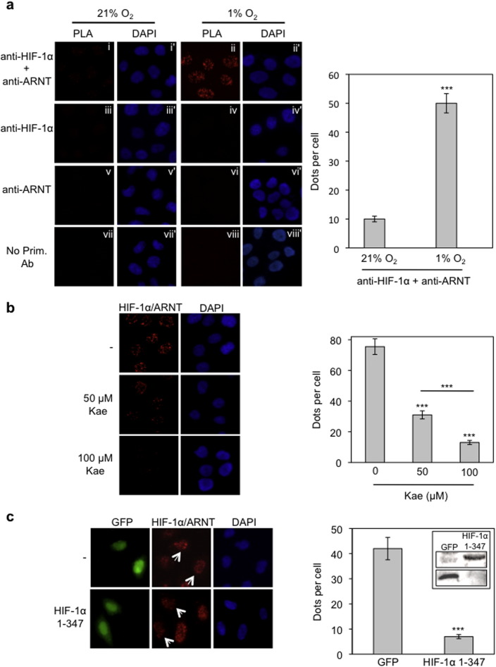
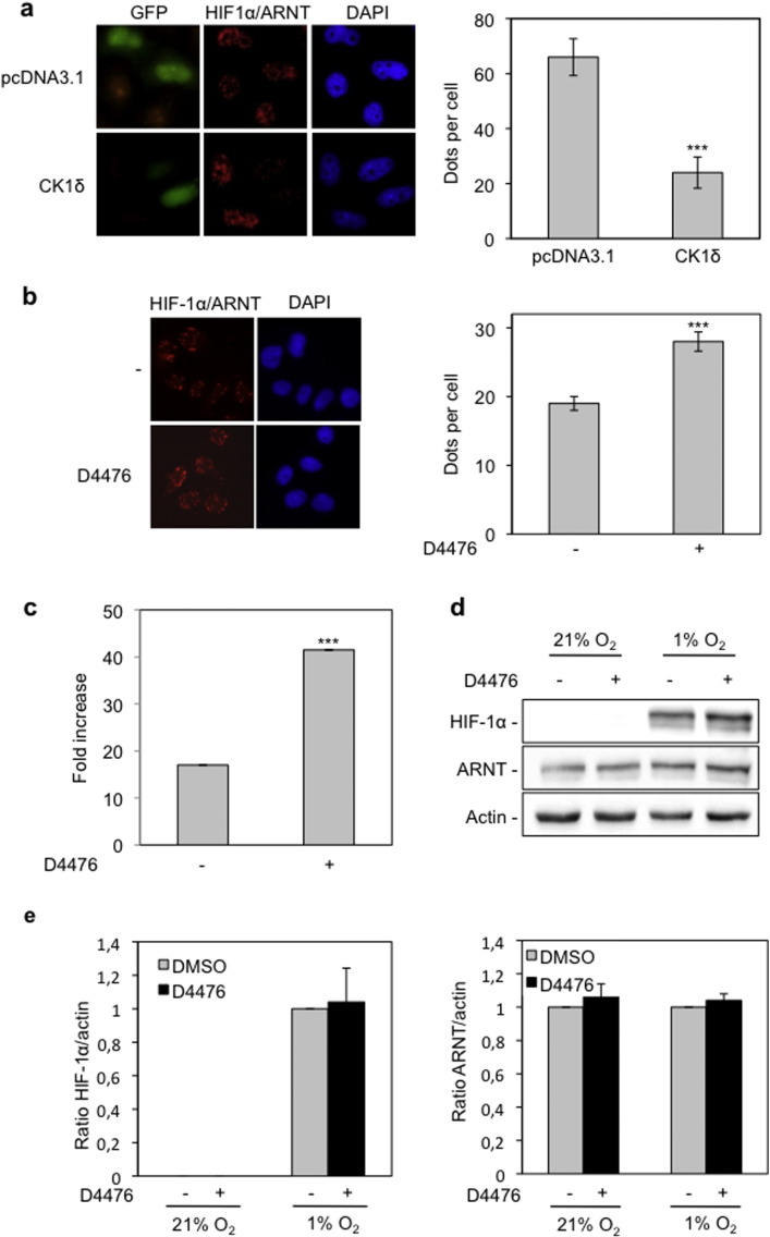
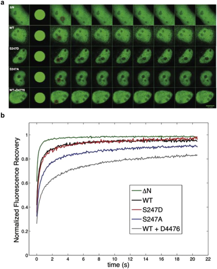
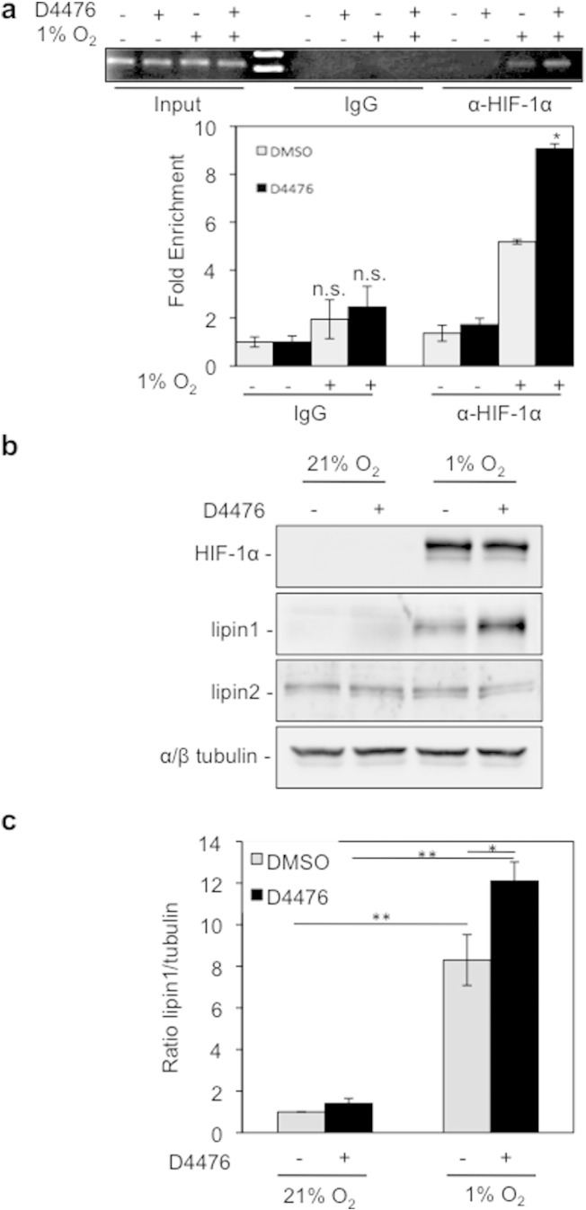
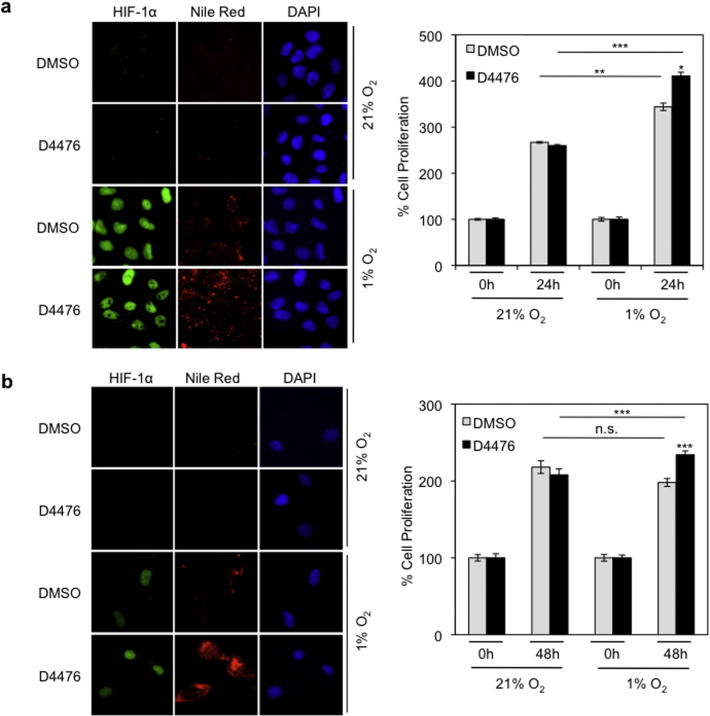
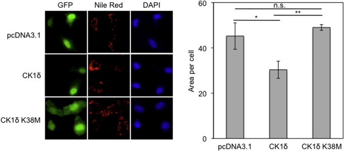
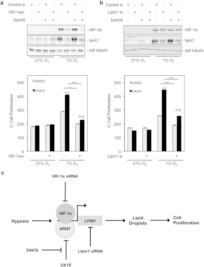
Proliferation of cells under hypoxia is facilitated by metabolic adaptation, mediated by the transcriptional activator Hypoxia Inducible Factor-1 (HIF-1). HIF-1α, the inducible subunit of HIF-1 is regulated by oxygen as well as by oxygen-independent mechanisms involving phosphorylation. We have previously shown that CK1δ phosphorylates HIF-1α in its N-terminus and reduces its affinity for its heterodimerization partner ARNT. To investigate the importance of this mechanism for cell proliferation under hypoxia, we visually monitored HIF-1α interactions within the cell nucleus using the in situ proximity ligation assay (PLA) and fluorescence recovery after photobleaching (FRAP). Both methods show that CK1δ-dependent modification of HIF-1α impairs the formation of a chromatin binding HIF-1 complex. This is confirmed by analyzing expression of lipin-1, a direct target of HIF-1 that mediates hypoxic neutral lipid accumulation. Inhibition of CK1δ increases lipid droplet formation and proliferation of both cancer and normal cells specifically under hypoxia and in an HIF-1α- and lipin-1-dependent manner. These data reveal a novel role for CK1δ in regulating lipid metabolism and, through it, cell adaptation to low oxygen conditions.

Keywords: CK1δ; HIF-1; Hypoxia; Lipid droplets; Lipid metabolism; Lipin1.


Figures

Fig. 1
Fig. 1
HIF-1α/ARNT complexes can be specifically detected and quantified by in situ PLA in HeLa cells. (a) Cells were incubated at normoxia or hypoxia (1% O2) for 4 h and processed for the detection of HIF-1α/ARNT interaction using simultaneous incubation with both primary anti-HIF-1 and anti-ARNT antibodies and the in situ PLA method (panels i and ii). Treatment of cells with a single primary antibody (panels iii–vi) or no primary antibodies (panels vii and viii) was used as negative controls. Panels i–viii show microscopic images of the PLA signal as nuclear dots while the corresponding panels i′–viii′ show the cell nuclei stained with DAPI. (b) Detection of HIF-1α/ARNT complexes by in situ PLA in HeLa cells incubated under hypoxia (1% Ο2) for 4 h in the absence or presence of kaempferol (50–100 μΜ). (c) Twenty hours post-transfection, HeLa cells expressing GFP or GFP-HIF-1α 1-347 were incubated for 4 h in hypoxia (1% O2) and HIF-1α/ARNT complexes were detected by in situ PLA. Arrows point to transfected cells. Left panels: microscopical images. Right panels: quantification of results presenting the average number of nuclear dots per cell ± SEM (n = 50). Inset in (c): immunobloting analysis of cell lysates with an anti-GFP antibody to show expression of GFP alone or GFP-HIF-1α 1-347.
Fig. 2
Fig. 2
Overexpression of CK1δ impairs and inhibition of CK1δ by D4476 increases formation of HIF-1α/ARNT complexes. (a) HeLa cells were co-transfected with pcDNA3.1 or pcDNA3.1-CK1δ and pEGFP plasmids. Twenty hours post-transfection cells were incubated under hypoxia (1% O2) for 4 h and HIF-1α/ARNT complexes were detected by in situ PLA. (b) Detection of HIF-1α/ARNT complexes, in the absence or presence of D4476 (10 μΜ) under hypoxia (1% O2) for 4 h, by in situ PLA. For (a) and (b): left panels: microscopical images. Right panels: quantification of results presenting the average number of nuclear dots per cell ± SEM (n = 50). (c) Determination of HIF-1 transcriptional activity in HeLa cells incubated for 16 h under normoxia or hypoxia (1% O2) in the absence or presence of D4476 (10 μΜ). Results are shown as fold increase in relation to the corresponding normoxic conditions and represent the mean of three independent experiments performed in triplicate ± SEM. (d) Western blot analysis of HeLa cells incubated for 4 h under normoxia or hypoxia (1% O2) in the absence or presence of D4476 (10 μΜ), for detection of HIF-1α and ARNT protein levels. (e) Histograms show the HIF-1α/actin (left) or ARNT/actin (right) protein levels ratio according to quantification of blots from three independent experiments performed as in (d).
Fig. 3
Fig. 3
Phospho-site mutation S247A and inhibition of CK1δ by D4476 reduce nuclear mobility of HIF-1α in living cells. (a) Transfected HeLa cells overexpressing the indicating HIF-1α forms tagged with GFP were processed for FRAP analysis 24 h post-transfection. Representative time-lapse images of cells are shown for each GFP-tagged protein. Circles indicate the bleached region. (b) Analysis of FRAP recoveries. Curves represent the mean corrected fluorescence intensities over time for GFP-tagged HIF-1α-ΔΝ, wt HIF-1α, HIF-1α S247A, HIF-1α S247D and wt HIF-1α in the presence of D4476, as indicated.
Fig. 4
Fig. 4
CK1δ inhibition stimulates lipin-1 expression under hypoxic conditions. (a) D4476 increases the interaction of HIF-1α with lipin1 promoter. Upper: gel electrophoresis of PCR products amplified from anti-HIF-1α or rabbit IgG chromatin immunoprecipitates of Huh7 cells, incubated for 8 h under normoxia or hypoxia (1% O2) in the absence or presence of D4476 (10 μΜ). Bottom: quantification of real-time PCR results. Data represent the mean (± SEM) of two independent experiments performed in triplicate. (b) Western blotting analysis of HeLa cells incubated for 24 h under normoxia or hypoxia (1% O2) in the absence or presence of D4476 (10 μΜ), for detection of HIF-1α and lipin protein levels. Tubulin was used as loading control. (c) Histogram shows the lipin-1/tubulin protein levels ratio according to quantification of blots from three independent experiments performed as in (b).
Fig. 5
Fig. 5
CK1δ inhibition increases lipid accumulation and cell proliferation under hypoxic conditions. Treatment with D4476 (10 μΜ) of HeLa (a) or hSMB (b) cells kept under normoxia or hypoxia (1% O2). Left: fluorescence microscope images of cells incubated under normoxia or hypoxia (1% O2) for 24 h in the absence or presence of D4476 (10 μΜ) and stained with Nile Red to visualize lipid droplets. Right: digitized graph of cell proliferation under normoxia or hypoxia (1% O2) in the absence or presence of D4476 (10 μΜ) after 24 h (a) or 48 h (b) treatment. Data represent the mean of three independent experiments performed in triplicate and expressed, as percent of the initial number of cells at time zero ± SEM.
Fig. 6
Fig. 6
Overexpression of active CK1δ impairs lipid accumulation under hypoxic conditions. Left: HeLa cells were co-transfected with pcDNA3.1 or pcDNA3.1-CK1δ wt or pcDNA3.1-CK1δ K38M and pEGFP plasmids. Twenty four hours post-transfection cells were incubated for 24 h under hypoxia (1% O2), stained with Nile Red to visualize lipid droplets and observed by fluorescence microscope. Right: quantification was performed using ImageJ software and represent the mean area ± SEM of Nile Red staining of 50 cells.
Fig. 7
Fig. 7
Stimulation of cancer cell proliferation by CΚ1δ inhibition is HIF-1α- and lipin-1-dependent. (a and b) Upper panels: results of western blotting analysis for HIF-1α and lipin-1 protein levels after HIF-1α or lipin-1 silencing, respectively. HeLa cells were transfected with control siRNA or siRNA against HIF-1α or lipin-1, and 24 h post-transfection, incubated for 24 h under normoxia or hypoxia (1% O2) in the absence or presence of D4476 (10 μΜ). Bottom panels: digitized graph of HeLa cell proliferation, treated in the same conditions as described above. Data represent the mean of three independent experiments performed in triplicate and expressed, as percent of the initial number of cells at time zero ± SEM. (c) Schematic model of the mechanism by which CK1δ impairs cell proliferation under hypoxia.

Similar articles

Cited by

References

    1. Semenza G.L. Trends Pharmacol. Sci. 2012;33:207–214. - PMC - PubMed
    1. Semenza G.L. N. Engl. J. Med. 2011;365:537–547. - PubMed
    1. Brahimi-Horn M.C., Bellot G., Pouyssegur J. Curr. Opin. Genet. Dev. 2011;21:67–72. - PubMed
    1. Sun R.C., Denko N.C. Cell Metab. 2014;19:285–292. - PMC - PubMed
    1. Huang D., Li T., Li X., Zhang L., Sun L., He X., Zhong X., Jia D., Song L., Semenza G.L., Gao P., Zhang H. Cell Rep. 2014;8:1930–1942. - PubMed

Publication types

MeSH terms

Substances

LinkOut - more resources