. 2015 Sep;93(8):716-26.
doi: 10.1038/icb.2015.38. Epub 2015 Mar 6.

Conserved and divergent aspects of human T-cell development and migration in humanized mice

Affiliations

Conserved and divergent aspects of human T-cell development and migration in humanized mice

Joanna Halkias et al. Immunol Cell Biol. 2015 Sep.

Abstract

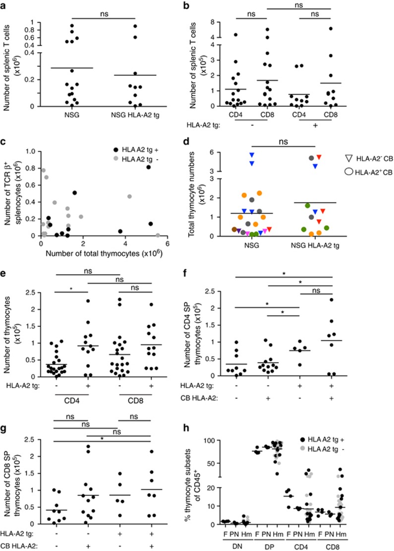
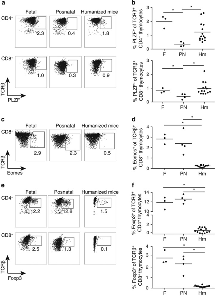
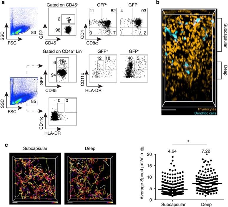
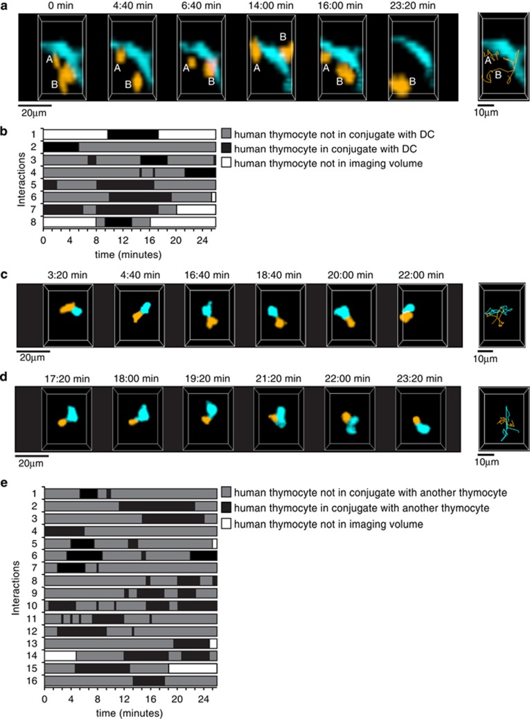
Humanized mice represent an important model to study the development and function of the human immune system. While it is known that mouse thymic stromal cells can support human T-cell development, the extent of interspecies cross-talk and the degree to which these systems recapitulate normal human T-cell development remain unclear. To address these questions, we compared conventional and non-conventional T-cell development in a neonatal chimera humanized mouse model with that seen in human fetal and neonatal thymus samples, and also examined the impact of a human HLA-A2 transgene expressed by the mouse stroma. Given that dynamic migration and cell-cell interactions are essential for T-cell differentiation, we also studied the intrathymic migration pattern of human thymocytes developing in a murine thymic environment. We found that both conventional T-cell development and intra-thymic migration patterns in humanized mice closely resemble human thymopoiesis. Additionally, we show that developing human thymocytes engage in short, serial interactions with other human hematopoietic-derived cells. However, non-conventional T-cell differentiation in humanized mice differed from both fetal and neonatal human thymopoiesis, including a marked deficiency of Foxp3(+) T-cell development. These data suggest that although the murine thymic microenvironment can support a number of aspects of human T-cell development, important differences remain, and additional human-specific factors may be required.


Figures

Figure 1
Figure 1
Development of conventional T cells in humanized mice is comparable to fetal and post-natal human thymus in the presence or absence of an HLA-A2 transgene. (a) Number of human TCRβ+ splenocytes in neonatal chimera NSG and NSG HLA-A2 tg mice at 11–13 weeks post reconstitution. Each dot represents an individual mouse. Line represents average. (b) Number of human CD4+ and CD8+ splenic T cells from neonatal chimera NSG and NSG HLA-A2 tg mice. (c) Number of splenic human T cells versus the number of thymocytes in individual humanized mice. (d) Quantification of total thymocytes in NSG and NSG HLA-A2 tg mice reconstituted with HLA-A2+ and HLA-A2 cord blood (CB). Each color corresponds to an individual CB donor. (e) Number of CD4+ and CD8+ SP thymocytes in neonatal chimera NSG and NSG HLA-A2 tg mice. P=0.2 for CD8+ SP thymocytes. (f) Number of CD4+ SP thymocytes in NSG and NSG HLA-A2 tg mice reconstituted with HLA-A2 or HLA-A2+ CB donors. (g) Number of CD8+ SP thymocytes in NSG and NSG HLA-A2 tg mice reconstituted with HLA-A2 or HLA-A2+ CB donors. P=0.0741 for NSG and NSG HLA-A2 tg mice reconstituted with HLA-A2 CB. (h) Proportion of thymocyte developmental intermediate subsets of CD45+ cells in human fetal (F) (18–20 weeks) and post-natal (PN) (1 week to 2.5 years) human thymic samples and in NSG and NSG HLA-A2 tg mice (Hm) (11–13 weeks post reconstitution). Each dot represents an individual thymus sample. For Hm samples, dots are color coded to distinguish NSG and NSG HLA-A2tg hosts. ns, not statistically significant, * indicates P<0.05. DN indicates CD4CD8 thymocytes, and DP indicates CD4+CD8+ thymocytes. All data shown in these graphs has been gated on human CD45+ cells.
Figure 2
Figure 2
Non-conventional T-cell development in humanized mice is distinct from fetal and postnatal human thymopoiesis. (a) Representative flow-cytometric analysis of PLZF expression in CD4+ (top panels) and CD8+ (bottom panels) human thymocytes gated on TCRβ+ cells in fetal, postnatal and humanized mice thymic samples. (b) Proportion of PLZF+ cells in CD4+ (top panel) and CD8+ (bottom panel) TCRβ+ human thymocytes in human fetal, postnatal and humanized mice thymic samples. Each dot represents one thymus. (c) Representative flow-cytometric analysis of Eomes expression in CD8+ thymocytes gated on human TCRβ+ cells in human fetal, postnatal and humanized mice thymic samples. (d) Proportion of Eomes+ cells in CD8+ TCRβ+ human thymocytes in human fetal, postnatal and humanized mice thymic samples. (e) Representative flow-cytometric analysis of Foxp3 expression in CD4+ (top panels) and CD8+ (bottom panels) human thymocytes gated on TCRβ+ cells in fetal, postnatal and humanized mice thymic samples. (f) Proportion of Foxp3+ cells in CD4+ (top panel) and CD8+ (bottom panel) TCRβ+ human thymocytes in human fetal, postnatal and humanized mice thymic samples. Line represents average. * indicates P<0.05.
Figure 3
Figure 3
2-photon imaging of the thymus of humanized mice reconstituted with GFP-expressing human CD34+ hematopoietic progenitors. NSG and NSG HLA-A2 tg mice were reconstituted with GFP-transduced HLA-A2 cord blood progenitors, and all data from NSG and NSG HLA-A2 tg thymi were combined for image analysis. (a) Flow-cytometric analysis of the thymus of a NSG humanized mouse 12 weeks post reconstitution depicting the gating strategy for identification of thymocyte developmental intermediates among human CD45+ cells (top panels), and HLA-DRint CD11c+ and HLA-DRhigh CD11c+ DC subsets among CD45+ Lin (CD3 CD19CD56) cells (bottom panels) within the GFP+ and GFP fractions of the collagenase digested thymus. (b) 2-photon imaging volume of GFP labeled human thymocytes and DCs within the thymus of an NSG humanized mouse at 12 weeks post reconstitution. For ease of visualization, thymocytes are artificially colored orange and DCs are colored turquoise. Depicted is the compartmentalization of the imaging volume based on distance from the capsule. Scale bar, 70 μm. Thymus was processed for flow cytometry after imaging and data from the same thymus sample are shown in (a). (c) Cell tracks from representative 2-photon time-lapse data sets of human thymocytes within the subcapsular (90 μm in depth, starting 30 μm below the capsule) and deep (30 μm in depth, starting 165 μm below the capsule) portions of the thymus of a humanized mouse (~27 min movies). Tracks are color-coded to represent the passage of time (blue>red>yellow>white). Scale bar 20  μm. (d) Average speed of human thymocytes in the thymus of humanized mice. Each dot represents average speed of an individual tracked cell, and the line represents the average value of compiled track averages for each location. Data represent analysis of two individual imaging volumes obtained from two individual mice. n=300 tracks (subcapsular cells); n=149 tracks (deep cells). * indicates P<0.05.
Figure 4
Figure 4
Contacts between human thymocytes and DCs in the thymus of humanized mice. NSG and NSG HLA-A2 tg mice were reconstituted with GFP-transduced HLA-A2 cord blood progenitors, and all data from NSG and NSG HLA-A2 tg thymi were combined for image analysis. (a) Cropped representation of 2-photon imaging volumes at indicated time points of human thymocytes contacting a human DC within the thymus of a humanized mouse. Thymocytes and DCs were distinguished based on size and morphology. The thymocytes are color-coded in orange, 'A' and 'B' identify two individual thymocytes. The DC is color-coded in turquoise. Far right panel, thymocyte tracks (orange) for the duration of the movie. Representative of a total of 8 interactions involving 8 different labeled thymocytes/150 thymocytes tracked with an approximate density of 8 DCs/imaging volume. (b) Timeline chart depicting the time of contact with DCs of 8 tracked thymocytes for a total of 40 time points (~27 min). Black boxes indicate periods of thymocyte:DC contacts. (c, d) Cropped representations of 2-photon imaging volumes at indicated time points of human thymocyte:thymocyte contacts within the thymus of a humanized mouse. Thymocytes are color-coded in orange and turquoise for improved visualization. Far right panels, thymocyte tracks for the duration of the movie. (e) Timeline chart depicting the time of contact with other thymocytes of 16 tracked thymocytes for a total of 40 time points (~27 min). For ease of representation, only thymocytes in contact with one other thymocyte are depicted. Black boxes indicate periods of contact between thymocytes.

Similar articles

Cited by

References

    1. 1Ishikawa F, Yasukawa M, Lyons B, Yoshida S, Miyamoto T, Yoshimoto G et al. Development of functional human blood and immune systems in NOD/SCID/IL2 receptor {gamma} chain(null) mice. Blood 2005; 106: 1565–1573. - PMC - PubMed
    1. 2Traggiai E, Chicha L, Mazzucchelli L, Bronz L, Piffaretti JC, Lanzavecchia A et al. Development of a human adaptive immune system in cord blood cell-transplanted mice. Science 2004; 304: 104–107. - PubMed
    1. 3Brehm MA, Cuthbert A, Yang C, Miller DM, DiIorio P, Laning J et al. Parameters for establishing humanized mouse models to study human immunity: analysis of human hematopoietic stem cell engraftment in three immunodeficient strains of mice bearing the IL2rgamma(null) mutation. Clin Immunol 2010; 135: 84–98. - PMC - PubMed
    1. 4Lepus CM, Gibson TF, Gerber SA, Kawikova I, Szczepanik M, Hossain J et al. Comparison of human fetal liver, umbilical cord blood, and adult blood hematopoietic stem cell engraftment in NOD-scid/gammac-/-, Balb/c-Rag1-/-gammac-/-, and C.B-17-scid/bg immunodeficient mice. Hum Immunol 2009; 70: 790–802. - PMC - PubMed
    1. 5Aryee KE, Shultz LD, Brehm MA. Immunodeficient mouse model for human hematopoietic stem cell engraftment and immune system development. Methods Mol Biol 2014; 1185: 267–278. - PMC - PubMed

Publication types

MeSH terms