. 2015 Feb 13:14:37.
doi: 10.1186/s12943-015-0315-3.

miR-663 attenuates tumor growth and invasiveness by targeting eEF1A2 in pancreatic cancer

Affiliations

miR-663 attenuates tumor growth and invasiveness by targeting eEF1A2 in pancreatic cancer

Wenqiao Zang et al. Mol Cancer. .

Retraction in

Abstract

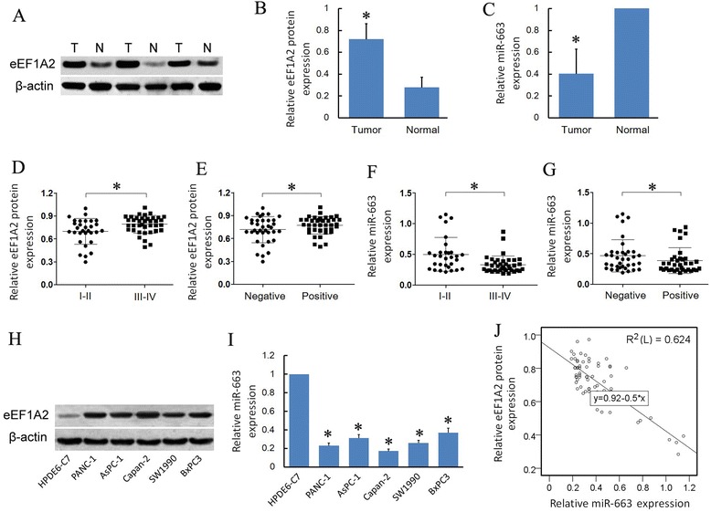
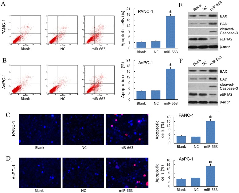
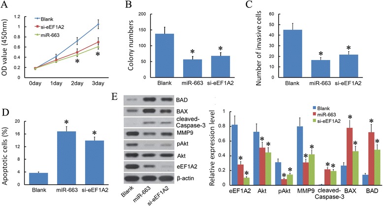
Background: miR-663 is associated with many important biologic processes, such as the evolution, development, viral infection, inflammatory response, and carcinogenesis among vertebrates. However, the molecular function and mechanism of miR-663 in pancreatic cancer growth and invasion is still unclear.

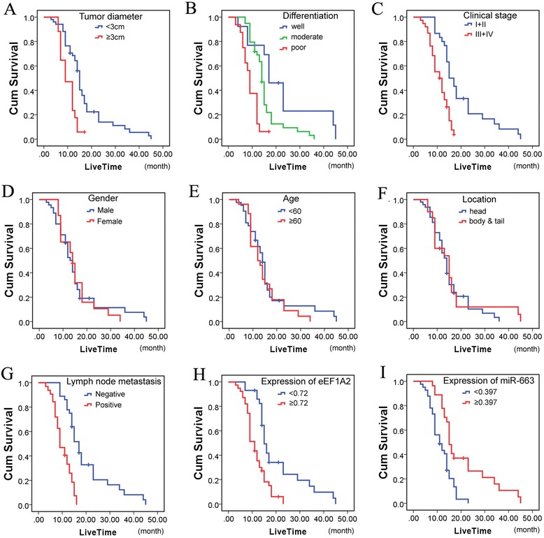
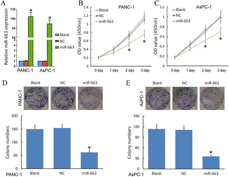
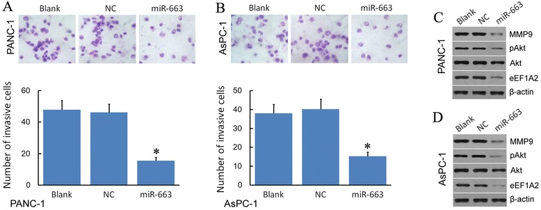
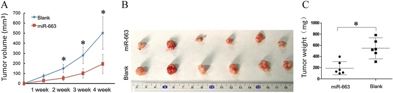
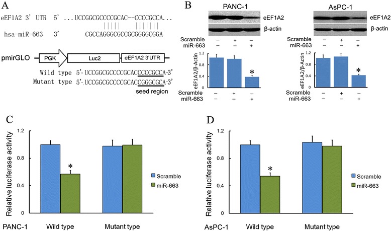
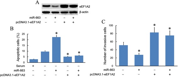
Methods: Western blot and real-time PCR were used to study the expression level of eEF1A2 protein and miR-663 in pancreatic cancer tissues and cell lines. The Pearson χ (2) test was used to determine the correlation between miR-663 expression and clinicopathologic features of patients. Patients' survival was analyzed using the Kaplan-Meier method, using the log-rank test for comparison. The biological function of miR-663 was examined by measuring cell growth, cell invasion and apoptosis analysis in vitro and in vivo. miR-663 target gene and signaling pathway was identified by luciferase activity assay and western blot.

Results: We found that, in pancreatic cancer, eEF1A2 was significantly upregulated but miR-663 was significantly downregulated. Further results showed that the expression level of eEF1A2 and miR-663 was strongly associated with TNM stage and node metastasis status of the patients. miR-663 and eEF1A2 were inversely correlated with each other, and the changes in the expression levels of each can also predict the survival of patients with pancreatic cancer. We identified miR-663 as a tumor attenuate molecular that attenuated the proliferation and invasion of pancreatic cancer cells both in vitro and in vivo. Finally, we confirmed that the expression of eEF1A2 can partially restore the pro-apoptotic and anti-invasion functions of miR-663.

Conclusions: miR-663 attenuated the proliferation and invasion of pancreatic cells both in vitro and in vivo by directly targeting eEF1A2. miR-663 and eEF1A2 might be potential targets for the treatment of pancreatic cancer in the future.


Figures

Figure 1
Figure 1
The expression levels of eEF1A2 protein and miR-663 in pancreatic cancer tissues and cell lines. A. The expression levels of eEF1A2 protein in paired pancreatic cancer tissues and adjacent non-tumor tissues. B. eEF1A2 protein expression level in pancreatic cancer tissues was higher than in adjacent non-cancerous tissues. C. miR-663 expression level in pancreatic cancer tissues was lower than in adjacent non-cancerous tissues. D. eEF1A2 protein expression levels in TNM stage I-II were lower than in TNM stage III-IV in pancreatic cancer tissues. E. eEF1A2 protein expression levels in pancreatic cancer tissues with positive node metastasis were higher than with negative node metastasis. F. miR-663 expression levels in TNM stageI-II were higher than in TNM stage III-IV in pancreatic cancer tissues. G. miR-663 expression levels in pancreatic cancer tissues with positive node metastasis were lower than with negative node metastasis. H. eEF1A2 protein expression level in pancreatic cancer cell lines were higher than in the normal pancreatic cell line. I. miR-663 expression level in pancreatic cancer cell lines were lower than in the normal pancreatic cell line. J. eEF1A2 and miR-663 have an inverse correlation.
Figure 2
Figure 2
The survival of patient in pancreatic cancer. Patients’ survival was analyzed using the Kaplan–Meier method, with the log-rank test for comparison. A statistically significant difference (P < 0.05) was observed. A. Tumor diameter was strongly associated with the survival of patients. B. Differentiation was significantly associated with the survival of patients. C. The clinical stage was significantly associated with the survival of patients. D. Gender was not significantly associated with the survival of patients. E. Age was not significantly associated with the survival of patients. F. Location was not significantly associated with the survival of patients. G. Lymph node metastasis was significantly associated with the survival of patients. H. eEF1A2 expression was significantly associated with the survival of patients. I. miR-663 expression was significantly associated with the survival of patients.
Figure 3
Figure 3
miR-663 attenuates pancreatic cancer cell proliferation. A. The expression of miR-663 was upregulated in the cells trasfected with miR-633 agomir. The expression of miR-663 was measured by RT-PCR. B. A statistically significant decrease in PANC-1 cell proliferation in the miR-663 group was observed compared to the Blank and NC groups. Effect of miR-663 on cell proliferation was measured by CCK-8 assay. C. A statistically significant decrease in AsPC-1 cell proliferation in the miR-663 group was observed compared to the Blank and NC groups. Effect of miR-663 on cell proliferation was measured by CCK-8 assay. D. miR-663 transfection of PANC-1 reduced growth of colonies. Colony formation in soft agar after 14 days using PANC-1 exposed to miR-663. Quantification of clone number per 10× microscopic field for cells exposed to miR-663 in duplicate experiments performed with triplicate wells. Compared to the Blank and NC groups, a significant reduction (*P < 0.05) in colony forming potential was observed after treatment with miR-663 as assessed by counting the number of clones. E. miR-663 transfection of AsPC-1 reduced growth of colonies. Colony formation in soft agar after 14 days using AsPC-1 exposed to miR-663. Quantification of clone number per 10× microscopic field for cells exposed to miR-663 in duplicate experiments performed with triplicate wells. Compared to the Blank and NC groups, a significant reduction (*P < 0.05) in colony forming potential was observed after treatment with miR-663 as assessed by counting the number of clones.
Figure 4
Figure 4
miR-663 attenuates pancreatic cancer cell invasiveness. A. miR-663 attenuates pancreatic cancer PANC-1 cell invasiveness. Invasive ability of PANC-1 after transfection was assessed using Transwell assays. The number of invasive miR-663-transfected PANC-1 cells was significantly lower than in the Blank and NC groups. *P < 0.05 compared to the control group. B. miR-663 attenuates pancreatic cancer AsPC-1 cell invasiveness. Invasive ability of AsPC-1 after transfection was assessed using transwell assays. The number of invasive miR-663-transfected AsPC-1cells was significantly lower than in the Blank and NC groups. *P < 0.05 compared to the control group. C. miR-663 attenuates protein expression of MMP9, Akt, pAkt and eEF1A2 in PANC-1 cells. Protein expression was measured by Western blot assay. miR-663 significantly attenuates protein expression of MMP9 , Akt and pAkt in PANC-1 cells compared to the Blank and NC groups. β-actin was used as a reference. D. miR-663 attenuates protein expression of MMP9, Akt, pAkt and eEF1A2 in AsPC-1 cells. Protein expression was measured by Western blot assay. miR-663 significantly attenuates protein expression of MMP9, Akt and pAkt in AsPC-1 cells compared to the Blank and NC groups. β-actin was used as a reference.
Figure 5
Figure 5
miR-663 induces cell apoptosis in PANC-1 and AsPC-1 cells. A. miR-663 induces PANC-1cell apoptosis. The apoptosis cells of PANC-1 were assessed using flow cytometry assay. The number of apoptosis miR-663-transfected PANC-1 cells was significantly higher than in the Blank and NC groups. *P < 0.05 compared to the control group. B. miR-663 induces AsPC-1 cell apoptosis. The apoptosis cells of AsPC-1 were assessed using flow cytometry assay. The number of apoptosis miR-663-transfected AsPC-1 cells was significantly higher than in the Blank and NC groups. *P < 0.05 compared to the control group. C. Results from Hoechst 33342 staining assay showed that transfection with the miR-663 agomir led to a significant increase in PANC-1 cell apoptosis compared to the Blank and NC groups (P < 0.05). D. Results from Hoechst 33342 staining assay showed that transfection with the miR-663 agomir led to a significant increase in AsPC-1 cell apoptosis compared to the Blank and NC groups (P < 0.05). E. miR-663 increased protein expression of BAD, BAX, and cleaved caspase-3 in PANC-1 cells. Protein expression was measured by Western blot assay. The protein levels of BAD, BAX and cleaved caspase-3 were enhanced in miR-663 group in PANC-1 cells compared to the Blank and NC groups. β-actin was used as a reference. F. miR-663 increased protein expression of BAD, BAX and cleaved caspase-3 in AsPC-1 cells. Protein expression was measured by Western blot assay. The protein levels of BAD, BAX and cleaved caspase-3 were enhanced in miR-663 group in AsPC-1 cells compared to the Blank and NC groups. β-actin was used as a reference.
Figure 6
Figure 6
MiR-663 inhibited tumor growth in vivo. A. The subcutaneous tumor volume growth curve of PANC-1 stably expressed miR-663 or Blank control in vivo was shown. The tumor volume was significantly lower in miR-663 nude mice as compared to the control nude mice. B. Tumors were harvested after 4 weeks. C. The total weight of the metastatic tumors in each group. Significant difference (*P < 0.05).
Figure 7
Figure 7
eEF1A2 is the direct target of miR-663 in PANC-1 and AsPC-1 cells. A. The putative miR-663 binding sequences for the eEF1A2 3′ UTRs. The 3′ untranslated region (3′UTR) of eEF1A2 contains a seed region for miR-663. B. Western blot analysed eEF1A2 expression in transfected cells. Transfection of miR-663 agomir resulted in significant reduction of eEF1A2 protein expression by western blot in PANC-1 and AsPC-1 cells. β-actin was used as a reference. C. miR-663 significantly decreased the luciferase activity of wild type in PANC-1. D. miR-663 significantly decreased the luciferase activity of wild type in AsPC-1 cells. (*P < 0.05).
Figure 8
Figure 8
Expression of eEF1A2 restored the pro-apoptotic function and anti-migration of miR-663. A. eEF1A2 protein level was detected by western blot assay. Cells were transfected with pcDNA3.1- eEF1A2 (not including 3′UTR) or (and) miR-663. Western blot assay showed that transfection of miR-663 agomir inhibited the expression of eEF1A2. Co-transfection of pcDNA3.1- eEF1A2 and miR-663 abrogated the effects of miR-663 on eEF1A2 expression. β-actin was used as a reference. B. Expression of eEF1A2 restored the pro-apoptotic function of miR-663. Cells were transfected with pcDNA3.1- eEF1A2 (not including 3′ UTR) or (and) miR-663. The cell apoptosis were assessed using flow cytometry assay. Co-transfected cells with pcDNA3.1- eEF1A2 and miR-663, the expression of eEF1A2 lacking the 3′ UTR sequence was found to restore the pro-apoptotic functions of miR-663. C. Expression of eEF1A2 restored the anti-migration function of miR-663. Cells were transfected with pcDNA3.1- eEF1A2 (not including 3′ UTR) or (and) miR-663. Invasive ability was assessed using transwell assays. Co-transfected cells with pcDNA3.1- eEF1A2 and miR-663, the expression of eEF1A2 lacking the 3′ UTR sequence was found to restore the anti-migration functions of miR-663.
Figure 9
Figure 9
eEF1A2 silencing and miR-663 overexpression exert anti-proliferative, anti-invasive and pro-apoptotic effects on pancreatic cancer cells. A. eEF1A2 silencing and miR-663 overexpression inhibited the proliferation. CCK8 array was used to assess PANC-1 proliferation. B. eEF1A2 silencing and miR-663 overexpression reduced growth of colonies of PANC-1 cells (*P < 0.05). Colony formation assay was used. C. eEF1A2 silencing and miR-663 overexpression suppressed PANC-1 cell invasiveness. Invasive ability of PANC-1 after transfection was assessed using Transwell assays. (*P < 0.05). D. eEF1A2 silencing and miR-663 overexpression induced PANC-1cell apoptosis. The apoptosis cells of PANC-1 were assessed using flow cytometry assay. (*P < 0.05). E. Protein level was detected by western blot assay. MMP9, pAKT and eEF1A2 were significantly reduced, and that BAD, BAX and cleaved caspase-3 were significantly increased in the miR-663 overexpressing and eEF1A2 silencing cells. β-actin was used as a reference.

References

    1. Siegel R, Naishadham D, Jemal A. Cancer statistics, 2012. CA Cancer J Clin. 2012;62(1):10–29. doi: 10.3322/caac.20138. - DOI - PubMed
    1. Hidalgo M. Pancreatic cancer. N Engl J Med. 2010;362(17):1605–17. doi: 10.1056/NEJMra0901557. - DOI - PubMed
    1. Rhim AD, Mirek ET, Aiello NM, Maitra A, Bailey JM, McAllister F, et al. EMT and dissemination even precede pancreatic tumor formation. Cell. 2012;148(1–2):349–61. doi: 10.1016/j.cell.2011.11.025. - DOI - PMC - PubMed
    1. Jian P, Li ZW, Fang TY, Jian W, Zhuan Z, Mei LX, et al. Retinoic acid induces HL-60 cell differentiation via the upregulation of miR-663. J Hematol Oncol. 2011;4:20. doi: 10.1186/1756-8722-4-20. - DOI - PMC - PubMed
    1. Ni CW, Qiu H, Jo H. MicroRNA-663 upregulated by oscillatory shear stress plays a role in inflammatory response of endothelial cells. Am J Physiol Heart Circ Physiol. 2011;300:1762–9. doi: 10.1152/ajpheart.00829.2010. - DOI - PMC - PubMed

Publication types

MeSH terms