. 2015 Mar 6;347(6226):1113-7.
doi: 10.1126/science.aaa5026.

Structural biology. Structural basis for chemokine recognition and activation of a viral G protein-coupled receptor

Affiliations

Structural biology. Structural basis for chemokine recognition and activation of a viral G protein-coupled receptor

John S Burg et al. Science. .

Abstract

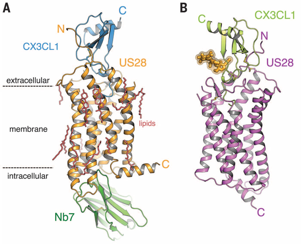
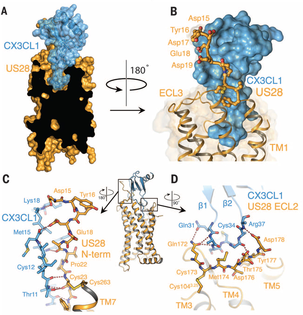
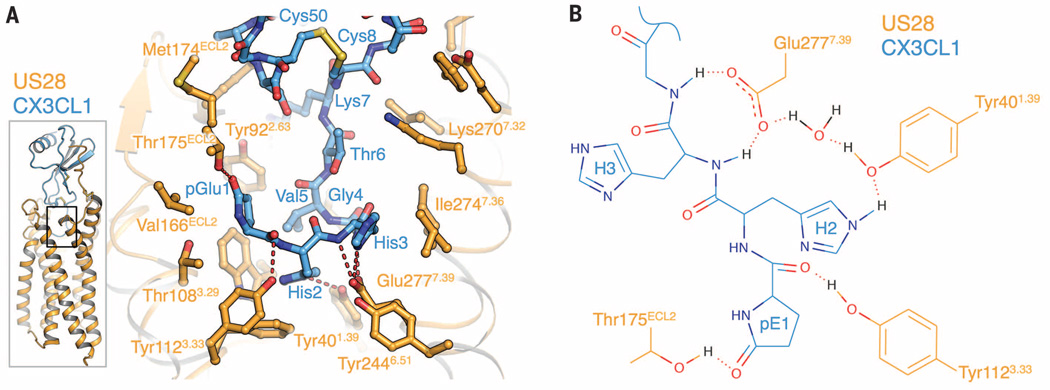
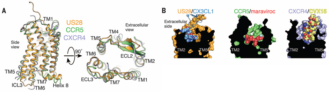
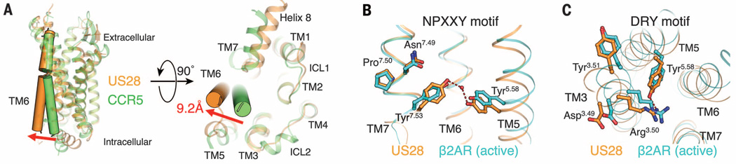
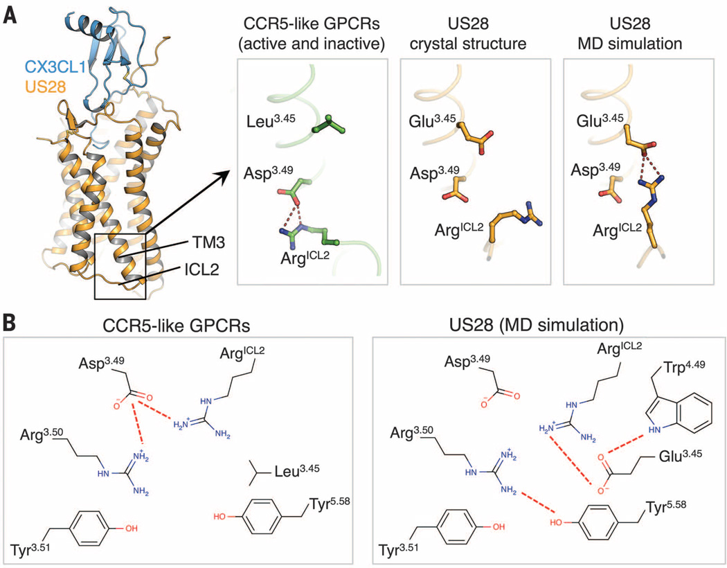
Chemokines are small proteins that function as immune modulators through activation of chemokine G protein-coupled receptors (GPCRs). Several viruses also encode chemokines and chemokine receptors to subvert the host immune response. How protein ligands activate GPCRs remains unknown. We report the crystal structure at 2.9 angstrom resolution of the human cytomegalovirus GPCR US28 in complex with the chemokine domain of human CX3CL1 (fractalkine). The globular body of CX3CL1 is perched on top of the US28 extracellular vestibule, whereas its amino terminus projects into the central core of US28. The transmembrane helices of US28 adopt an active-state-like conformation. Atomic-level simulations suggest that the agonist-independent activity of US28 may be due to an amino acid network evolved in the viral GPCR to destabilize the receptor's inactive state.


Figures

Fig. 1
Fig. 1. Structure of US28 in complex with CX3CL1
(A) Ternary complex of CX3CL1 (blue), US28 (orange), and nanobody (green) at 2.9 Å. (B) Binary complex of US28 (magenta) bound to CX3CL1 (light green). Asn-linked glycans are shown in yellow. C, C terminus; N, N terminus.
Fig. 2
Fig. 2. Interaction of the US28 N terminus with CX3CL1 (site 1)
(A) Cutaway surface representation of CX3CL1 (blue) bound to US28 (orange). (B) The N-terminal region of US28 forms a large contact surface with CX3CL1. Side chains of US28 interacting with CX3CL1 are shown as sticks. (C) Amino acid interactions between CX3CL1 and US28 at chemokine binding site 1. The entire US28-CX3CL1 complex is shown for reference with the nanobody removed for clarity. (D) Amino acid interactions between US28 ECL2 and the β1–β2 loop of fractalkine.
Fig. 3
Fig. 3. Interaction of the CX3CL1 N terminus with the US28 ligand binding pocket (site 2)
(A) Side chain contacts between CX3CL1 site 2 region (blue) and US28 (orange). (B) Two-dimensional plot of side-chain contacts between the CX3CL1 N-terminal hook and US28.
Fig. 4
Fig. 4. Comparison of US28-CX3CL1 with chemokine receptor small-molecule and peptide complexes
(A) Overall superposition of the US28 (orange), CCR5 (green; PDB ID: 4MBS), and CXCR4 (purple; PDB ID: 3ODU) TM helices from the side (left) and as viewed from extracellular space (right). (B) Surface cutaway side views comparing ligand binding modes for US28-CX3CL1 (orange-blue), CCR5-maraviroc (green-red; PDB ID: 4MBS), and CXCR4-CVX15 (purple-yellow; PDB ID: 3OE0).
Fig. 5
Fig. 5. Active-state hallmarks of US28 bound to CX3CL1
(A) Comparison between the TM6 conformations of US28 (orange) and CCR5 (green; PDB ID: 4MBS). (B) The NPXXY motif of US28 (orange) forms side-chain contacts resembling the active-state conformation of β2AR (light blue; PDB ID: 3SN6). (C) The DRY motif of US28 (orange) forms side-chain contacts resembling the active-state conformation of β2AR (light blue).
Fig. 6
Fig. 6. Structural basis for the constitutive activity of US28
(A) Conformations of ArgICL2 in CCR5 (green; PDB ID: 4MBS; left), the US28 crystal structure (orange; center) and the US28 MD simulations (orange; right). (B) Schematic diagram of the network of side-chain interactions surrounding the DRY motif in CCR5 (left panel) and US28 (MD simulation; right).

Comment in

References

    1. Katritch V, Cherezov V, Stevens RC. Annu. Rev. Pharmacol. Toxicol. 2013;53:531–556. - PMC - PubMed
    1. Tan Q, et al. Science. 2013;341:1387–1390. - PMC - PubMed
    1. Wu B, et al. Science. 2010;330:1066–1071. - PMC - PubMed
    1. Rosenbaum DM, Rasmussen SG, Kobilka BK. Nature. 2009;459:356–363. - PMC - PubMed
    1. Charo IF, Ransohoff RM. Engl. N. J. Med. 2006;354:610–621. - PubMed

Publication types