

Differences in functional brain connectivity alterations associated with cerebral amyloid deposition in amnestic mild cognitive impairment
- PMID: 25745400
- PMCID: PMC4333804
- DOI: 10.3389/fnagi.2015.00015
Differences in functional brain connectivity alterations associated with cerebral amyloid deposition in amnestic mild cognitive impairment
Abstract
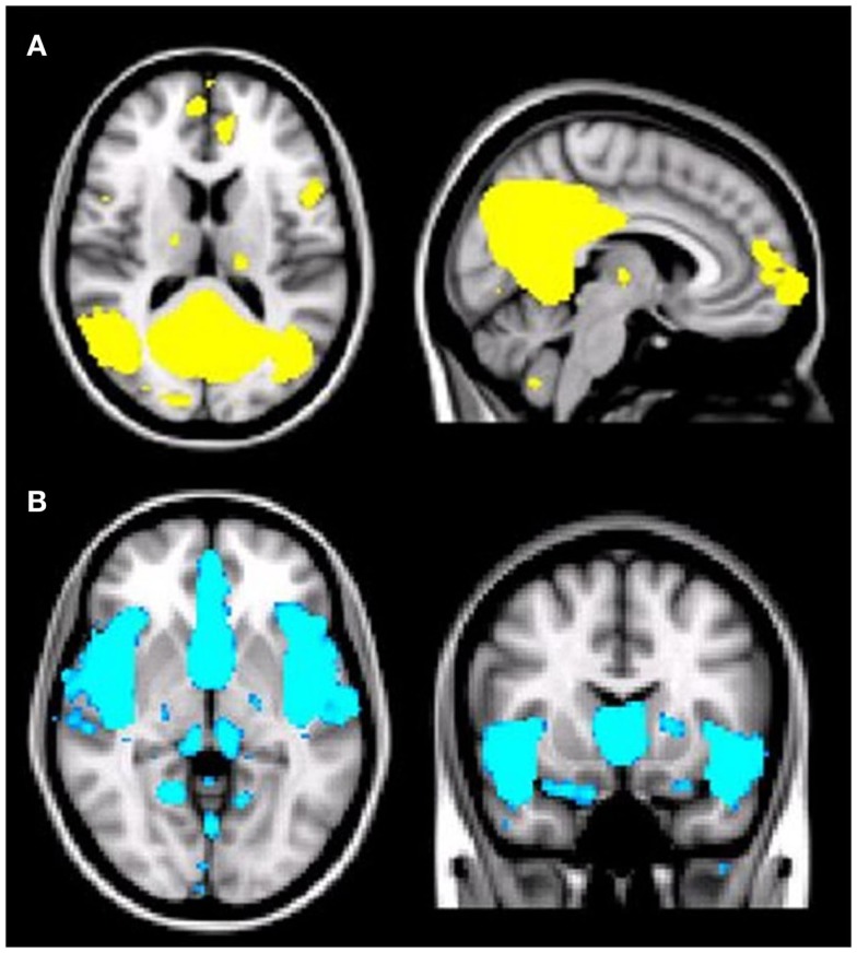
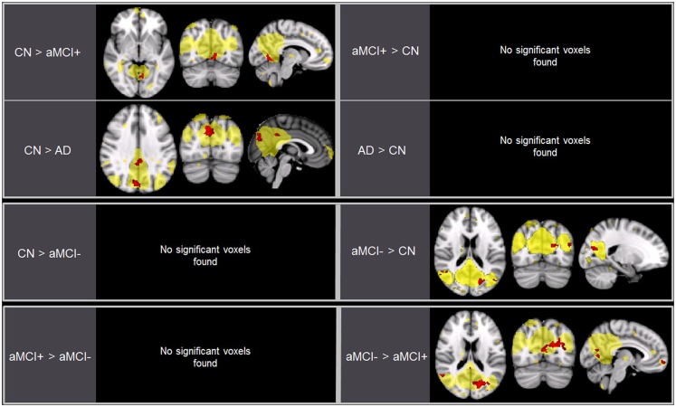
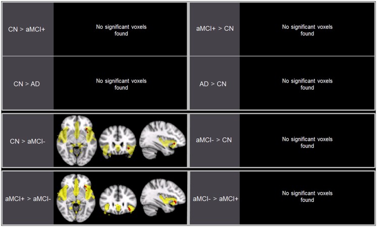
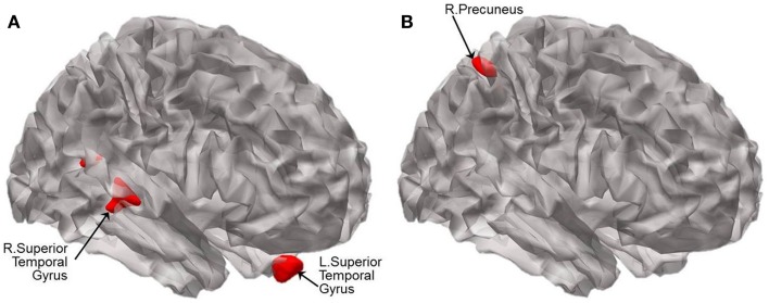
Despite potential implications for the early detection of impending Alzheimer's disease (AD), very little is known about the differences of large-scale brain networks between amnestic mild cognitive impairment (aMCI) with high cerebral amyloid-beta protein (Aβ) deposition (i.e., aMCI+) and aMCI with no or very little Aβ deposition (i.e., aMCI-). We first aimed to extend the current literature on altering intrinsic functional connectivity (FC) of the default mode network (DMN) and salience network (SN) from cognitively normal (CN) to AD dementia. Second, we further examined the differences of the DMN and the SN between aMCI-, aMCI+, and CN. Forty-three older adult (12 CN, 10 aMCI+, 10 aMCI-, and 11 AD dementia) subjects were included. All participants received comprehensive clinical and neuropsychological assessment, resting-state functional magnetic resonance imaging, structural MRI, and Pittsburgh compound-B-PET scans. FC data were preprocessed using multivariate exploratory linear optimized decomposition into independent components of FMRIB's Software Library. Group comparisons were carried out using the "dual-regression" approach. In addition, to verify presence of gray matter volume changes with intrinsic functional network alterations, voxel-based morphometry was performed on the acquired T1-weighted data. As expected, AD dementia participants exhibited decreased FC in the DMN compared to CN (particularly in the precuneus and cingulate gyrus). The degree of alteration in the DMN in aMCI+ compared to CN was intermediate to that of AD. In contrast, aMCI- exhibited increased FC in the DMN compared to CN (primarily in the precuneus) as well as aMCI+. In terms of the SN, aMCI- exhibited decreased FC compared to both CN and aMCI+ particularly in the inferior frontal gyrus. FC within the SN in aMCI+ and AD did not differ from CN. Compared to CN, aMCI- showed atrophy in bilateral superior temporal gyri whereas aMCI+ showed atrophy in right precuneus. The results indicate that despite the similarity in cross-sectional cognitive features, aMCI- has quite different functional brain connectivity compared to aMCI+.
Keywords: amnestic mild cognitive impairment; amyloid-beta deposition; brain functional connectivity; default mode network; salience network.
Figures
Similar articles
-
Interhemispheric functional connectivity for Alzheimer's disease and amnestic mild cognitive impairment based on the triple network model.J Zhejiang Univ Sci B. 2018 Dec.;19(12):924-934. doi: 10.1631/jzus.B1800381. J Zhejiang Univ Sci B. 2018. PMID: 30507076 Free PMC article.
-
Abnormal insula functional network is associated with episodic memory decline in amnestic mild cognitive impairment.Neuroimage. 2012 Oct 15;63(1):320-7. doi: 10.1016/j.neuroimage.2012.06.062. Epub 2012 Jul 6. Neuroimage. 2012. PMID: 22776459 Free PMC article.
-
Distinct Disruptive Patterns of Default Mode Subnetwork Connectivity Across the Spectrum of Preclinical Alzheimer's Disease.Front Aging Neurosci. 2019 Nov 13;11:307. doi: 10.3389/fnagi.2019.00307. eCollection 2019. Front Aging Neurosci. 2019. PMID: 31798440 Free PMC article.
-
Functional MRI-Specific Alterations in Salience Network in Mild Cognitive Impairment: An ALE Meta-Analysis.Front Aging Neurosci. 2021 Jul 26;13:695210. doi: 10.3389/fnagi.2021.695210. eCollection 2021. Front Aging Neurosci. 2021. PMID: 34381352 Free PMC article.
-
Three Large-Scale Functional Brain Networks from Resting-State Functional MRI in Subjects with Different Levels of Cognitive Impairment.Psychiatry Investig. 2016 Jan;13(1):1-7. doi: 10.4306/pi.2016.13.1.1. Epub 2015 Nov 20. Psychiatry Investig. 2016. PMID: 26766941 Free PMC article. Review.
Cited by
-
Resting-state default mode network connectivity in young individuals with Down syndrome.Brain Behav. 2021 Jan;11(1):e01905. doi: 10.1002/brb3.1905. Epub 2020 Nov 12. Brain Behav. 2021. PMID: 33179859 Free PMC article.
-
Inflammatory insult during pregnancy accelerates age-related behavioral and neurobiochemical changes in CD-1 mice.Age (Dordr). 2016 Jun;38(3):59. doi: 10.1007/s11357-016-9920-3. Epub 2016 May 19. Age (Dordr). 2016. PMID: 27194408 Free PMC article.
-
Differential effects of Down's syndrome and Alzheimer's neuropathology on default mode connectivity.Hum Brain Mapp. 2019 Oct 15;40(15):4551-4563. doi: 10.1002/hbm.24720. Epub 2019 Jul 26. Hum Brain Mapp. 2019. PMID: 31350817 Free PMC article.
-
Transcranial direct current stimulation and neuronal functional connectivity in MCI: role of individual factors associated to AD.Front Psychiatry. 2024 Aug 19;15:1428535. doi: 10.3389/fpsyt.2024.1428535. eCollection 2024. Front Psychiatry. 2024. PMID: 39224475 Free PMC article.
-
A feasibility trial of gamma sensory flicker for patients with prodromal Alzheimer's disease.Alzheimers Dement (N Y). 2021 May 13;7(1):e12178. doi: 10.1002/trc2.12178. eCollection 2021. Alzheimers Dement (N Y). 2021. PMID: 34027028 Free PMC article.
References
LinkOut - more resources
Full Text Sources
Other Literature Sources
Research Materials