Review
. 2015 Feb 19:6:28.
doi: 10.3389/fneur.2015.00028. eCollection 2015.

The importance of structural anisotropy in computational models of traumatic brain injury

Affiliations
Review

The importance of structural anisotropy in computational models of traumatic brain injury

Rika W Carlsen et al. Front Neurol. .

Abstract

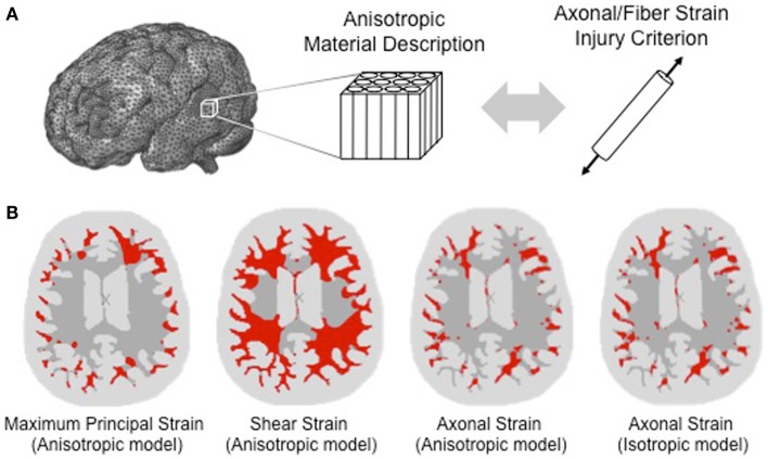
Understanding the mechanisms of injury might prove useful in assisting the development of methods for the management and mitigation of traumatic brain injury (TBI). Computational head models can provide valuable insight into the multi-length-scale complexity associated with the primary nature of diffuse axonal injury. It involves understanding how the trauma to the head (at the centimeter length scale) translates to the white-matter tissue (at the millimeter length scale), and even further down to the axonal-length scale, where physical injury to axons (e.g., axon separation) may occur. However, to accurately represent the development of TBI, the biofidelity of these computational models is of utmost importance. There has been a focused effort to improve the biofidelity of computational models by including more sophisticated material definitions and implementing physiologically relevant measures of injury. This paper summarizes recent computational studies that have incorporated structural anisotropy in both the material definition of the white matter and the injury criterion as a means to improve the predictive capabilities of computational models for TBI. We discuss the role of structural anisotropy on both the mechanical response of the brain tissue and on the development of injury. We also outline future directions in the computational modeling of TBI.

Keywords: axonal strain; computational model; diffuse axonal injury; injury criterion; traumatic brain injury.


Figures

Figure 1
Figure 1
Accounting for structural anisotropy in a computational head model. (A) Structural anisotropy can be incorporated into computational head models either through an anisotropic material description for the white matter or through an injury criterion that accounts for the fiber orientation, such as stretch along the fiber direction (Brain model image courtesy of Dr. Reuben Kraft). (B) Predicted regions of axonal injury for an axial slice of brain are highlighted in red using a finite element analysis of a head impact that resulted in concussive injury. Starting from the left, the first three images show the difference in the predicted location of injury for different injury criteria. Injury thresholds of 31 and 25% were adopted for the maximum principal strain and shear strain, respectively (6), and a strain threshold of 18% was used for the axonal strain (7). With a structurally based injury criterion such as axonal strain, the locations of injury do not differ significantly between anisotropic and isotropic material definitions of the white matter as shown in the third and fourth image of the same axial slice [adapted from Ref. (8) with permission].

References

    1. Smith DH, Meaney DF. Axonal damage in traumatic brain injury. Neuroscientist (2000) 6(6):483–9510.1177/107385840000600611 - DOI
    1. Ji S, Zhao W, Ford J, Beckwith J. Group-wise evaluation and comparison of white matter fiber strain and maximum principal strain in sports-related concussion. J Neurotrauma (2014).10.1089/neu.2013.3268 - DOI - PMC - PubMed
    1. Bain AC, Meaney DF. Tissue-level thresholds for axonal damage in an experimental model of central nervous system white matter injury. J Biomech Eng (2000) 122(6):615–22.10.1115/1.1324667 - DOI - PubMed
    1. Pan Y, Sullivan D, Shreiber D, Pelegri A. Finite element modeling of CNS white matter kinematics: use of a 3D RVE to determine material properties. Front Bioeng Biotechnol (2013) 1(19):19.10.3389/fbioe.2013.00019 - DOI - PMC - PubMed
    1. Le Bihan D, Mangin J-F, Poupon C, Clark CA, Pappata S, Molko N, et al. Diffusion tensor imaging: concepts and applications. J Magn Reson Imaging (2001) 13(4):534–46.10.1002/jmri.1076 - DOI - PubMed

LinkOut - more resources