. 2015 Dec 3;34(49):5997-6006.
doi: 10.1038/onc.2015.48. Epub 2015 Mar 9.

Expression of miR-200c in claudin-low breast cancer alters stem cell functionality, enhances chemosensitivity and reduces metastatic potential

Affiliations

Expression of miR-200c in claudin-low breast cancer alters stem cell functionality, enhances chemosensitivity and reduces metastatic potential

J Knezevic et al. Oncogene. .

Abstract

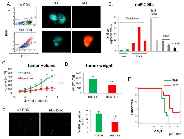
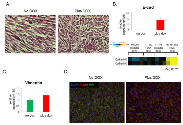
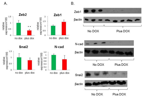
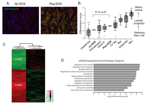
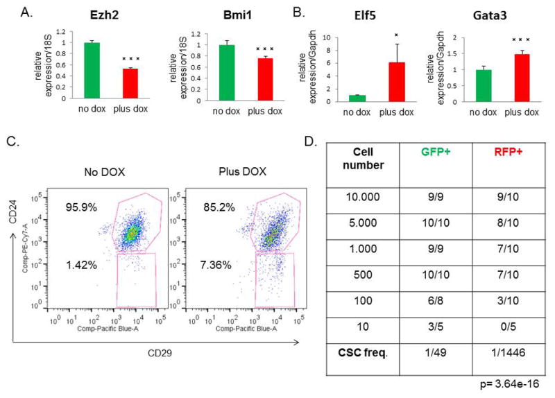
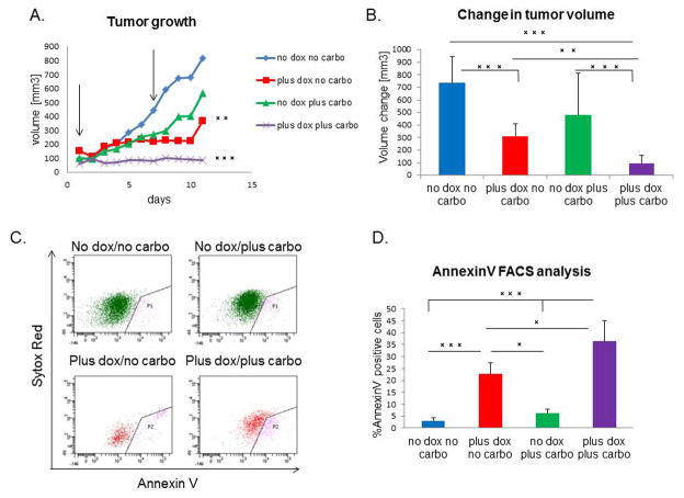
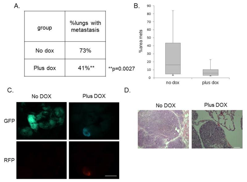
Claudin-low tumors are a highly aggressive breast cancer subtype with no targeted treatments and a clinically documented resistance to chemotherapy. They are significantly enriched in cancer stem cells (CSCs), which makes claudin-low tumor models particularly attractive for studying CSC behavior and developing novel approaches to minimize CSC therapy resistance. One proposed mechanism by which CSCs arise is via an epithelial-mesenchymal transition (EMT), and reversal of this process may provide a potential therapeutic approach for increasing tumor chemosensitivity. Therefore, we investigated the role of known EMT regulators, miR-200 family of microRNAs in controlling the epithelial state, stem-like properties and therapeutic response in an in vivo primary, syngeneic p53(null) claudin-low tumor model that is normally deficient in miR-200 expression. Using an inducible lentiviral approach, we expressed the miR-200c cluster in this model and found that it changed the epithelial state, and consequently, impeded CSC behavior in these mesenchymal tumors. Moreover, these state changes were accompanied by a decrease in proliferation and an increase in the differentiation status. miR-200c expression also forced a significant reorganization of tumor architecture, affecting important cellular processes involved in cell-cell contact, cell adhesion and motility. Accordingly, induced miR200c expression significantly enhanced the chemosensitivity and decreased the metastatic potential of this p53(null) claudin-low tumor model. Collectively, our data suggest that miR-200c expression in claudin-low tumors offers a potential therapeutic application to disrupt the EMT program on multiple fronts in this mesenchymal tumor subtype, by altering tumor growth, chemosensitivity and metastatic potential in vivo.


Conflict of interest statement

Authors declare no conflict of interest.

Figures

Figure 1
Figure 1. miR-200c decelerates tumor growth
(A) RFP was induced in tumors after DOX administration, as seen by FACS analysis and fluorescent microscopy. Scale bar represents 5mm. (B) miR-200c was induced to a physiological level, compared with its levels in different subtypes of p53null tumors (20), as shown by qPCR. (C) Tumor growth, as measured by the daily increase in tumor volume, was significantly lower in the DOX-treated group compared with untreated controls. (D) Tumor weight at the end of the treatment showed that the miR-200c-expressing tumors were much smaller than untreated controls. (E) The Ki67 proliferation marker was significantly decreased in miR-200c-induced tumors, revealing that the tumor growth deceleration was due, in part, to a lower proliferation rate. Scale bar represents 50μm. (F) Tumor latency increased with miR-200c upregulation. Equal numbers (5 000) of GFP+ and RFP+ cells were injected into the cleared fat pads of wild-type Balb/c recipient mice, and tumors were palpated every day to establish their latency, while maintaining vehicle or DOX treatment.
Figure 2
Figure 2. miR-200c alters claudin-low tumor morphology and causes MET
(A) Significant morphological changes were observed in tumors with induced miR-200c, as shown by standard H&E staining. Scale bars represents 50μm. (B) qPCR showed that E-cadherin mRNA was highly induced post-DOX. Reverse phase protein array (RPPA) analysis showed a significant upregulation of E-cadherin protein in miR-200c-induced tumors, but not in tumors containing luciferase (FF3). The two bands represent runs with two different validated antibodies, according to the RPPA core. (C) There was no change in mRNA levels of vimentin following DOX administration, as shown by qPCR. (D) Images of immunofluorescent staining show that vimentin protein expression was decreased in cells that were positive for E-cadherin staining. Scale bar is 50μm.
Figure 3
Figure 3. miR-200c upregulation leads to a decrease in protein expression of several mesenchymal markers
(A) Other mesenchymal markers showed either a significant decrease (Zeb2), or no significant change (Zeb1, Snai2, and N-cad) by qPCR. (B) Western blots show a dramatic reduction in Zeb1 and N-cad, as well as Snai2 protein levels in DOX-treated cells. Protein was isolated from primary tumor cells that were sorted for GFP or RFP positivity to isolate untreated and DOX-treated cells, respectively.
Figure 4
Figure 4. miR-200c induction alters the differentiation state of claudin-low tumors
(A) Images of immunofluorescent staining show that luminal and basal markers, specifically K8 and K14, were upregulated after miR-200c induction, suggesting a change in their differentiation profile. Scale bar represents 50μm. (B) Microarray analysis confirmed this enhanced differentiation by revealing a significant increase in the differentiation score of miR-200c-expressing primary tumors compared with our no dox treated tumors. Note that the miR-200c tumors had a differentiation score close to the basal tumors, and no longer fell into the claudin-low category. The classes of murine tumors are as described in detail previously (51), where Myc, Erbb2 and Neu tumors are of luminal subtype clustering, and p53null-basal, class 14 and C3-tag cluster into the basal subtype. (C) Microarray analysis also revealed a significant change in 1457 genes, with 562 being down-regulated, and 895 being up-regulated after treatment with DOX. The microarray analysis was performed on RNA extracted from GFP+ or RFP+ cells sorted from primary tumors, and the heatmap depicts genes with a 0% FDR. The analysis was consistent with a reversal of EMT, as many of the genes found to be downregulated are associated with the mesenchymal state, such as FoxC2, and also confirmed the induction of several keratin markers. (D) The graph indicates the top 12 most significantly enriched gene ontology terms in the 895 miR-200c upregulated gene set. Importantly, most of them were related to terms describing cell-to-cell adhesion and motility.
Figure 5
Figure 5. Stem cell functionality is compromised after miR-200c induction
(A) There was a significant decrease in the stem cell markers Ezh2 and Bmi1 by qPCR post-DOX treatment. (B) Conversely, the mRNA levels of the luminal progenitor marker Elf5 and the mature luminal marker Gata3 were increased in RFP+ cells, compared with GFP+ cells. (C) A representative FACS profile for CD24+/CD29+ cells shows a change in the overall profile of miR-200c tumors, with the number of double-positive cells decreased relative to untreated controls. (D) Table depicts the results from the limiting dilution transplantation assay. As decreasing numbers of either GFP+ or RFP+ sorted cells were transplanted back into the recipient mouse, fewer tumors arose in each transplantation group. The RFP+ miR-200c-expressing cells showed a significantly reduced CSC frequency, as shown in the bottom row, confirming that miR-200c leads to a reduction in stem cell functionality.
Figure 6
Figure 6. miR-200c sensitizes tumor cells to chemotherapy
(A) DOX and carboplatin combination treatment caused tumor stasis, as seen by a significant difference in the growth rate of combination group, compared to all other treatment groups. Arrows point to the time of carboplatin injection. (B) There is a significant decrease in the average change in volume after treatment (n=20 for carboplatin+DOX group, n=15 for DOX only, n=8 for vehicle and carboplatin alone groups) (C–D) AnnexinV FACS analysis shows a significant increase in the number of cells undergoing early stages of apoptosis in the combination treatment group, as compared to all other groups. Sytox red staining represents dead cells, therefore cells positive only for AnnexinV were considered currently undergoing apoptosis. Analysis included only GFP+ or RFP+ cells, therefore AnnexinV positive cells are represented only through these gates, and quantified to the left.
Figure 7
Figure 7. Metastatic potential is severely impaired following miR-200c induction
(A) Table shows that the percentage of tumor-bearing lungs was significantly reduced in the miR-200c-induced group. Data was analyzed using a Chi-squared test for two populations. (B) The average areas of metastasis were also lower in the miR-200c-induced group compared with the vehicle-treated group. (C) Fluorescent images of GFP and RFP expression shows metastasis present in the lung of both groups. Scale bar equals 5mm. (D) Representative H&E images of lungs are shown following 8 weeks of DOX treatment after tail-vein injection of 30 000 primary GFP+ cells. Scale bar equals 100μm.

References

    1. Prat A, Parker JS, Karginova O, Fan C, Livasy C, Herschkowitz JI, et al. Phenotypic and molecular characterization of the claudin-low intrinsic subtype of breast cancer. Breast Cancer Res. 2010 Jan;12(5):R68. - PMC - PubMed
    1. Thiery JP. Epithelial-mesenchymal transitions in tumour progression. Nat Rev Cancer. 2002;2:442–54. - PubMed
    1. Thiery JP, Acloque H, Huang RYJ, Nieto MA. Epithelial-Mesenchymal Transitions in Development and Disease. Cell. 2009;139:871–90. - PubMed
    1. Hennessy BT, Gonzalez-Angulo A-M, Stemke-Hale K, Gilcrease MZ, Krishnamurthy S, Lee J-S, et al. Characterization of a naturally occurring breast cancer subset enriched in epithelial-to-mesenchymal transition and stem cell characteristics. Cancer Res. 2009;69:4116–24. - PMC - PubMed
    1. Taube JH, Herschkowitz JI, Komurov K, Zhou AY, Gupta S, Yang J, et al. Core epithelial-to-mesenchymal transition interactome gene-expression signature is associated with claudin-low and metaplastic breast cancer subtypes. Proc Natl Acad Sci U S A. 2010;107:15449–54. - PMC - PubMed

Publication types

MeSH terms