. 2015 Mar 6;10(3):e0119257.
doi: 10.1371/journal.pone.0119257. eCollection 2015.

Evaluation of iron oxide nanoparticle micelles for magnetic particle imaging (MPI) of thrombosis

Affiliations

Evaluation of iron oxide nanoparticle micelles for magnetic particle imaging (MPI) of thrombosis

Lucas W E Starmans et al. PLoS One. .

Abstract

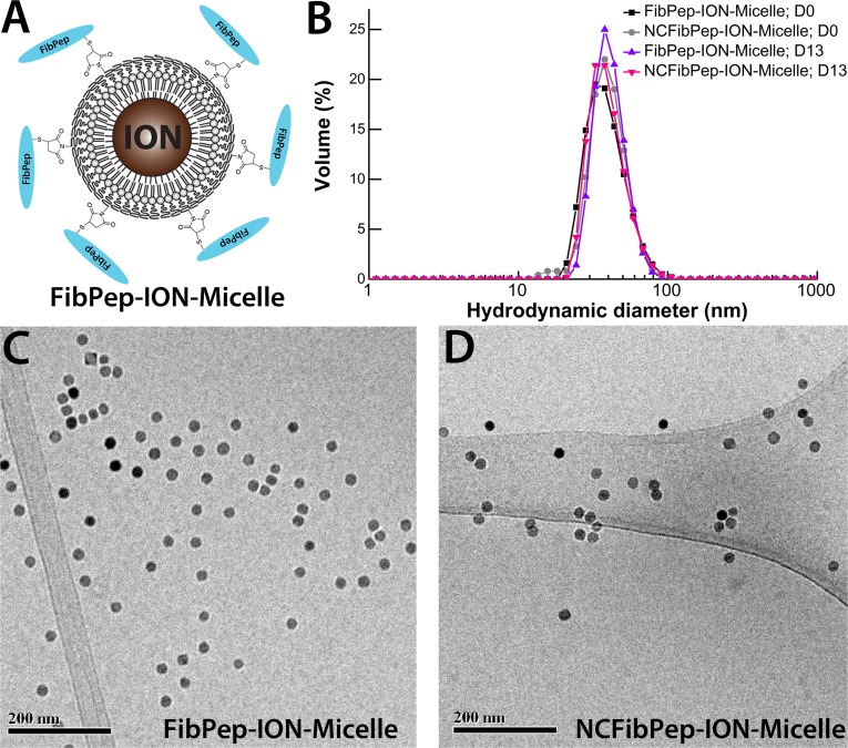
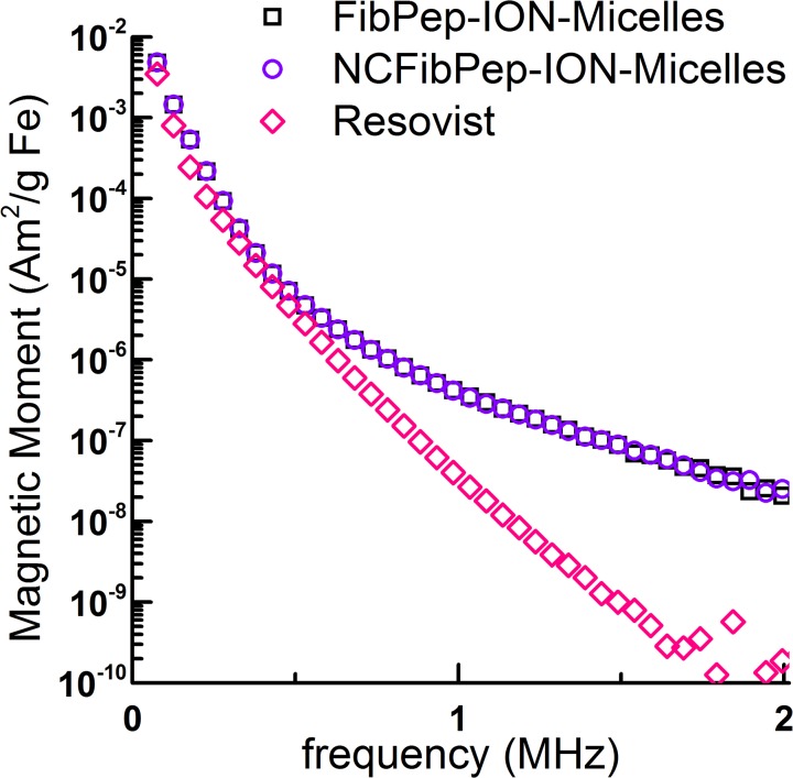
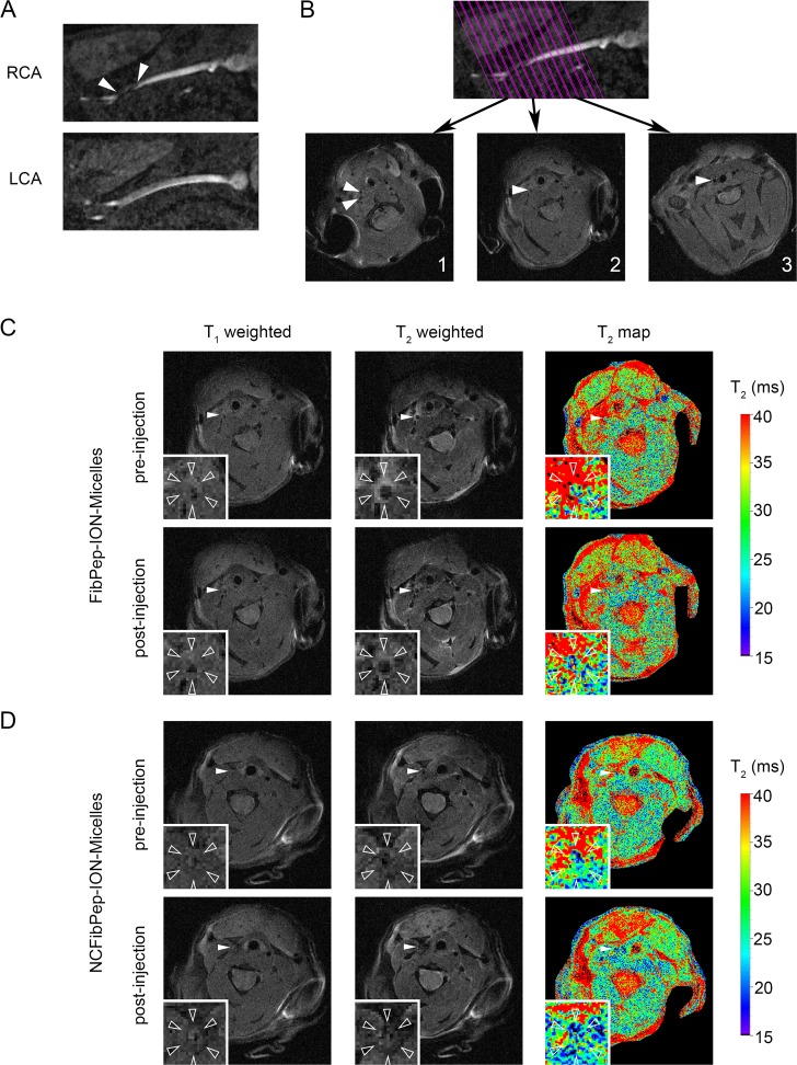
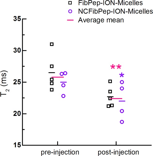
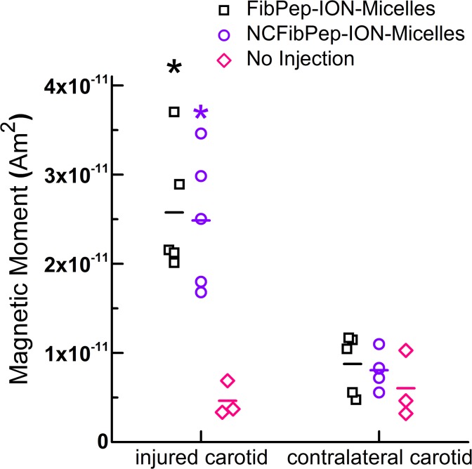
Magnetic particle imaging (MPI) is an emerging medical imaging modality that directly visualizes magnetic particles in a hot-spot like fashion. We recently developed an iron oxide nanoparticle-micelle (ION-Micelle) platform that allows highly sensitive MPI. The goal of this study was to assess the potential of the ION-Micelles for MPI-based detection of thrombi. To this aim, an in vivo carotid artery thrombosis mouse model was employed and ex vivo magnetic particle spectrometer (MPS) measurements of the carotid arteries were performed. In addition, we studied the effect of functionalization of the ION-Micelle nanoplatform with fibrin-binding peptides (FibPeps) with respect to nanoparticle thrombus uptake and hence thrombus detection. In vivo quantitative MR imaging pre- and post-ION-Micelle injection was performed as reference for visualization of ION-micelle uptake. ION-Micelles significantly decreased T2 values in the thrombi with respect to pre-injection T2 values (p < 0.01) and significantly increased ex vivo MPS thrombus signal with respect to the noninjured, contralateral carotid (p < 0.01). Functionalization of the ION-Micelles with the FibPep peptides did not result in an increased MPS thrombus signal with respect to the non-fibrin binding ION-Micelles. The lack of a significant increased thrombus uptake for the FibPep-ION-Micelles indicates that (non-fibrin-specific) entrapment of nanoparticles in the mesh-like thrombi is the key contributor to thrombus nanoparticle uptake. Therefore, (nontargeted) ION-Micelles might be of value for noninvasive MPI-based diagnosis, characterization and treatment monitoring of thrombosis.


Conflict of interest statement

Competing Interests: Authors Erica Aussems-Custers and Holger Grüll are employed by Philips Research. This does not alter the authors' adherence to PLOS ONE policies on sharing data and materials.

Figures

Fig 1
Fig 1. FibPep-ION-Micelle nanoplatform.
(A) Schematic representation of the FibPep-ION-Micelle nanoplatform. Reproduced from Starmans and coworkers [10]. (B) Volume-weighted size-distribution profiles of the FibPep-ION-Micelles and NCFibPep-ION-Micelles at the day of synthesis (D0) and at the final day of the in vivo experiments (13 days post synthesis, D13). (C, D) Representative cryo-TEM images of (C) FibPep-ION-Micelles and (D) NCFibPep-ION-Micelles.
Fig 2
Fig 2. MPS of FibPep-ION-Micelles, NCFibPep-ION-Micelles and Resovist.
Data is plotted as magnetic moment (normalized for iron content) versus frequency. Note that the curves of FibPep-ION-Micelles and NCFibPep-ION-Micelles largely overlap.
Fig 3
Fig 3. In vivo MRI.
(A) Images of right and left carotid arteries (RCA & LCA) reconstructed from the 3D time of flight (TOF) image. The thrombus can be observed in the RCA just proximal of the bifurcation (arrowheads). Presence of blood flow in the thrombosed carotid was confirmed for all animals. (B) On the TOF image 13 parallel slices were planned perpendicular to the RCA. T1-weighted images from three slices are shown: (1) distal from the bifurcation, (2) in the thrombus and (3) proximal to the thrombus (arrowheads: RCA). (C, D) Pre- and post-injection T1- and T2-weighted images and T2 maps of (C) FibPep-ION-Micelles and (D) NCFibPep-ION-Micelles (arrowheads: RCA). The insets show magnifications of the RCA (open arrowheads: outer vessel wall).
Fig 4
Fig 4. Quantitative analysis in vivo MRI scans.
In vivo T2 of the thrombus pre- and post-injection with FibPep-ION-Micelles (n = 5) and NCFibPep-ION-Micelles (n = 4). The horizontal lines represent the group means and the average mean for the two groups combined (n = 9). Mean T2 values were significantly decreased post-injection compared to pre-injection for the NCFibPep-ION-Micelles group (p < 0.05; marked with *) and combined group (p < 0.01; marked with **).
Fig 5
Fig 5. Ex vivo MPS of carotid arteries.
MPS measurements of excised injured and contralateral, noninjured carotid arteries following injection with FibPep-ION-Micelles (n = 5) and NCFibPep-ION-Micelles (n = 5) or without injection (n = 3). Data is expressed as magnetic moment of the third harmonic (76 kHz), horizontal lines represent group means. * P < 0.01 versus contralateral carotid (all three groups) and injured carotid of mice which had not received an injection of ION-Micelles.
Fig 6
Fig 6. Ex vivo confocal microscopy of carotid arteries.
Representative autofluorescence confocal microscopy images of transversal histological sections of the (A) injured carotid and (B) noninjured, contralateral carotid artery.

Similar articles

Cited by

References

    1. Lu Y, Lorenzoni A, Fox JJ, Rademaker J, Els N Vander, Grewal RK, et al. Noncontrast perfusion single-photon emission CT/CT scanning. Chest. 2014;145: 1079–1088. 10.1378/chest.13-2090 - DOI - PubMed
    1. Omran H, Jung W, Rabahieh R, Wirtz P, Becher H, Illien S, et al. Imaging of thrombi and assessment of left atrial appendage function: a prospective study comparing transthoracic and transoesophageal echocardiography. Heart. 1999;81: 192–198. - PMC - PubMed
    1. Srichai MB, Junor C, Rodriguez LL, Stillman AE, Grimm RA, Lieber ML, et al. Clinical, imaging, and pathological characteristics of left ventricular thrombus: A comparison of contrast-enhanced magnetic resonance imaging, transthoracic echocardiography, and transesophageal echocardiography with surgical or pathological validation. Am Heart J. 2006;152: 75–84. 10.1016/j.ahj.2005.08.021 - DOI - PubMed
    1. Litmanovich D, Bankier AA, Cantin L, Raptopoulos V, Boiselle PM. CT and MRI in diseases of the aorta. Am J Roentgenol. 2009;193: 928–940. 10.2214/AJR.08.2166 - DOI - PubMed
    1. Markel A, Weich Y, Gaitini D. Doppler ultrasound in the diagnosis of venous thrombosis. Angiology. 1995;46: 65–73. 10.1177/000331979504600109 - DOI - PubMed

Publication types