

Increased Vesicular Monoamine Transporter 2 (VMAT2; Slc18a2) Protects against Methamphetamine Toxicity
- PMID: 25746685
- PMCID: PMC4489556
- DOI: 10.1021/acschemneuro.5b00010
Increased Vesicular Monoamine Transporter 2 (VMAT2; Slc18a2) Protects against Methamphetamine Toxicity
Abstract
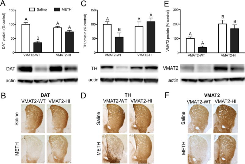
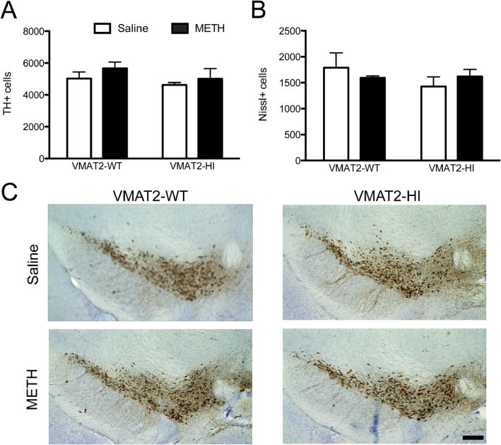
The psychostimulant methamphetamine (METH) is highly addictive and neurotoxic to dopamine terminals. METH toxicity has been suggested to be due to the release and accumulation of dopamine in the cytosol of these terminals. The vesicular monoamine transporter 2 (VMAT2; SLC18A2) is a critical mediator of dopamine handling. Mice overexpressing VMAT2 (VMAT2-HI) have an increased vesicular capacity to store dopamine, thus augmenting striatal dopamine levels and dopamine release in the striatum. Based on the altered compartmentalization of intracellular dopamine in the VMAT2-HI mice, we assessed whether enhanced vesicular function was capable of reducing METH-induced damage to the striatal dopamine system. While wildtype mice show significant losses in striatal levels of the dopamine transporter (65% loss) and tyrosine hydroxylase (46% loss) following a 4 × 10 mg/kg METH dosing regimen, VMAT2-HI mice were protected from this damage. VMAT2-HI mice were also spared from the inflammatory response that follows METH treatment, showing an increase in astroglial markers that was approximately one-third of that of wildtype animals (117% vs 36% increase in GFAP, wildtype vs VMAT2-HI). Further analysis also showed that elevated VMAT2 level does not alter the ability of METH to increase core body temperature, a mechanism integral to the toxicity of the drug. Finally, the VMAT2-HI mice showed no difference from wildtype littermates on both METH-induced conditioned place preference and in METH-induced locomotor activity (1 mg/kg METH). These results demonstrate that elevated VMAT2 protects against METH toxicity without enhancing the rewarding effects of the drug. Since the VMAT2-HI mice are protected from METH despite higher basal dopamine levels, this study suggests that METH toxicity depends more on the proper compartmentalization of synaptic dopamine than on the absolute amount of dopamine in the brain.
Keywords: Methamphetamine; VMAT2; dopamine; inflammation; neurodegeneration; vesicle.
Conflict of interest statement
The authors declare no competing financial interest.
Figures


References
-
- Ricaurte GA, Guillery RW, Seiden LS, Schuster CR, Moore RY. Dopamine nerve terminal degeneration produced by high doses of methylamphetamine in the rat brain. Brain Res. 1982;235:93–103. - PubMed
-
- LaVoie MJ, Card JP, Hastings TG. Microglial activation precedes dopamine terminal pathology in methamphetamine-induced neurotoxicity. Exp Neurol. 2004;187:47–57. - PubMed
-
- Kuhn DM, Francescutti-Verbeem DM, Thomas DM. Dopamine quinones activate microglia and induce a neurotoxic gene expression profile: relationship to methamphetamine-induced nerve ending damage. Ann N Y Acad Sci. 2006;1074:31–41. - PubMed
-
- Ricaurte GA, Seiden LS, Schuster CR. Further evidence that amphetamines produce long-lasting dopamine neurochemical deficits by destroying dopamine nerve fibers. Brain Res. 1984;303:359–364. - PubMed
Publication types
MeSH terms
Substances
Grants and funding
- T32ES012870/ES/NIEHS NIH HHS/United States
- F31DA037652/DA/NIDA NIH HHS/United States
- P30ES019776/ES/NIEHS NIH HHS/United States
- T32 ES012870/ES/NIEHS NIH HHS/United States
- 210296/Canadian Institutes of Health Research/Canada
- P50NS071669/NS/NINDS NIH HHS/United States
- F31NS084739/NS/NINDS NIH HHS/United States
- F31 NS084739/NS/NINDS NIH HHS/United States
- ES023839/ES/NIEHS NIH HHS/United States
- R01 ES023839/ES/NIEHS NIH HHS/United States
- F31 DA037652/DA/NIDA NIH HHS/United States
- P30 ES019776/ES/NIEHS NIH HHS/United States
- P50 NS071669/NS/NINDS NIH HHS/United States
LinkOut - more resources
Full Text Sources
Other Literature Sources
Medical
Miscellaneous
