. 2015 May;1853(5):1219-28.
doi: 10.1016/j.bbamcr.2015.02.020. Epub 2015 Mar 4.

IL4 receptor α mediates enhanced glucose and glutamine metabolism to support breast cancer growth

Affiliations

IL4 receptor α mediates enhanced glucose and glutamine metabolism to support breast cancer growth

Katherine T Venmar et al. Biochim Biophys Acta. 2015 May.

Abstract

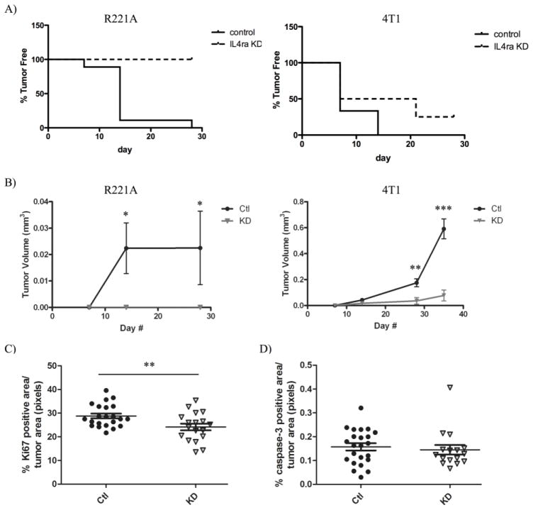
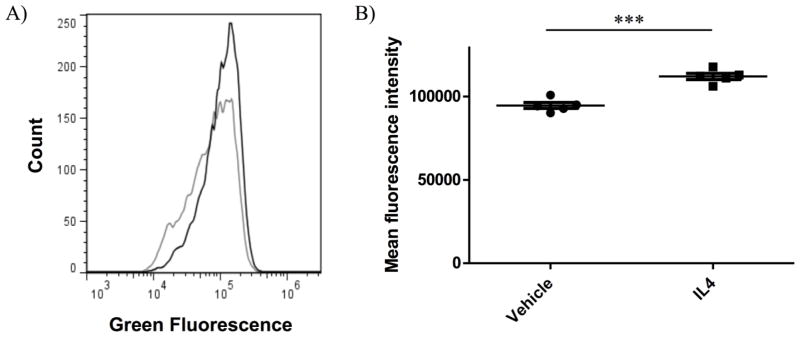
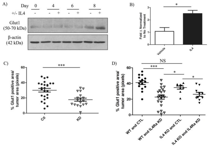
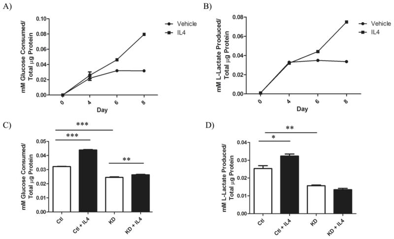
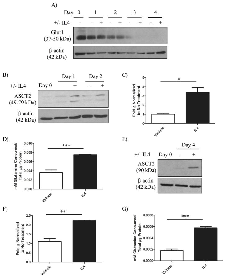
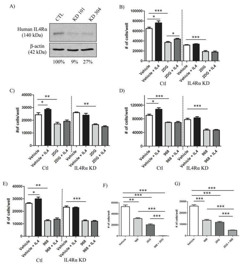
The type II interleukin-4 receptor (IL4R) is expressed in human breast cancer, and in murine models thereof. It is activated by interleukin-4 (IL4), a cytokine produced predominantly by immune cells. Previously, we showed that expression of IL4Rα, a signaling component of IL4R, mediates enhanced metastatic growth through promotion of tumor cell survival and proliferation. In lymphocytes, these processes are supported by increased glucose and glutamine metabolism, and B lymphocyte survival is dependent upon IL4/IL4R-induced glucose metabolism. However, it is unknown whether IL4R-mediated metabolic reprogramming could support tumor growth. Here, we show that IL4Rα expression increases proliferation thus enhancing primary mammary tumor growth. In vitro, IL4-enhanced glucose consumption and lactate production in 4T1 cells was mediated by IL4Rα. Expression of the glucose transporter GLUT1 increased in response to IL4 in vitro, and enhanced GLUT1 expression was associated with the presence of IL4Rα in 4T1 mammary tumors in vivo. Although IL4 treatment did not induce changes in glucose metabolism in MDA-MB-231 human breast cancer cells, it increased expression of the main glutamine transporter, ASCT2, and enhanced glutamine consumption in both MDA-MB-231 and 4T1 cells. Pharmacologic inhibition of glutamine metabolism with compound 968 blocked IL4/IL4Rα-increased cell number in both cell lines. Our results demonstrate that IL4R mediates enhanced glucose and glutamine metabolism in 4T1 cancer cells, and that IL4-induced growth is supported by IL4/IL4R-enhanced glutamine metabolism in both human and murine mammary cancer cells. This highlights IL4Rα as a possible target for effective breast cancer therapy.

Keywords: Cytokine; Glucose; Metabolism; Proliferation; Survival.


Conflict of interest statement

No conflicts of interest to declare.

Figures

Figure 1
Figure 1. IL4Rα promotes the growth of mammary tumors at the primary site
R221a or 4T1 sh-control (Ctl) or IL4Rα knockdown (KD) cells were orthotopically injected into the 4th mammary gland of mice. A) Representation of tumor latency for R221a (left, p < .0001) and 4T1 (right, p = NS) mammary tumors (R221a n = 16; 4T1 n = 14). B) Graphs of R221a (left) and 4T1 (right) mammary tumor volume over time calculated from tumor dimensions (R221a n = 16; 4T1 n = 14). C) Quantification of total Ki67 D) or cleaved caspase-3 positive area per 4T1 IL4Rα KD or sh-control mammary tumor area (n = 11). Murine tumor growth data represented in A&B was repeated with similar results obtained.
Figure 2
Figure 2. Glucose uptake in mammary cancer cells is increased following short-term IL4 exposure
4T1 parental cells were treated with vehicle or 10 ng/mL murine IL4 for 22 hours prior to a 2 hour exposure to 2-NBDG +/− 10 ng/mL IL4, and immediate analysis by flow cytometry. A) Flow cytometry trace of 2-NBDG uptake in 4T1 cells treated with IL4 (black) or vehicle control (grey). B) Quantification of Mean fluorescence intensity by flow cytometry representative of 2-NBDG uptake in 4T1 cells with IL4 or vehicle control.
Figure 3
Figure 3. IL4Rα mediates enhanced expression of GLUT1 in mammary cancer cells
4T1 sh-control clones were treated with vehicle or 20 ng/mL murine IL4 every other day for 8 days. A) Western blot analysis of deglycosylated glucose transporter 1 (GLUT1) upregulation in 4T1 sh-control clones over time. B) Quantification of GLUT1 mRNA expression normalized to murine ribosomal S18 in 4T1 sh-control clones 6 days after initial IL4 treatment. Quantification of IHC staining for GLUT1 in C) 4T1 sh-control (Ctl) and IL4Rα knockdown (KD) orthotopic mammary tumors (n = 8) and D) in metastatic lung tumors originating from sh-control (Ctl) or IL4Rα knockdown (IL4Ra KD) 4T1 cells in wild-type (WT) or IL4 knockout (IL4 KO) mice (n = 3–6). The upregulation of GLUT1 expression in response to IL4 was reproducible in three independent experiments, and IHC analysis of GLUT1 in 4T1 mammary tumors in two separate experiments.
Figure 4
Figure 4. IL4Rα mediates enhanced glucose metabolism long-term in mammary cancer cells
A) Glucose consumption (P < .0001) and B) lactate production (P < .0001) from 4T1 sh-control cells treated with vehicle or 20 ng/mL IL4 every other day for 8 days. C) Comparison of glucose consumption and D) lactate production from 4T1 sh-control (Ctl) or IL4Rα knockdown (KD) clones 6 days after initial IL4 treatment. Glucose consumption and lactate production were measured using enzyme based assays and normalized to total cellular protein content measured by BCA assay. Increased glucose uptake and lactate production in response to IL4 using the enzymatic assays was confirmed in three independent experiments.
Figure 5
Figure 5. IL4 induces the expression of glutamine transporters and long-term glutamine uptake in breast cancer cells
MDA-MB-231 parental cells were treated with vehicle or 10 ng/mL human IL4 every other day for 4 days. A) Western blot analysis of deglycosylated glucose transporter 1 (GLUT1) protein expression in MDA-MB-231 cells in response to IL4 over time. B) Western blot analysis of glutamine transporter ASC-amino acid transporter 2 (ASCT2) upregulation in MDA-MB-231 cells in response to IL4. C) Quantification of mRNA expression of ASCT2 normalized to ribosomal S18 in MDA-MB-231 cells treated with vehicle or 10 ng/mL IL4 for 24 hours. D) Quantification of glutamine consumption in MDA-MB-231 cells 3 days after initial IL4 exposure compared to a vehicle treated control. 4T1 sh-control clones were treated with vehicle or 20 ng/mL murine IL4 every other day for 6 days. E) Western blot analysis of ASCT2 upregulation in 4T1 cells. F) Quantification of mRNA expression of ASCT2 normalized to ribosomal S18 in 4T1 cells treated with vehicle or 20 ng/mL IL4 for 2 days. G) Quantification of glutamine consumption 6 days after initial IL4 exposure compared to a vehicle treated control. Glutamine consumption was measured using an enzyme-based assay and normalized to total cellular protein content by BCA assay. Upregulation of ASCT2 protein in response to IL4 was confirmed in two independent experiments, and the upregulation of glutamine metabolism in each cell line was reproducible in three independent experiments per cell line.
Figure 6
Figure 6. Increased breast cancer growth in response to IL4/IL4Rα is dependent upon glutamine but not glucose metabolism in vitro
A) Western blot analysis illustrating the percent knockdown of IL4Rα protein expression in MDA-MB-231 cells. MDA-MB-231 or 4T1 sh-control (Ctl) or IL4Rα knockdown (IL4Rα KD) clones were cultured +/− IL4 (MDA-MB-231 cells: 10 ng/mL human IL4; 4T1 cells: 20 ng/mL murine IL4) and +/− metabolic inhibitors for 48 hours before cell number was analyzed using a CyQUANT® proliferation kit and standard curves. Graphs in B–E represent data from individual experiments, and each of these experiments was repeated twice mores with similar results obtained. Treatment with the glycolysis inhibitor, 2DG, did not inhibit IL4-induced increases in cell number in B) 4T1 clones (+/− 1.5 mM 2DG) or C) MDA-MB-231 clones (+/− 12 mM 2DG). Treatment with the glutaminase 1 inhibitor, 968, blocked IL4-induced increases in cell number in both D) 4T1 clones (+/− 10 μM 968) and E) MDA-MB-231 clones (+/− 20 μM 968). F) The combined treatment of both 4T1 cells (1.5 mM 2DG and 10μM 968) and G) MDA-MB-231 cells (12 mM 2DG and 15 μM 968) with 2DG and 968 had an additive effect in reducing cell number compared to either drug or vehicle alone. Experiments represented in F–G were repeated with similar results obtained.

References

    1. Cancer Facts and Figures 2013. American Cancer Society; Atlanta: 2013.
    1. Ben-Baruch A. Host microenvironment in breast cancer development: inflammatory cells, cytokines and chemokines in breast cancer progression: reciprocal tumor-microenvironment interactions. Breast Cancer Res. 2003;5:31–6. - PMC - PubMed
    1. Nelms K, Keegan AD, Zamorano J, Ryan JJ, Paul WE. The IL-4 receptor: signaling mechanisms and biologic functions. Annu Rev Immunol. 1999;17:701–38. - PubMed
    1. Todaro M, Lombardo Y, Francipane MG, Alea MP, Cammareri P, Iovino F, et al. Apoptosis resistance in epithelial tumors is mediated by tumor-cell-derived interleukin-4. Cell Death Differ. 2008;15:762–72. - PubMed
    1. LaPorte SL, Juo ZS, Vaclavikova J, Colf LA, Qi X, Heller NM, et al. Molecular and structural basis of cytokine receptor pleiotropy in the interleukin-4/13 system. Cell. 2008;132:259–72. - PMC - PubMed

Publication types