Review
. 2015 May:479-480:418-33.
doi: 10.1016/j.virol.2015.02.029. Epub 2015 Mar 6.

Ultrastructure of the replication sites of positive-strand RNA viruses

Affiliations
Review

Ultrastructure of the replication sites of positive-strand RNA viruses

Christian Harak et al. Virology. 2015 May.

Abstract

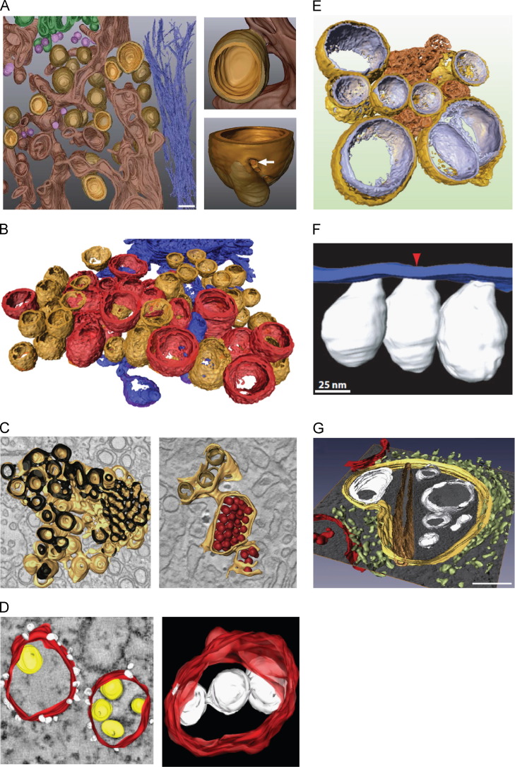
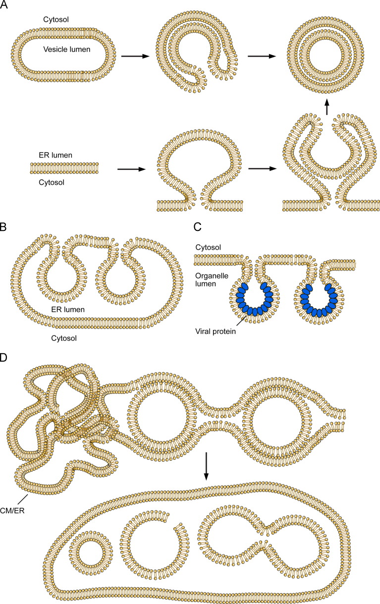
Positive strand RNA viruses replicate in the cytoplasm of infected cells and induce intracellular membranous compartments harboring the sites of viral RNA synthesis. These replication factories are supposed to concentrate the components of the replicase and to shield replication intermediates from the host cell innate immune defense. Virus induced membrane alterations are often generated in coordination with host factors and can be grouped into different morphotypes. Recent advances in conventional and electron microscopy have contributed greatly to our understanding of their biogenesis, but still many questions remain how viral proteins capture membranes and subvert host factors for their need. In this review, we will discuss different representatives of positive strand RNA viruses and their ways of hijacking cellular membranes to establish replication complexes. We will further focus on host cell factors that are critically involved in formation of these membranes and how they contribute to viral replication.

Keywords: Host factor; Membrane; Positive strand RNA virus; RNA replication; RNA synthesis; Replication factory.


Figures

Fig. 1
Fig. 1
Ultrastructure of membrane alterations induced by different positive strand RNA viruses. (A) Left: Membranous web induced by HCV. The ER is shown in dark brown, the inner membrane of DMVs and double membrane tubules as yellow brown and their outer membrane as semi-transparent light brown. Top right: View into the lumen of a DMV connected to ER membranes. Bottom right: View of a pore (white arrow) connecting the DMV lumen to the cytosol (Romero-Brey et al., 2012). (B) Late replication complexes of CVB3-infected cells. DMVs are shown in orange, multilamellar structures in red and parts of the neighboring ER in blue (Limpens et al., 2011). (C) Left: Interconnected reticular network induced by DENV infection. The cytosolic face of the membrane network is shown in brown, the ER lumen in black. Right: Viral particles were found in continuous ER cisternae and are depicted in red. ER membranes are colored in light bown and inner vesicle membranes in dark brown (Welsch et al., 2009). (D) Left: Surface model of replication complexes induced by Kunjin virus showing ER membranes in red, ribosomes in white and viral RNA in yellow. Right: Vesicles (white) were found to be connected to each other and to ER membranes (red) (Gillespie et al., 2010). (E) Cluster of heterogeneous DMVs induced by SARS-CoV infection. The outer DMV membrane is shown in gold, the inner membrane in silver and CMs in bronze (Knoops et al., 2008). (F) Surface rendering of FHV replication complexes. Virally-induced spherules into the mitochondrial lumen are shown in white, mitochondrial membranes in blue. A red arrow depicts an opening of a spherule towards the cytosol (Kopek et al., 2007). (G) 3D model of a tomographic slice of Rubella virus replication factories. Invaginated vesicles are shown in white, rigid membrane sheets in dark brown, the cytopathic vacuole in yellow, the ER in light-green and mitochondria in red (Fontana et al., 2010). Figures were reproduced with permission from the respective journals.
Fig. 2
Fig. 2
Schematic illustration depicting the diversity of membrane alterations caused by a selection of different viruses. (A) Hypothetical model of double membrane vesicle (DMV) biogenesis originating from a single membrane vesicle (SMV, top), typically seen for Enteroviruses. The DMV is generated by an invagination event engulfing cytosol and creating a confined protected luminal space that can be connected to the cytosol by a narrow channel. Upon membrane merging, a closed DMV is created. Alternatively, DMVs can arise from exvagination of membranes into the cytosol accompanied by a secondary invagination event (bottom), as it has been suggested for HCV. (B) Replication complexes induced by DENV. An interconnected network is formed by ER membranes including SMVs that originate from invaginations into the ER. (C) Invaginated spherules as observed for BMV or FHV. A single oligomerizing viral protein (blue) is responsible for membrane alterations and forms a protein shell inside the spherules keeping them in shape. (D) Complexity of membrane alterations induced by SARS-CoV. Early stages of infection show DMVs interconnected by their outer membrane to each other as well as to convoluted membranes (CM) or ER membranes (top). Later stages of infection show membranous vesicle packets containing one or several SMVs, which can be fused to each other or exhibit membrane discontinuities (bottom) (Fontana et al., 2010).

References

    1. Acosta E.G., Kumar A., Bartenschlager R. Revisiting dengue virus–host cell interaction: new insights into molecular and cellular virology. Adv.Virus Res. 2014;88:1–109. - PubMed
    1. Ahlquist P. Parallels among positive-strand RNA viruses, reverse-transcribing viruses and double-stranded RNA viruses. Nat. Rev. Microbiol. 2006;4:371–382. - PMC - PubMed
    1. Ahola T., Lampio A., Auvinen P., Kaariainen L. Semliki Forest virus mRNA capping enzyme requires association with anionic membrane phospholipids for activity. EMBO J. 1999;18:3164–3172. - PMC - PubMed
    1. Aizaki H., Lee K.J., Sung V.M., Ishiko H., Lai M.M. Characterization of the hepatitis C virus RNA replication complex associated with lipid rafts. Virology. 2004;324:450–461. - PubMed
    1. Aldabe R., Irurzun A., Carrasco L. Poliovirus protein 2BC increases cytosolic free calcium concentrations. J. Virol. 1997;71:6214–6217. - PMC - PubMed

Publication types

MeSH terms

LinkOut - more resources