Review
. 2015 Jun;185(6):1528-36.
doi: 10.1016/j.ajpath.2014.08.030. Epub 2015 Mar 5.

The role of viral, host, and secondary bacterial factors in influenza pathogenesis

Affiliations
Review

The role of viral, host, and secondary bacterial factors in influenza pathogenesis

John C Kash et al. Am J Pathol. 2015 Jun.

Abstract

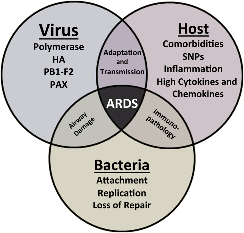
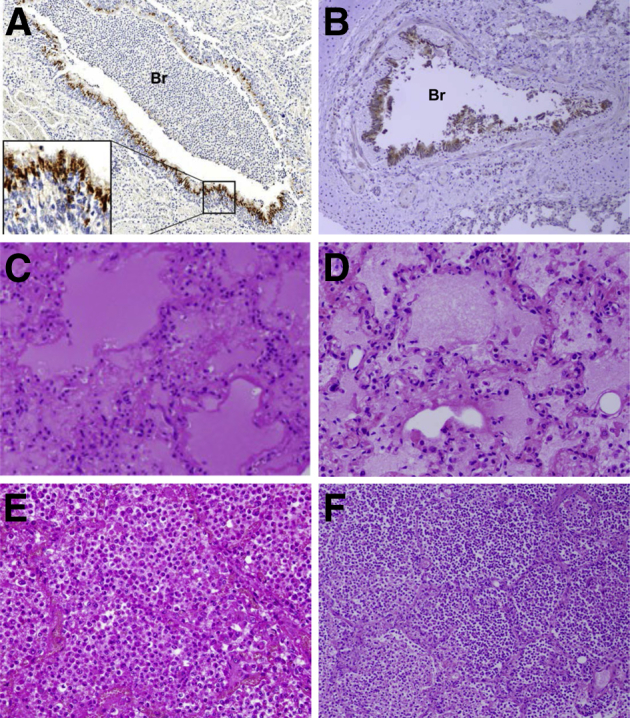
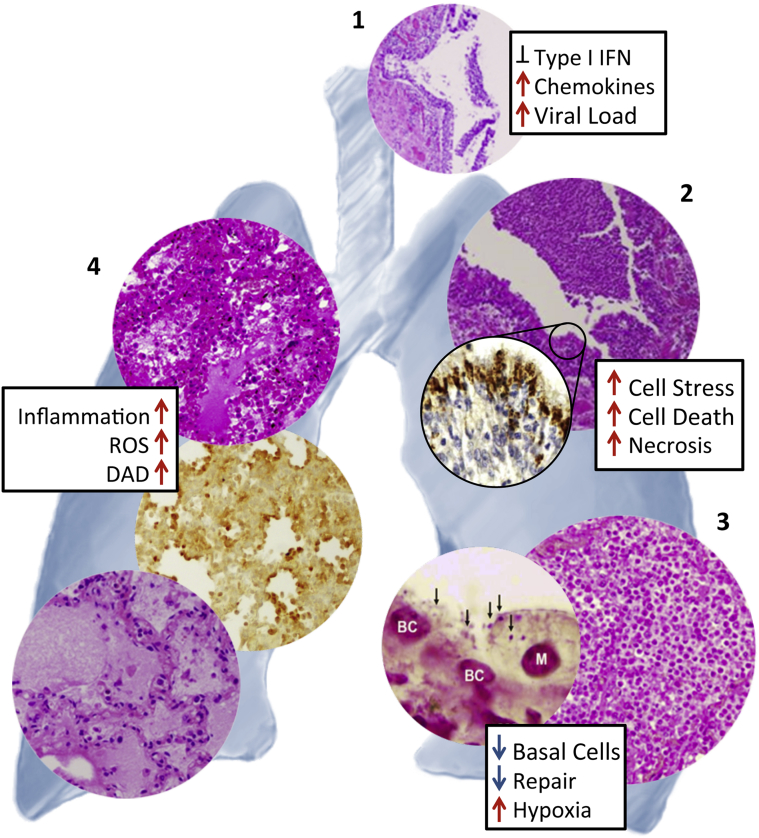
Influenza A virus infections in humans generally cause self-limited infections, but can result in severe disease, secondary bacterial pneumonias, and death. Influenza viruses can replicate in epithelial cells throughout the respiratory tree and can cause tracheitis, bronchitis, bronchiolitis, diffuse alveolar damage with pulmonary edema and hemorrhage, and interstitial and airspace inflammation. The mechanisms by which influenza infections result in enhanced disease, including development of pneumonia and acute respiratory distress, are multifactorial, involving host, viral, and bacterial factors. Host factors that enhance risk of severe influenza disease include underlying comorbidities, such as cardiac and respiratory disease, immunosuppression, and pregnancy. Viral parameters enhancing disease risk include polymerase mutations associated with host switch and adaptation, viral proteins that modulate immune and antiviral responses, and virulence factors that increase disease severity, which can be especially prominent in pandemic viruses and some zoonotic influenza viruses causing human infections. Influenza viral infections result in damage to the respiratory epithelium that facilitates secondary infection with common bacterial pneumopathogens and can lead to secondary bacterial pneumonias that greatly contribute to respiratory distress, enhanced morbidity, and death. Understanding the molecular mechanisms by which influenza and secondary bacterial infections, coupled with the role of host risk factors, contribute to enhanced morbidity and mortality is essential to develop better therapeutic strategies to treat severe influenza.


Figures

Figure 1
Figure 1
Diagram showing interrelationships between viral genes, host factors, and secondary bacterial infections associated with development of severe pathology and disease. ARDS, acute respiratory distress syndrome; HA, hemagglutinin; PAX, polymerase acidic frameshift protein; PB1, polymerase basic protein 1 frame 2 protein; SNP, single-nucleotide polymorphism.
Figure 2
Figure 2
Representative pathological changes during fatal primary influenza and secondary bacterial infections in human autopsies from the 1918 (A, C, and E) and 2009 (B, D, and F) pandemics. A: Immunohistochemical stained section of lung from a 1918 pandemic influenza fatal case showing acute influenza viral bronchiolitis with infiltration of neutrophils and other inflammatory cells in the lumen of a bronchiole (Br). Influenza viral antigen (reddish-brown stain) is readily apparent in the apical cells of the bronchiolar respiratory epithelium (inset), on a hematoxylin-stained background.B: Immunohistochemical stained section of lung from a 2009 pandemic influenza fatal case showing acute influenza viral bronchiolitis (Br). Influenza viral antigen (reddish-brown stain) is readily apparent in the apical cells of the bronchiolar respiratory epithelium, on a hematoxylin-stained background.C: Hematoxylin and eosin (H&E)–stained section of lung from a 1918 pandemic influenza fatal case showing diffuse alveolar damage with hyaline membranes lining alveoli. The alveolar airspaces contain edema fluid, strands of fibrin, desquamated epithelial cells, and inflammatory cells.D: H&E-stained section of lung from a 2009 pandemic influenza fatal case showing diffuse alveolar damage with hyaline membranes lining alveoli. The alveolar air spaces contain edema fluid, strands of fibrin, desquamated epithelial cells, and inflammatory cells.E: H&E-stained section of lung from a 1918 pandemic influenza fatal case showing a massive infiltrate of neutrophils that fills the alveolar air spaces associated with a secondary bacterial bronchopneumonia. Alveolar capillary congestion is prominent.F: H&E-stained section of lung from a 2009 pandemic influenza fatal case showing a massive infiltration of neutrophils in the airspaces of alveoli associated with a secondary bacterial bronchopneumonia.A and C were modified from Sheng et al (published by College of American Pathologists),E from Taubenberger and Morens (published by Annual Reviews), and B, D, and F from Gill et al (published by National Academy of Sciences of the United States of America). All images used with permission of the publishers. Original magnifications: ×40 (A and B); ×100 (E and F); ×200 (C and D).
Figure 3
Figure 3
Relationship between pathological changes and host responses during severe primary influenza and secondary bacterial infections in human autopsies and experimental animal infections. Necrotizing tracheitis (1) in a 2009 pandemic H1N1 fatality.Inset shows the relative expression of inflammatory and type I interferon (IFN) gene expression responses in fatal 1918 influenza infection compared to seasonal virus in cynomolgus macaques. Severe necrotizing bronchitis/bronchiolitis (2) in a 1918 influenza autopsy sample showing intense viral antigen staining in the ciliated and goblet cells of the respiratory epithelium, but not in basal cells.Inset shows the relative expression of inflammatory response and cell death genes in lungs of mice infected with 1918 compared to seasonal H1N1. Secondary bacterial pneumonias (3) with massive infiltration of neutrophils into alveolar airspaces in a 1918 pandemic influenza fatal case (right panel). Left panel: Gram stain showing presence of Streptococcus pneumoniae bacteria (arrows) on remaining basal epithelial cells (nuclei labeled BC) and a macrophage (M) in mouse lung after coinfection of 2009 pandemic H1N1 and S. pneumoniae.Inset shows loss of lung repair and regeneration gene expression responses in mice infected with 2009 pandemic H1N1 with a secondary S. pneumoniae infection compared to primary 2009 pandemic H1N1 infection. Development of diffuse alveolar damage (DAD) (4) with alveolar hyaline membrane formation, varying degrees of acute intra-alveolar edema, acute hemorrhage, interstitial and airspace inflammatory infiltrates, and small-vessel thromboses in 1918 pandemic influenza (top panel) and 2009 pandemic influenza (bottom panel) autopsies, along with pronounced alveolar epithelial immunostaining for reactive oxygen species (ROS) damage in mouse tissue infected with 1918 influenza virus.Inset shows relative expression of inflammatory and ROS response genes in lungs of mice infected with 1918 compared to seasonal H1N1. Photomicrograph in 1 was modified from Gill et al (published by National Academy of Sciences of the United States of America); in 2 from Sheng et al (published by College of American Pathologists); in 3 from Morens et al (published by Oxford University Press) (right panel) and Kash et al (published by American Society for Microbiology) (left panel); and in 4 from Taubenberger and Morens (published by Annual Reviews) (top panel), Kash et al (published by Elsevier B.V.) (middle panel), and Gill et al (published by National Academy of Sciences of the United States of America) (bottom panel). All images used with permission of the publishers.

References

    1. Wright P.F., Neumann G., Kawaoka Y. Orthomyxoviruses. In: Knipe D.M., Howley P.M., editors. Lippincott Williams & Wilkins; Philadelphia: 2007. pp. 1691–1740.
    1. Thompson M.G., Shay D.K., Zhou H., Bridges C.B., Cheng P.Y., Burns E., Bresee J.S., Cox N.J. Estimates of deaths associated with seasonal influenza: United States, 1976-2007. MMWR Morb Mortal Wkly Rep. 2010;59:1057–1062. - PubMed
    1. Johnson N.P., Mueller J. Updating the accounts: global mortality of the 1918-1920 “Spanish” influenza pandemic. Bull Hist Med. 2002;76:105–115. - PubMed
    1. Taubenberger J.K., Morens D.M. 1918 Influenza: the mother of all pandemics. Emerg Infect Dis. 2006;12:15–22. - PMC - PubMed
    1. Peiris J.S.M. Avian influenza viruses in humans. Rev Sci Tech. 2009;28:161–173. - PubMed

Publication types