. 2015 Mar 6;10(3):e0119346.
doi: 10.1371/journal.pone.0119346. eCollection 2015.

SD-208, a novel protein kinase D inhibitor, blocks prostate cancer cell proliferation and tumor growth in vivo by inducing G2/M cell cycle arrest

Affiliations

SD-208, a novel protein kinase D inhibitor, blocks prostate cancer cell proliferation and tumor growth in vivo by inducing G2/M cell cycle arrest

Manuj Tandon et al. PLoS One. .

Abstract

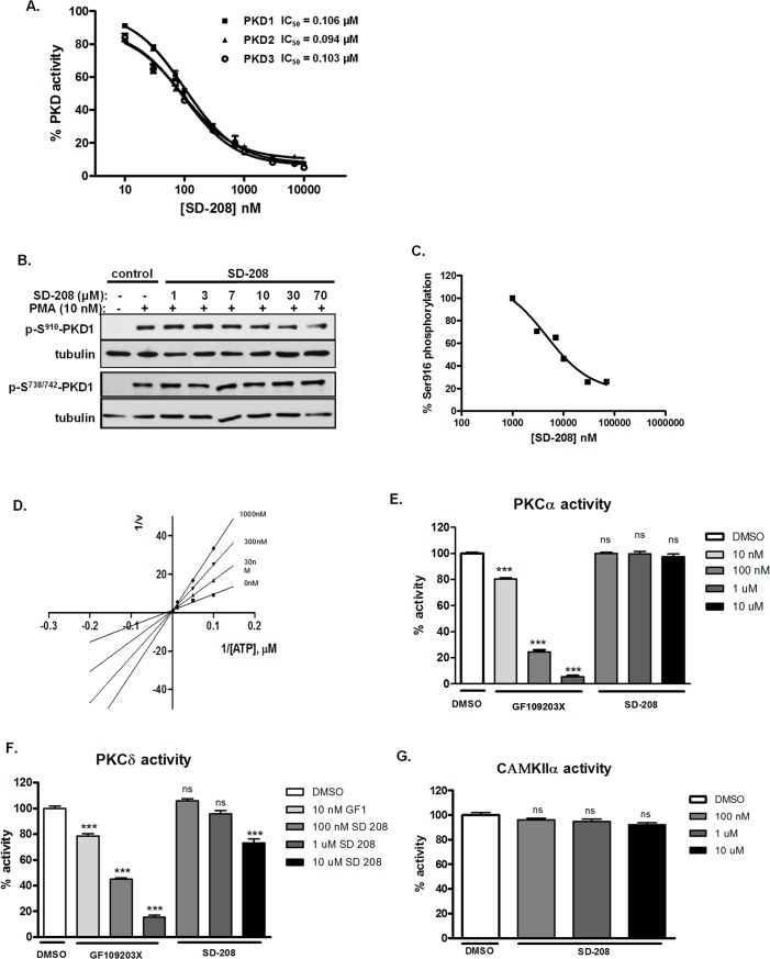
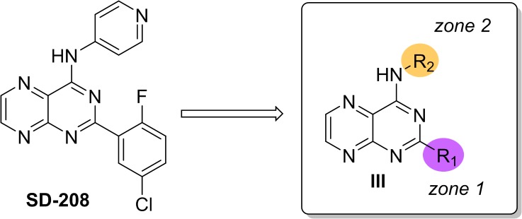
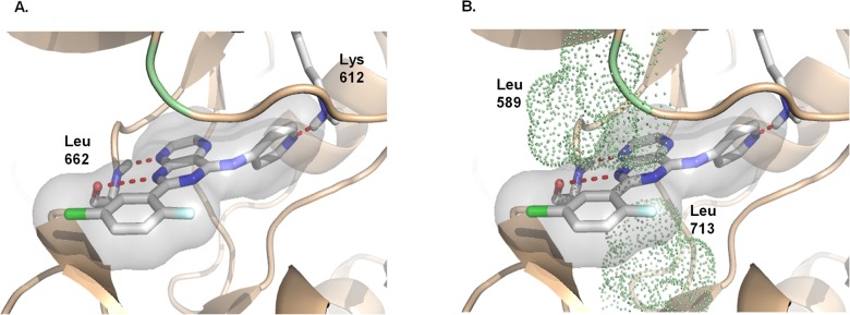
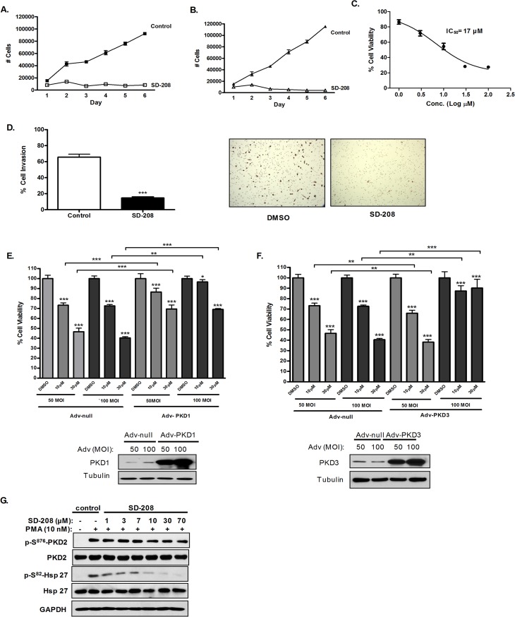
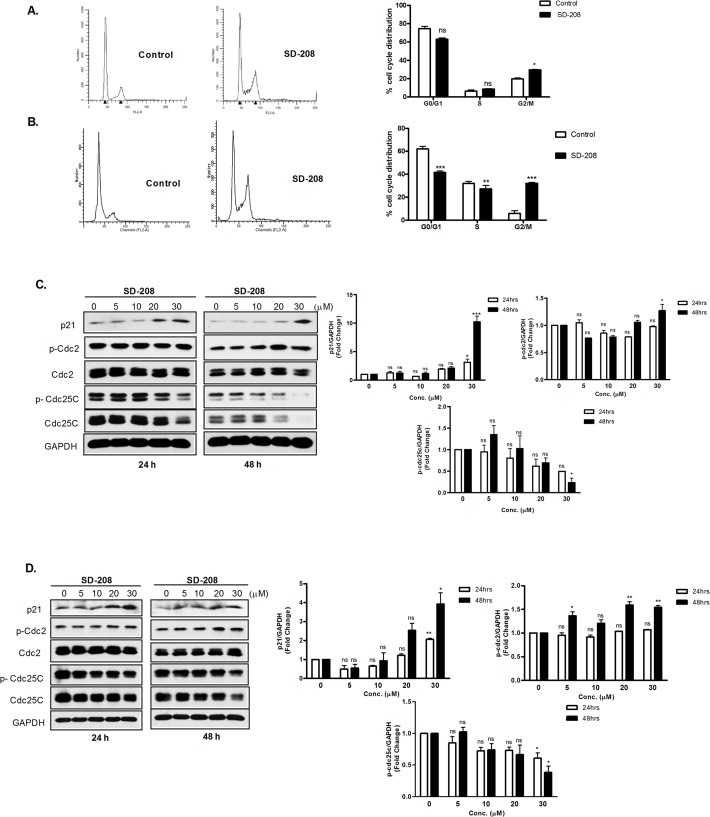
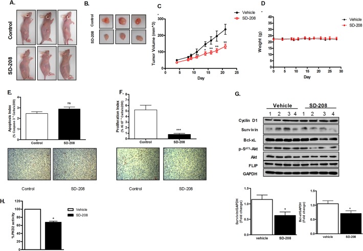
Protein kinase D (PKD) has been implicated in many aspects of tumorigenesis and progression, and is an emerging molecular target for the development of anticancer therapy. Despite recent advancement in the development of potent and selective PKD small molecule inhibitors, the availability of in vivo active PKD inhibitors remains sparse. In this study, we describe the discovery of a novel PKD small molecule inhibitor, SD-208, from a targeted kinase inhibitor library screen, and the synthesis of a series of analogs to probe the structure-activity relationship (SAR) vs. PKD1. SD-208 displayed a narrow SAR profile, was an ATP-competitive pan-PKD inhibitor with low nanomolar potency and was cell active. Targeted inhibition of PKD by SD-208 resulted in potent inhibition of cell proliferation, an effect that could be reversed by overexpressed PKD1 or PKD3. SD-208 also blocked prostate cancer cell survival and invasion, and arrested cells in the G2/M phase of the cell cycle. Mechanistically, SD-208-induced G2/M arrest was accompanied by an increase in levels of p21 in DU145 and PC3 cells as well as elevated phosphorylation of Cdc2 and Cdc25C in DU145 cells. Most importantly, SD-208 given orally for 24 days significantly abrogated the growth of PC3 subcutaneous tumor xenografts in nude mice, which was accompanied by reduced proliferation and increased apoptosis and decreased expression of PKD biomarkers including survivin and Bcl-xL. Our study has identified SD-208 as a novel efficacious PKD small molecule inhibitor, demonstrating the therapeutic potential of targeted inhibition of PKD for prostate cancer treatment.


Conflict of interest statement

Competing Interests: Co-author Qiming Jane Wang is a PLOS ONE Editorial Board member. This does not alter the authors' adherence to PLOS ONE Editorial policies and criteria.

Figures

Fig 1
Fig 1. SD-208 was a cell-active, ATP-competitive PKD inhibitor and did not inhibit PKC and CAMK.
A. Determination of PKD kinase activity in vitro. Inhibition of recombinant human PKD1, 2 and 3 was assayed in the presence of 10 different concentrations of SD-208 by an in vitro radiometric PKD kinase assay. The IC50 values were calculated as the mean ± SEM of at least three independent experiments with triplicate determinations at each compound concentration in each experiment. The data were plotted as a function of inhibitor concentration and a representative graph is shown. B. SD-208 inhibited PMA-induced PKD1 activation in prostate cancer cells. LNCaP cells were pretreated with different doses of inhibitors for 45 min, followed by PMA stimulation at 10 nM for 20 min. Cell lysates were subjected to immunoblotting for p-S910-PKD1 and p-S738/742-PKD1. Tubulin was blotted as loading control. The experiment was repeated three times and the representative blots are shown. C. Determination of cellular IC 50 . Western blots from ‘B” were quantified using densitometry analysis. The data were plotted and IC50 values were derived from the concentration-response curves using GraphPad. One of the three concentration-response curves is shown. D. SD-208 is an ATP-competitive kinase inhibitor. PKD1 kinase activity was measured as a function of increasing concentrations of ATP in the presence of varying concentrations of SD-208. Lineweaver-Burke plots of the data are shown. Data presented were the mean ± S.E. of three independent experiments with triplicate determinations at each data point in each experiment. E-F. Selectivity of SD-208 against related kinases. Inhibition of PKCα (B) or PKCδ (C) was determined at 10 nM, 100 nM, 1μM, and 10 μM. As controls, the PKC inhibitor GF109203X potently inhibited PKCα and PKCδ activity. Data are the mean ± SEM of two independent experiments. G. Inhibition of CAMKIIα was measured by the radiometric CAMK kinase assay. Data are the mean ± S.E. of two independent experiments with triplicate determinations at each data point in each experiment. Statistical significance was determined using the unpaired t-test. ns, not significantly significant; *, p<0.05; **, p<0.01; ***, p<0.001.
Fig 2
Fig 2. Scope of first-generation SAR studies on SD-208.
2-Zone model for SD-208 analog synthesis.
Fig 3
Fig 3. Molecular modeling of SD-208 in the active site of a PKD1 homology model.
This depiction demonstrated a potential binding mode of SD208 in the active site of PKD1. Red dotted lines represent key interactions between SD-208 and PKD1.
Fig 4
Fig 4. SD-208 inhibited prostate cancer cells proliferation, survival, and invasion and the anti-proliferative effect of SD-208 was mediated through the inhibition of PKD.
A-B. SD-208 inhibited PC3 (A) and LNCaP (B) prostate cancer cell proliferation. PC3 and LNCaP cells were plated in triplicates in 24-well plates. Cells were allowed to attach overnight. A cell count at day 1 was made, and then either a vehicle (DMSO) or SD-208 at 30 μM was added. Cells were counted daily for a total of 6 days. Data are the mean ± S.E. of two independent experiments with triplicate determinations at each data point in each experiment. C. SD-208 inhibited PC3 prostate cancer cell survival. PC3 cells were seeded into 96-well plates (3000 cells/well) and were then incubated in media containing 0.3–100 μM inhibitors for 72 h. MTT solution was added to each well and incubated for 4 h. Optical density was read at 570 nm to determine cell viability. The IC50 was determined as the mean of three independent experiments for each compound. D. SD-208 inhibited prostate cancer cell invasion. DU145 cells were incubated with 30 μM SD 208 in Matrigel inserts. After 20 h, noninvasive cells were removed and invasive cells were fixed in 100% methanol, stained in 0.4% hematoxylin solution, and photographed. The number of cells that invaded the Matrigel matrix was determined by cell counts in 6 fields relative to the number of cells that migrated through the control insert. Percentage invasion was calculated as the percent of the cells invaded through Matrigel inserts vs. the total cells migrated through the control inserts. Data are the mean ± S.E. of three independent experiments with triplicate determinations at each data point in each experiment. Statistical significance was determined using the unpaired t-test. ***, p<0.001. E-F. Overexpression of PKD1 and PKD3 in prostate cancer cells rescued the anti-proliferative effects of SD-208. PC3 (0.5 million) cells were seeded in a 60 mm dish and infected the next day with 50 and 100 MOI of PKD1 and PKD3 adenoviruses (Adv-PKD1 and Adv-PKD3). Empty adenovirus (Adv-null) was used as control. After 24 h, 3000 cells/well were plated in 96-well plates and treated with and without 10 and 30 μM SD-208 for 72 h. MTT solution was added to each well and incubated for 4 h. Optical density was read at 570 nm to determine cell viability. The overexpression of PKD1 and PKD3 was confirmed by Western blotting analysis. This experiment was repeated three times and data are the mean ± S.E. of all three independent experiments. Statistical significance between DMSO and inhibitor treatment was determined using the unpaired t-test.*, p<0.05; **, p<0.01; ***, p<0.001. G. PKD mediated Hsp27 activity in prostate cancer cells was inhibited by SD-208. DU145 cells were pretreated with different doses of inhibitors for 45 min, followed by PMA stimulation at 10 nM for 20 min. Cell lysates were subjected to immunoblotting for p-S910-PKD1 and p-S738/742-PKD1. GAPDH was blotted as loading control. The experiment was repeated two times and the representative blots are shown.
Fig 5
Fig 5. SD-208 arrested cells in G2/M and regulated the levels and activities of cell cycle regulatory proteins at the G2/M phase of cell cycle in prostate cancer cells.
A-B. SD-208 induced G2/M cell cycle arrest in prostate cancer cells. DU145 cells (A) and PC3 cells (B) were treated with either vehicle (DMSO) or 30 μM SD 208 for 48 h. Cell cycle distribution was determined by flow cytometry after propidium iodide labeling of fixed cells. The cell cycle plots are representative of three independent experiments (left). Data in the bar graph are the mean ± SEM of three independent experiments (right). Statistical significance was determined using the unpaired t-test and is indicated. **, p<0.01; ***, p<0.001. C-D. Effects of SD-208 on the expression and activities of G2/M cell cycle regulatory proteins. DU145 (C) and PC3 (D) cells were treated with DMSO or 5–30 µM SD-208 for 24 and 48 h. At the end of each treatment, cells were harvested and subjected to immunoblotting for the cell cycle regulatory proteins as indicated. GAPDH was blotted as loading control. The densitometry data (mean ± SEM from four experiments) were plotted as ‘fold change’ over the control after normalization with GAPDH. The experiments were repeated four times and representative blots from one experiment are shown.
Fig 6
Fig 6. SD-208 inhibited the growth of PC3 tumor xenografts in vivo.
A-D. Effects of SD-208 on the growth of PC3 tumor xenograft. PC3 cells (1.4 X 10⁶) were injected s.c. to both flanks of nude mice. Once tumors were palpable, mice were randomized into two groups (5 mice/group) and were administered 0.2 ml twice daily doses of vehicle [1% (w/v) methylcellulose] or SD-208 (60 mg/kg) by oral gavage. Tumor volume (C) and mouse weight (D) were measured every 2 to 3 days. Twenty-four days after treatment, mice were euthanized by CO2 inhalation, and tumors were excised. Images of representative mice (A) and dissected tumors (B) are shown. All animal studies were conducted in accordance with the Institutional Animal Care and Use Committee (IACUC). Statistical significance was determined using the unpaired t-test. **, p<0.01; ***, p<0.001. E, Analysis of apoptotic marker cleaved caspase-3 by IHC. Quantification of in situ cleaved caspase-3-positive cells as described in Materials and Methods. Representative images of cleaved caspase-3 staining (x200) are shown. F, Analysis of proliferative marker Ki67 by IHC. Quantification of Ki-67-positive cells or proliferation index as described in Materials and Methods. Representative images of Ki-67 staining (x200) are shown. G. Analysis of PKD-regulated biomarkers in PC3 tumor xenografts. Cell lysates were prepared from tumor explants obtained from vehicle and SD-208-treated mice. Levels of PKD-regulated proteins were analyzed by Western blotting analysis. The densitometry data represents ‘fold change’ as compared with vehicle control after normalization with GAPDH as loading control. H. Effect of SD-208 on the PKD2 activity in PC3 tumor xenografts. Cell lysates from tumor explants obtained from vehicle and SD-208-treated mice were immunoprecipitated with PKD2 antibody followed by in vitro kinase assay as described in Material and Methods. The experiment was repeated three times and data are the mean ± S.E. of all three independent experiments with triplicate determinations at each data point in each experiment.

Similar articles

Cited by

References

    1. Jemal A, Murray T, Samuels A, Ghafoor A, Ward E, Thun MJ (2003) Cancer statistics, 2003. CA Cancer J Clin 53: 5–26. - PubMed
    1. Karan D, Lin MF, Johansson SL, Batra SK (2003) Current status of the molecular genetics of human prostatic adenocarcinomas. Int J Cancer 103: 285–293. - PubMed
    1. Wang QJ (2006) PKD at the crossroads of DAG and PKC signaling. Trends Pharmacol Sci 27: 317–323. - PubMed
    1. Valverde AM, Sinnett-Smith J, Van Lint J, Rozengurt E (1994) Molecular cloning and characterization of protein kinase D: a target for diacylglycerol and phorbol esters with a distinctive catalytic domain. Proc Natl Acad Sci U S A 91: 8572–8576. - PMC - PubMed
    1. Sturany S, Van Lint J, Muller F, Wilda M, Hameister H, Hocker M, et al. (2001) Molecular cloning and characterization of the human protein kinase D2. A novel member of the protein kinase D family of serine threonine kinases. J Biol Chem 276: 3310–3318. - PubMed

Publication types

MeSH terms