Review
. 2015 May 15;308(10):H1177-91.
doi: 10.1152/ajpheart.00007.2015. Epub 2015 Mar 6.

Chasing cardiac physiology and pathology down the CaMKII cascade

Affiliations
Review

Chasing cardiac physiology and pathology down the CaMKII cascade

Alicia Mattiazzi et al. Am J Physiol Heart Circ Physiol. .

Abstract

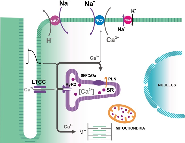
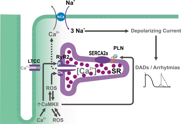
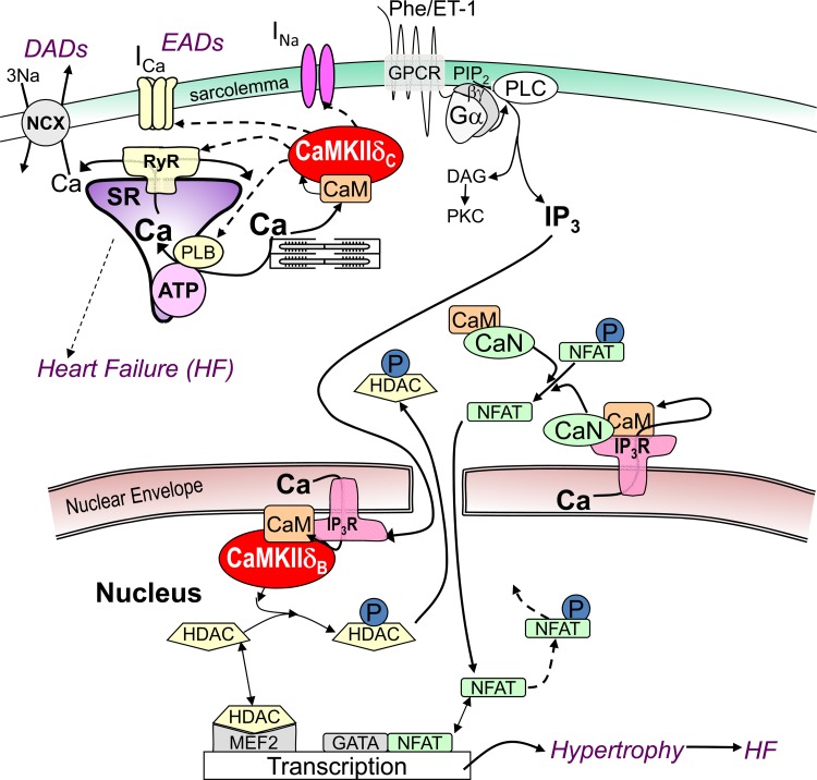
Calcium dynamics is central in cardiac physiology, as the key event leading to the excitation-contraction coupling (ECC) and relaxation processes. The primary function of Ca(2+) in the heart is the control of mechanical activity developed by the myofibril contractile apparatus. This key role of Ca(2+) signaling explains the subtle and critical control of important events of ECC and relaxation, such as Ca(2+) influx and SR Ca(2+) release and uptake. The multifunctional Ca(2+)-calmodulin-dependent protein kinase II (CaMKII) is a signaling molecule that regulates a diverse array of proteins involved not only in ECC and relaxation but also in cell death, transcriptional activation of hypertrophy, inflammation, and arrhythmias. CaMKII activity is triggered by an increase in intracellular Ca(2+) levels. This activity can be sustained, creating molecular memory after the decline in Ca(2+) concentration, by autophosphorylation of the enzyme, as well as by oxidation, glycosylation, and nitrosylation at different sites of the regulatory domain of the kinase. CaMKII activity is enhanced in several cardiac diseases, altering the signaling pathways by which CaMKII regulates the different fundamental proteins involved in functional and transcriptional cardiac processes. Dysregulation of these pathways constitutes a central mechanism of various cardiac disease phenomena, like apoptosis and necrosis during ischemia/reperfusion injury, digitalis exposure, post-acidosis and heart failure arrhythmias, or cardiac hypertrophy. Here we summarize significant aspects of the molecular physiology of CaMKII and provide a conceptual framework for understanding the role of the CaMKII cascade on Ca(2+) regulation and dysregulation in cardiac health and disease.

Keywords: Ca2+; CaMKII; arrhythmias; cell death; hypertrophy; ischemia/reperfusion.


Figures

Fig. 1.
Fig. 1.
Ca2+ fluxes associated with excitation-contraction coupling in mammalian cardiac myocytes. During the action potential, Ca2+ influx via L-type Ca2+ channels (LTCC) triggers Ca2+ release from the sarcoplasmic reticulum (SR) by Ca2+ binding to the ryanodine receptors (RyR2) in the SR membrane. In addition to interacting with the myofilaments (MF), Ca2+ is removed from the cytosol mainly by the SR Ca2+-ATPase (SERCA2a), which is regulated by phospholamban (PLN), and by the electrogenic sarcolemmal Na+/Ca2+ exchanger (NCX1), which is driven by the Na+ electrochemical gradient across the membrane. This gradient is maintained by the Na+-K+-ATPase (NKA). Intracellular [Na+] may also be affected by the operation of the Na+-H+ exchanger (NHE).
Fig. 2.
Fig. 2.
A: schematic representation of CaMKII structure and regulation. See text for description. Note the proximity of sites involved in the sustained regulation of CaMKII. The question mark in Cys290 indicates a computationally predicted site of CaMKII nitrosylation [modified from Erickson et al. (35)]. B: effects of CaMKIIδC on excitation-contraction coupling (ECC). See text for description. INa, Na+ current; Ito, transient outward K+ current; IK1, inward rectifier K+ current.
Fig. 3.
Fig. 3.
A: increase in diastolic [Ca2+]i at the onset of reperfusion. At the onset of reperfusion, there is an abrupt increase in diastolic [Ca2+]i (Ca2+ bump) associated with a mirror-like image of the decrease in SR Ca2+ content. Mean values are from individual signals recorded at the epicardial layer of intact hearts loaded with Rhod-2 and Mag-Fluo-4, respectively [modified from Valverde et al. (132)]. B: typical records showing the decrease in SR Ca2+ content associated with a diminished Ca2+ transient amplitude, after the Ca2+ bump. AU, arbitrary units.
Fig. 4.
Fig. 4.
A–C: Ca2+ sparks increase during ischemia and turn into Ca2+ waves during reperfusion. Typical examples and overall results are shown [modified from Mattiazzi et al. (74)].
Fig. 5.
Fig. 5.
Scheme of the possible mechanisms of CaMKII-dependent arrhythmias. Increases in [Ca2+]i and/or reactive oxygen species (ROS) can activate CaMKII, which in turn result in further increase in ROS, leading to the phosphorylation and oxidation of the RyR2 and phosphorylation of phospholamban, which, in concert, would enhance SR Ca2+ load and favor SR Ca2+ leak, resulting in an NCX-dependent depolarizing current (transient inward current, or Iti), which generates arrhythmogenic delayed afterdepolarization (DADs).
Fig. 6.
Fig. 6.
Ca2+-dependent signaling in excitation-transcription coupling via Ca-CaM. CaMKIIδ can acutely regulate ion channels (that carry INa and ICa) and Ca2+ handling proteins (RyR2, IP3R, PLN), contributing to triggered arrhythmias such as early and delayed afterdepolarization (EADs and DADs). G protein-coupled receptor (GPCR) agonists endothelin-1 (ET-1) and phenylephrine (PE) activate Gαq/βγ and phospholipase C (PLC) to produce diacylglycerol (DAG), which can activate protein kinases C and D. PKC, CaMKII, and PKD can phosphorylate (P) HDAC, and calcineurin (CaN) can dephosphorylate nuclear factor of activated T cells (NFAT), altering nuclear MEF2- and GATA-dependent transcription.

References

    1. Anderson ME. Multiple downstream proarrhythmic targets for calmodulin kinase II: moving beyond an ion channel-centric focus. Cardiovasc Res 73: 657–666, 2007. - PubMed
    1. Ather S, Wang W, Wang Q, Li N, Anderson ME, Wehrens XH. Inhibition of CaMKII phosphorylation of RyR2 prevents inducible ventricular arrhythmias in mice with Duchenne muscular dystrophy. Heart Rhythm 10: 592–599, 2013. - PMC - PubMed
    1. Backs J, Olson EN. Control of cardiac growth by histone acetylation/deacetylation. Circ Res 98: 15–24, 2006. - PubMed
    1. Bai Y, Jones PP, Guo J, Zhong X, Clark RB, Zhou Q, Wang R, Vallmitjana A, Benitez R, Hove-Madsen L, Semeniuk L, Guo A, Song LS, Duff HJ, Chen SR. Phospholamban knockout breaks arrhythmogenic Ca2+ waves and suppresses catecholaminergic polymorphic ventricular tachycardia in mice. Circ Res 113: 517–526, 2013. - PMC - PubMed
    1. Bare DJ, Kettlun CS, Liang M, Bers DM, Mignery GA. Cardiac type 2 inositol 1,4,5-trisphosphate receptor: interaction and modulation by calcium/calmodulin-dependent protein kinase II. J Biol Chem 280: 15912–15920, 2005. - PubMed

Publication types

MeSH terms

Substances