. 2015 Mar 6;11(3):e1004729.
doi: 10.1371/journal.ppat.1004729. eCollection 2015 Mar.

Visualization of HIV-1 interactions with penile and foreskin epithelia: clues for female-to-male HIV transmission

Affiliations

Visualization of HIV-1 interactions with penile and foreskin epithelia: clues for female-to-male HIV transmission

Minh H Dinh et al. PLoS Pathog. .

Abstract

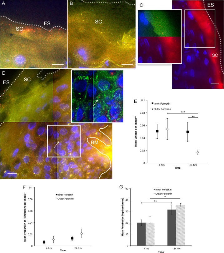
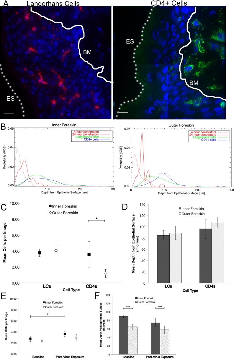
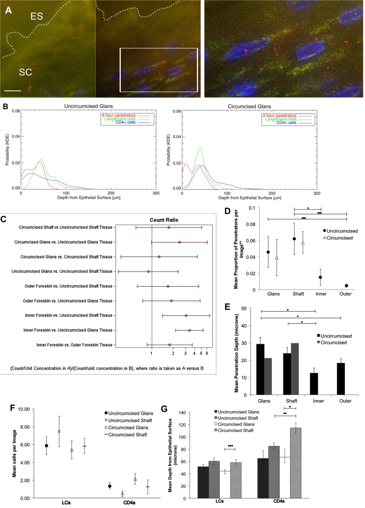
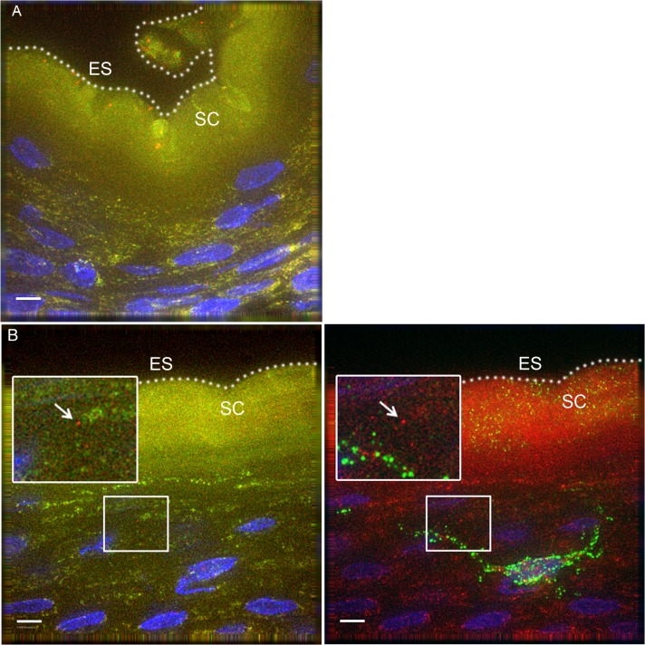
To gain insight into female-to-male HIV sexual transmission and how male circumcision protects against this mode of transmission, we visualized HIV-1 interactions with foreskin and penile tissues in ex vivo tissue culture and in vivo rhesus macaque models utilizing epifluorescent microscopy. 12 foreskin and 14 cadaveric penile specimens were cultured with R5-tropic photoactivatable (PA)-GFP HIV-1 for 4 or 24 hours. Tissue cryosections were immunofluorescently imaged for epithelial and immune cell markers. Images were analyzed for total virions, proportion of penetrators, depth of virion penetration, as well as immune cell counts and depths in the tissue. We visualized individual PA virions breaching penile epithelial surfaces in the explant and macaque model. Using kernel density estimated probabilities of localizing a virion or immune cell at certain tissue depths revealed that interactions between virions and cells were more likely to occur in the inner foreskin or glans penis (from local or cadaveric donors, respectively). Using statistical models to account for repeated measures and zero-inflated datasets, we found no difference in total virions visualized at 4 hours between inner and outer foreskins from local donors. At 24 hours, there were more virions in inner as compared to outer foreskin (0.0495 +/- 0.0154 and 0.0171 +/- 0.0038 virions/image, p = 0.001). In the cadaveric specimens, we observed more virions in inner foreskin (0.0507 +/- 0.0079 virions/image) than glans tissue (0.0167 +/- 0.0033 virions/image, p<0.001), but a greater proportion was seen penetrating uncircumcised glans tissue (0.0458 +/- 0.0188 vs. 0.0151 +/- 0.0100 virions/image, p = 0.099) and to significantly greater mean depths (29.162 +/- 3.908 vs. 12.466 +/- 2.985 μm). Our in vivo macaque model confirmed that virions can breach penile squamous epithelia in a living model. In summary, these results suggest that the inner foreskin and glans epithelia may be important sites for HIV transmission in uncircumcised men.


Conflict of interest statement

I have read the journal's policy and the authors of this manuscript have the following competing interests: MHD received funding from Bristol Meyers Squibb through a Virology Fellows Grant for this work. They had no role in study design, data collection and analysis, decision to publish, or preparation of the manuscript. This does not alter our adherence to all PLOS Pathogens policies on sharing data and materials.

Figures

Fig 1
Fig 1. Ex vivo PA GFP HIV-1 interactions with adult human foreskin tissues.
Foreskins obtained from consenting adult donors and inoculated with R5-tropic PA GFP-Vpr HIV-1 for 4 (n = 10) or 24 hours (n = 12) in culture. (A) and (B) Representative images of virion interactions with inner (A) and outer (B) foreskins after 4 hours of HIV exposure ex vivo. When seen, virions (red) were found predominantly on the surface or in the stratum corneum (SC). ES, dotted line, epithelial surface. (C) When co-inoculated with fluorescently labeled bovine serum albumin (BSA, red, right panel), virions (red, top half of inset, pseudo-colored to reveal PA GFP) were seen diffusing to depths that BSA also reached. (D) The majority of penetrating virions (virions seen below the SC) were found interstitially, as determined by tissues stained with fluorescent wheat germ agglutinin (WGA, green, inset). All images: white bar = 10 μm, blue = cell nuclei. (E-G) Estimated means of total virion counts (E), ** = adjusted for virus stock concentrations; proportion of penetrators (F); depths of penetration (G). Dark squares and bars represent inner foreskin; open diamonds and bars represent outer foreskin. *p<0.05, **p<0.01, ***p<0.001
Fig 2
Fig 2. Tissue resident immune cells in foreskin tissues.
Tissue cryosections immunofluorescently stained with OKT6 or α-CD4 antibodies to detect Langerhans cells (LCs) or CD4+ cells, respectively. (A) Representative images of LCs (red, left panel) and CD4+ cells (green, right panel) shown. White bar = 10 μm. Cell nuclei stained with DAPI (blue). Only cells within the epithelium (above the basement membrane, denoted with white solid line and BM) were used in analysis. ES, dotted line, epithelial surface. (B) Probability density distributions using kernel density estimations of viral penetration depths from the epithelial surface after 4 hours (dotted red) and 24 hours (solid red) of exposure in inner (left) and outer (right) foreskins. Overlap of 24 hour penetrators and CD4+ cells (blue) in inner 2X greater than outer foreskin. (C) Cell count analysis shows greater numbers of CD4+ cells in inner (black squares) as compared to outer (white diamonds) foreskin (* p<0.05). (D) Analysis of cell depths show no difference between inner and outer foreskin. (E) Analysis of LCs in foreskin tissue before and after virus exposure in a subset of 4 donor samples. No difference seen in cell counts between inner and outer foreskin, but marginally more cells/image seen in inner foreskin after 24 hours of virus exposure (*p<0.05). (F) No difference in depths of cells before and after virus exposure, but this subset did have differences in LC depths between inner and outer foreskin at both time points. ***p<0.001
Fig 3
Fig 3. HIV-1 and immune cells in cadaveric penile epithelia.
Penile tissues obtained from tissue donation organization banks inoculated with R5-tropic PA GFP-Vpr HIV-1 for 4 hours. (A) Representative image of glans tissue from uncircumcised donor after exposure in culture to HIV-1. Most virions were found on the epithelial surface (ES, white dotted line) in the SC. White bar = 10 μm. Cell nuclei stained with DAPI (blue). (B) Probability density distributions using kernel density estimations of viral penetration depths and tissue resident immune cells in uncircumcised glans (left) and circumcised glans (right). Overlap of 4 hour penetrators (red) and CD4+ cells (blue) appear different between tissues. (C) Interactions of estimated means of virions/image between tissue types and circumcision status, with log ratios presented for ease of reporting. Count ratios with CI >1 are considered statistically significant. (D) Estimated means of proportion of penetrators in tissues from uncircumcised (black circles) and circumcised donors (triangles). (E) Mean depth of virion penetration from uncircumcised (dark bars) and circumcised (gray bars) donors. Uncircumcised glans tissue allows higher proportion of penetrators than foreskin tissues and to greater depths. (F) Analysis of tissue resident immune cell counts shows more LCs found in epithelium than CD4+ cells. (G) Analysis of mean depths of cells shows LCs located more superficially in circumcised glans (white bar) versus shaft (gray dotted bar) and CD4+ cells more superficial in uncircumcised (gray hatched bar) as compared to circumcised shaft (gray dotted bar) tissues and in circumcised glans (white bar) versus shaft (gray dotted bar). *p<0.05, **p<0.01, ***p<0.001.
Fig 4
Fig 4. PA GFP HIV-1 interactions with rhesus macaque male penile tissues in vivo.
Seven adult male rhesus macaques were inoculated with PA GFP HIV-1 in vivo. Penile tissues were obtained 4 hours after inoculation, cryosections immunostained for Langerhans cells, and imaged using epifluorescent microscopy. These experiments validated observations made with tissue explants using these techniques: similar to the ex vivo tissue culture model, most virions were seen attached to the epithelial surface (ES, dotted white line) or in the SC of the penile tissues, and relatively few virions were seen penetrating into the tissue. Representative images of (A) PA GFP HIV-1 in stratified squamous epithelium of macaque inner foreskin and (B) penetrating virion (red, left panel) near a superficial Langerhans cell (green, right panel) in inner foreskin tissue. Cell nuclei stained with DAPI (blue). White bar = 10 μm. Table shows summary data from images analyzed from all animals. Overall, significant inter-animal differences noted and hair follicles contributing to larger number of virions visualized in outer foreskin of these animals.

Similar articles

Cited by

References

    1. UNAIDS (2013) UNAIDS Report on the global AIDS epidemic 2013. http://www.unaids.org/sites/default/files/en/media/unaids/contentassets/...
    1. Boily MC, Baggaley RF, Wang L, Masse B, White RG, et al. (2009) Heterosexual risk of HIV-1 infection per sexual act: systematic review and meta-analysis of observational studies. Lancet Infect Dis 9: 118–129. 10.1016/S1473-3099(09)70021-0 - DOI - PMC - PubMed
    1. Auvert B, Taljaard D, Lagarde E, Sobngwi-Tambekou J, Sitta R, et al. (2005) Randomized, controlled intervention trial of male circumcision for reduction of HIV infection risk: the ANRS 1265 Trial. PLoS Med 2: e298 - PMC - PubMed
    1. Bailey RC, Moses S, Parker CB, Agot K, Maclean I, et al. (2007) Male circumcision for HIV prevention in young men in Kisumu, Kenya: a randomised controlled trial. Lancet 369: 643–656. - PubMed
    1. Gray RH, Kigozi G, Serwadda D, Makumbi F, Watya S, et al. (2007) Male circumcision for HIV prevention in men in Rakai, Uganda: a randomised trial. Lancet 369: 657–666. - PubMed

Publication types