

Constitutively active Akt1 cooperates with KRas(G12D) to accelerate in vivo pancreatic tumor onset and progression
- PMID: 25748236
- PMCID: PMC4351297
- DOI: 10.1016/j.neo.2014.12.006
Constitutively active Akt1 cooperates with KRas(G12D) to accelerate in vivo pancreatic tumor onset and progression
Abstract
Background and aims: Pancreatic adenocarcinoma is a deadly disease characterized by metastatic progression and resistance to conventional therapeutics. Mutation of KRAS is the most frequent early event in pancreatic tumor progression. AKT isoforms are frequently activated in pancreatic cancer, and reports have implicated hyperactivation of AKT1, as well as AKT2, in pancreatic tumor formation. The objective here is to delineate the role of AKT in facilitating in vivo pancreatic tumor progression in the context of KRAS mutation and predisposition to pancreatic cancer.
Methods: Mice with Akt1 and KRas mutant alleles expressed using the pancreas Pdx promoter were mated to characterize the incidence and frequency of histologic and genetic alterations known to occur commonly in human pancreatic ductal adenocarcinoma.
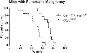
Results: Active Akt1 (Akt1(Myr), containing a myristoylation sequence) cooperated with active mutant KRas(G12D) to accelerate pancreatic carcinoma onset and progression and increase phosphorylation of downstream effectors in the Akt pathway. Mucin and smooth muscle actin expression was found in and around pancreatic intraepithelial neoplasms (PanINs), and accelerated time to metastasis was found in Akt1(Myr)/KRas(G12D) mice.
Conclusions: In contrast to prior reports of pancreatic KRas mutant mice mated with mice deficient for various tumor suppressor genes, which resulted in aggressive disease within a few months of age, Akt1(Myr)/KRas(G12D) mice enabled the study of PanINs and spontaneous pancreatic transformation more characteristic of human pancreatic progression in elderly individuals. The Akt1(Myr)/KRas(G12D) model holds promise for delineating the tumor biology and biomarkers critical for understanding their cooperation in cancer oncogenesis and future targeting in therapeutic strategies.
Copyright © 2014 The Authors. Published by Elsevier Inc. All rights reserved.
Figures


References
-
- Hansel D.E., Kern S.E., Hruban R.H. Molecular pathogenesis of pancreatic cancer. Annu Rev Genomics Hum Genet. 2003;4:237–256. - PubMed
-
- Gu G., Dubauskaite J., Melton D.A. Direct evidence for the pancreatic lineage: NGN3 + cells are islet progenitors and are distinct from duct progenitors. Development. 2002;129:2447–2457. - PubMed
-
- Hingorani S.R., Petricoin E.F., Maitra A., Rajapakse V., King C., Jacobetz M.A., Ross S., Conrads T.P., Veenstra T.D., Hitt B.A. Preinvasive and invasive ductal pancreatic cancer and its early detection in the mouse. Cancer Cell. 2003;4:437–450. - PubMed
MeSH terms
Substances
Grants and funding
LinkOut - more resources
Full Text Sources
Other Literature Sources
Medical
Molecular Biology Databases
Research Materials
Miscellaneous
