Review
. 2015 Jun:32:73-9.
doi: 10.1016/j.gde.2015.02.004. Epub 2015 Mar 3.

Watching eyes take shape

Affiliations
Review

Watching eyes take shape

Naiara Bazin-Lopez et al. Curr Opin Genet Dev. 2015 Jun.

Abstract

Vertebrate eye formation is a multistep process requiring coordinated inductive interactions between neural and non-neural ectoderm and underlying mesendoderm. The induction and shaping of the eyes involves an elaborate cellular choreography characterized by precise changes in cell shape coupled with complex cellular and epithelial movements. Consequently, the forming eye is an excellent model to study the cellular mechanisms underlying complex tissue morphogenesis. Using examples largely drawn from recent studies of optic vesicle formation in zebrafish and in cultured embryonic stem cells, in this short review, we highlight some recent advances in our understanding of the events that shape the vertebrate eye.


Figures

Figure 1
Figure 1
Eye field cells have different behaviours to, and do not intermix with, cells in adjacent neural plate domains. Left: Schematic representation of prospective forebrain territories at neural plate stage highlighting the eye field-telencephalon boundary. Eye field cells start to evaginate laterally (small green arrows) at the same time that most anterior neural plate cells are still converging towards the midline (large green arrows). The inset highlights the eye field-telencephalon boundary: Rx3 regulates genes that influence cell behaviours in the eye field. For instance, it restricts the expression of at least two eph genes to neural plate territories surrounding the eye and Eph/ephrin signalling subsequently maintains segregation between eye field cells and adjacent neural plate territories. Rx3 also controls the expression of genes that mediate discrete cell behaviours in the eye field.
Figure 2
Figure 2
The optic vesicle is progressively epithelialized during morphogenesis in fish. Left schematic is a frontal view of the brain showing the evaginating optic vesicles. The blue dashes indicate the internal position of ventricular space within the optic vesicles and orange dots indicate abundant Laminin. The inset shows the organization of neuroepithelial cells in the evaginating optic vesicles. Cells at the margin of the eye field (pale green) show coordinated epithelial organization dependent upon contact with a Laminin-1 enriched basal lamina. Core cells (yellow) intercalate into the nascent epithelium aligning their apicobasal polarity and shape with their polarized neighbours during this process. Based on Ivanovitch et al. [5••].
Figure 3
Figure 3
Generation of the choroid fissure during optic cup formation. Schematic frontal view of the forebrain showing the eyes and (inset) polarized retinal neuroepithelial cells during optic cup morphogenesis. The left side of the main figure shows a surface view of the optic vesicle and ventrally positioned choroid fissure. POM cells (green) surround the optic cup, invade the choroid fissure and later form the blood vessels of the eye (blue). The right side of the main figure shows a slice through the optic cup at the level of the choroid fissure. Retinal ganglion cell axons (red) use the fissure as a route out of the eye as they navigate towards central targets in the brain. The inset shows a higher resolution view of neuroepithelial cells in the neural retina. The basal end feet of these cells localize Integrin in an Ojoplano and Numb/Numbl dependent mechanism, and their constriction (green arrows) is thought to contribute to neuroepithelial bending and the invagination process. Based largely on Martinez-Morales et al. [48] and Bogdanović et al. [49]. Abbreviations: CF, choroid fissure; Hyp, hypothalamus; L, Lens; NR, neural retina; OS, optic stalk; POM, periocular mesenchyme; RPE, retinal pigment epithelium; RGC, retinal ganglion cells; Tel, telencephalon; VRV ventral retinal blood vessels.

References

    1. Beccari L., Marco-Ferreres R., Bovolenta P. The logic of gene regulatory networks in early vertebrate forebrain patterning. Mech Dev. 2013;130:95–111. - PubMed
    1. Cavodeassi F., Houart C. Brain regionalization: of signaling centers and boundaries. Dev Neurobiol. 2012;72:218–233. - PubMed
    1. Kiecker C., Lumsden A. The role of organizers in patterning the nervous system. Annu Rev Neurosci. 2012;35:347–367. - PubMed
    1. Zuber M.E., Gestri G., Viczian A.S., Barsacchi G., Harris W.A. Specification of the vertebrate eye by a network of eye field transcription factors. Dev Camb Engl. 2003;130:5155–5167. - PubMed
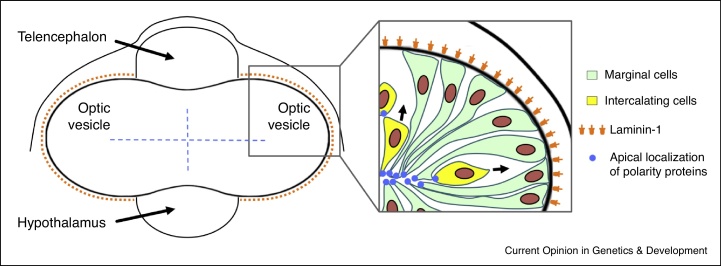
    1. Ivanovitch K., Cavodeassi F., Wilson S.W. Precocious acquisition of neuroepithelial character in the eye field underlies the onset of eye morphogenesis. Dev Cell. 2013;27:293–305. - PMC - PubMed
    2. Using 4-dimensional imaging in zebrafish, this study shows that eye field cells polarize and adopt neuroepithelial character prior to other regions of the neural plate in a process mediated by Laminin1.

LinkOut - more resources