. 2015 Mar 9:5:8883.
doi: 10.1038/srep08883.

Human iPSC-based cardiac microphysiological system for drug screening applications

Affiliations

Human iPSC-based cardiac microphysiological system for drug screening applications

Anurag Mathur et al. Sci Rep. .

Abstract

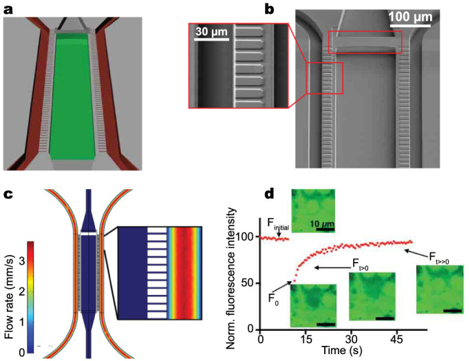
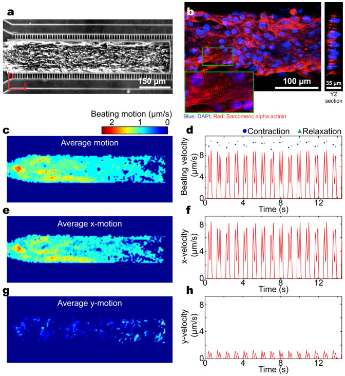
Drug discovery and development are hampered by high failure rates attributed to the reliance on non-human animal models employed during safety and efficacy testing. A fundamental problem in this inefficient process is that non-human animal models cannot adequately represent human biology. Thus, there is an urgent need for high-content in vitro systems that can better predict drug-induced toxicity. Systems that predict cardiotoxicity are of uppermost significance, as approximately one third of safety-based pharmaceutical withdrawals are due to cardiotoxicty. Here, we present a cardiac microphysiological system (MPS) with the attributes required for an ideal in vitro system to predict cardiotoxicity: i) cells with a human genetic background; ii) physiologically relevant tissue structure (e.g. aligned cells); iii) computationally predictable perfusion mimicking human vasculature; and, iv) multiple modes of analysis (e.g. biological, electrophysiological, and physiological). Our MPS is able to keep human induced pluripotent stem cell derived cardiac tissue viable and functional over multiple weeks. Pharmacological studies using the cardiac MPS show half maximal inhibitory/effective concentration values (IC₅₀/EC₅₀) that are more consistent with the data on tissue scale references compared to cellular scale studies. We anticipate the widespread adoption of MPSs for drug screening and disease modeling.


Figures

Figure 1
Figure 1. The cardiac microphysiological system (MPS).
(a) Schematic of the MPS nutrient channels (red) and cell-loading channel (green). (b) Scanning electron micrograph of the MPS showing in inset the 2 μm endothelial-like barriers connecting the nutrient channel and the cell channel. Red rectangular box shows the weir that enables efficient and consistent loading of singularized high density hiPSC-CMs. (c) Schematic of the cardiac MPS showing nutrient inlet and outlet ports connected to tubes. (d) Simulated velocity profile of flow in the MPS, inset shows the magnified view. Note the lack of convection within the diffusive barriers and predominant convective flow through the nutrient channels. Thus, mass transport to the tissue is exclusively diffusive. (e) Diffusion dynamics in the microphysiological system. Normalized fluorescence recovery of 4 kDa FITC–dextran (0.2 mg/mL). Insets are confocal microscopy images corresponding to the FRAP experiment. Finitial is the time regime that corresponds to the initial fluorescence before bleaching; F0 is the fluorescence measurement immediately after photobleaching; Ft > 0 corresponds to the recovery of fluorescence after photobleaching; Ft ≫ 0 corresponds to maximal recovery of fluorescence at the end of the experiment. Scale bar: 10 μm.
Figure 2
Figure 2. Characterization of the 3D cardiac tissue in the microphysiological system (MPS).
(a) Optical microscopy image of a cardiac tissue in the MPS. (b) Confocal fluorescence microscopy imaging of the microtissue in the MPS reveals an aligned multiple cell layer thick structure. Inset shows a magnified view of the sarcomeric alpha actinin and DAPI staining. (c, d) Heat map of the time-averaged beating motion and corresponding average beating kinetics in the MPS, (e, f) along the channel axis, and (g, h) perpendicular to the channel axis.
Figure 3
Figure 3. Cardiac tissue derived from a genetically engineered hiPS cell line expressing a GCaMP6 reporter in the MPS.
(a) Frames from a fluorescence movie (GFP channel) showing the switching from dim to bright during activity of Ca2+ channels. (b) Time-course of the normalized fluorescence intensity (green) and the beating motion (red) obtained by computational analysis of the movie. This combination allows high throughput analysis of mechanical and electrophysiological properties.
Figure 4
Figure 4. Dose-dependent study of different drugs on the cardiac MPS.
(a) Isoproterenol causes a dose-dependent increase in beat rate and an EC50 value of 315 nM. (b) Verapamil induces a dose-dependent decrease in beat rate with incidences of arrhythmia observed at higher concentrations and an IC50 value of 950 nM. (c, d) Metoprolol and E4031 induce a dose-dependent decrease in beat rate with IC50 values of 2.3 μM and 1.9 nM respectively. Grey areas indicate unbound estimated therapeutic plasma concentration in patients (ETPC). Note there is not an ETPC highlighted for E-4031 because the drug is used solely for research purposes and only one clinical trial was conducted. Error bars indicate mean ± S.D.

References

    1. Herper M. The Cost Of Creating A New Drug Now $5 Billion, Pushing Big Pharma To Change <http://www.forbes.com/sites/matthewherper/2013/08/11/how-the-staggering-...> (2013).
    1. Mathur A. et al. Human induced pluripotent stem cell-based microphysiological tissue models of myocardium and liver for drug development. Stem cell research & therapy 4 Suppl 1, S14 (2013). - PMC - PubMed
    1. Paul S. M. et al. How to improve R&D productivity: the pharmaceutical industry's grand challenge. Nature reviews. Drug discovery 9, 203–214 (2010). - PubMed
    1. Scott C. W., Peters M. F. & Dragan Y. P. Human induced pluripotent stem cells and their use in drug discovery for toxicity testing. Toxicol Lett 219, 49–58 (2013). - PubMed
    1. Chi K. R. Revolution dawning in cardiotoxicity testing. Nature reviews. Drug discovery 12, 565–567 (2013). - PubMed

Publication types

MeSH terms

Substances