

Prenatal stress enhances excitatory synaptic transmission and impairs long-term potentiation in the frontal cortex of adult offspring rats
- PMID: 25749097
- PMCID: PMC4352064
- DOI: 10.1371/journal.pone.0119407
Prenatal stress enhances excitatory synaptic transmission and impairs long-term potentiation in the frontal cortex of adult offspring rats
Abstract
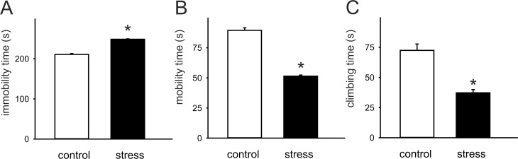
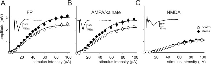
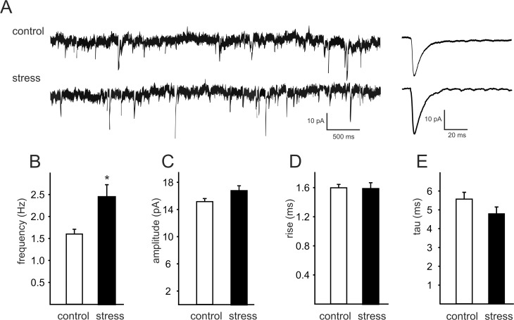
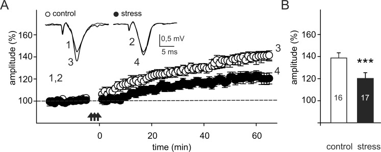
The effects of prenatal stress procedure were investigated in 3 months old male rats. Prenatally stressed rats showed depressive-like behavior in the forced swim test, including increased immobility, decreased mobility and decreased climbing. In ex vivo frontal cortex slices originating from prenatally stressed animals, the amplitude of extracellular field potentials (FPs) recorded in cortical layer II/III was larger, and the mean amplitude ratio of pharmacologically-isolated NMDA to the AMPA/kainate component of the field potential--smaller than in control preparations. Prenatal stress also resulted in a reduced magnitude of long-term potentiation (LTP). These effects were accompanied by an increase in the mean frequency, but not the mean amplitude, of spontaneous excitatory postsynaptic currents (sEPSCs) in layer II/III pyramidal neurons. These data demonstrate that stress during pregnancy may lead not only to behavioral disturbances, but also impairs the glutamatergic transmission and long-term synaptic plasticity in the frontal cortex of the adult offspring.
Conflict of interest statement
Figures
Similar articles
-
Imipramine counteracts corticosterone-induced enhancement of glutamatergic transmission and impairment of long-term potentiation in the rat frontal cortex.Pharmacol Rep. 2011;63(6):1404-12. doi: 10.1016/s1734-1140(11)70704-6. Pharmacol Rep. 2011. PMID: 22358088
-
Prenatal administration of morphine decreases CREBSerine-133 phosphorylation and synaptic plasticity range mediated by glutamatergic transmission in the hippocampal CA1 area of cognitive-deficient rat offspring.Hippocampus. 2003;13(8):915-21. doi: 10.1002/hipo.10137. Hippocampus. 2003. PMID: 14750654
-
β2-adrenoceptor stimulation restores frontal cortex plasticity and improves visuospatial performance in hidden-prenatally-malnourished young-adult rats.Neurobiol Learn Mem. 2015 Mar;119:1-9. doi: 10.1016/j.nlm.2014.11.003. Epub 2014 Nov 20. Neurobiol Learn Mem. 2015. PMID: 25464009
-
The 5-HT(7) receptor antagonist SB 269970 counteracts restraint stress-induced attenuation of long-term potentiation in rat frontal cortex.J Physiol Pharmacol. 2011 Dec;62(6):663-7. J Physiol Pharmacol. 2011. PMID: 22314569
-
Impaired in vivo synaptic plasticity in dentate gyrus and spatial memory in juvenile rats induced by prenatal morphine exposure.Hippocampus. 2009 Jul;19(7):649-57. doi: 10.1002/hipo.20540. Hippocampus. 2009. PMID: 19115391
Cited by
-
Restraint Stress and Repeated Corticosterone Administration Differentially Affect Neuronal Excitability, Synaptic Transmission and 5-HT7 Receptor Reactivity in the Dorsal Raphe Nucleus of Young Adult Male Rats.Int J Mol Sci. 2022 Nov 18;23(22):14303. doi: 10.3390/ijms232214303. Int J Mol Sci. 2022. PMID: 36430779 Free PMC article.
-
Psychosocial Crowding Stress-Induced Changes in Synaptic Transmission and Glutamate Receptor Expression in the Rat Frontal Cortex.Biomolecules. 2021 Feb 16;11(2):294. doi: 10.3390/biom11020294. Biomolecules. 2021. PMID: 33669305 Free PMC article.
-
Role of Polyinosinic:Polycytidylic Acid-Induced Maternal Immune Activation and Subsequent Immune Challenge in the Behaviour and Microglial Cell Trajectory in Adult Offspring: A Study of the Neurodevelopmental Model of Schizophrenia.Int J Mol Sci. 2021 Feb 4;22(4):1558. doi: 10.3390/ijms22041558. Int J Mol Sci. 2021. PMID: 33557113 Free PMC article.
-
Depression-like effects induced by chronic unpredictable mild stress in mice are rapidly reversed by a partial negative allosteric modulator of mGlu5 receptor, M-5MPEP.Psychopharmacology (Berl). 2025 Jun;242(6):1259-1273. doi: 10.1007/s00213-024-06724-4. Epub 2024 Nov 30. Psychopharmacology (Berl). 2025. PMID: 39615019 Free PMC article.
-
The Antidepressant-like Activity and Cognitive Enhancing Effects of the Combined Administration of (R)-Ketamine and LY341495 in the CUMS Model of Depression in Mice Are Related to the Modulation of Excitatory Synaptic Transmission and LTP in the PFC.Pharmaceuticals (Basel). 2023 Feb 14;16(2):288. doi: 10.3390/ph16020288. Pharmaceuticals (Basel). 2023. PMID: 37083635 Free PMC article.
References
-
- Basta-Kaim A, Szczesny E, Glombik K, Stachowicz K, Slusarczyk J, Nalepa I, et al. Prenatal stress affects insulin-like growth factor-1 (IGF-1) level and IGF-1 receptor phosphorylation in the brain of adult rats. Eur Neuropsychopharmacol. 2014;24: 1546–1556. 10.1016/j.euroneuro.2014.07.002 - DOI - PubMed
Publication types
MeSH terms
LinkOut - more resources
Full Text Sources
Other Literature Sources
Miscellaneous
