Review
. 2015 Jun;185(6):1518-27.
doi: 10.1016/j.ajpath.2014.11.030. Epub 2015 Mar 4.

Pathogenesis of Staphylococcus aureus abscesses

Affiliations
Review

Pathogenesis of Staphylococcus aureus abscesses

Scott D Kobayashi et al. Am J Pathol. 2015 Jun.

Abstract

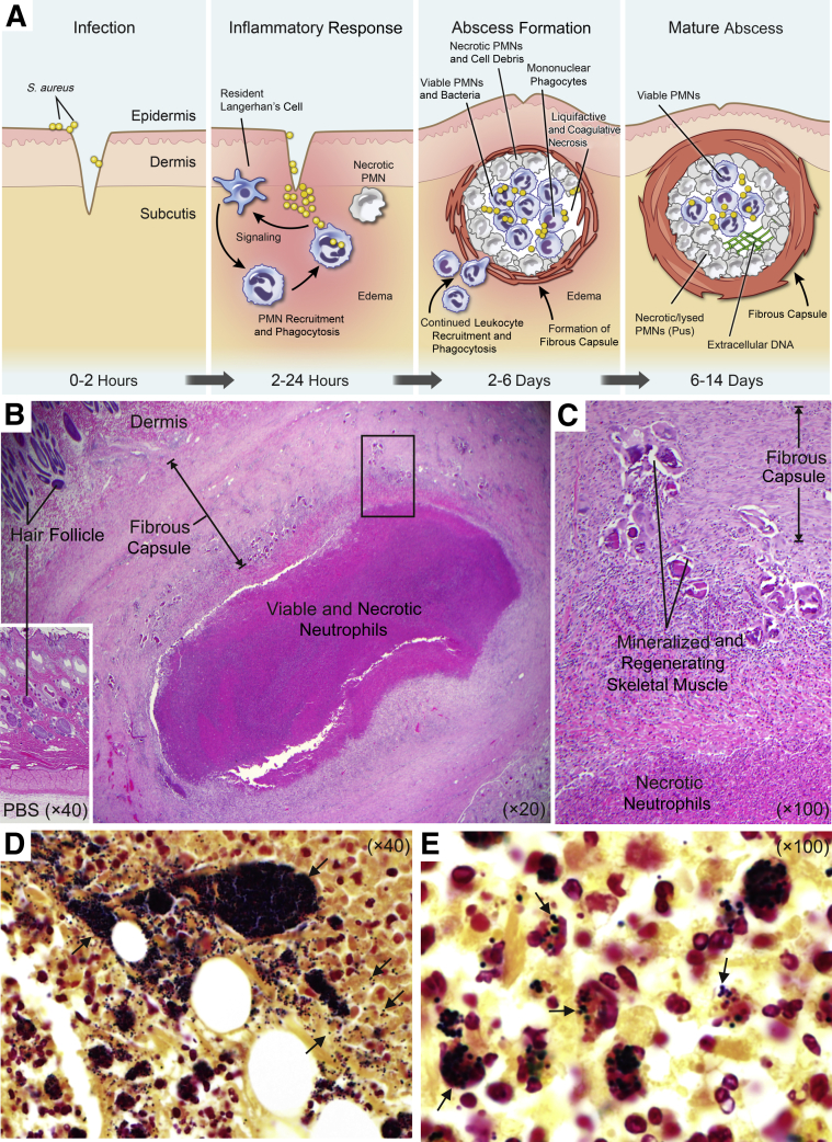
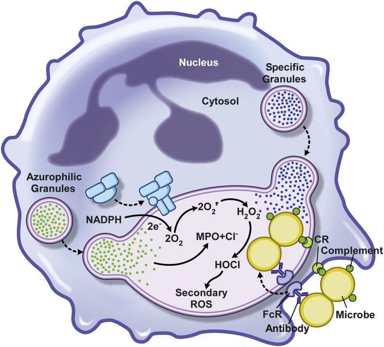
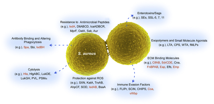
Staphylococcus aureus causes many types of human infections and syndromes-most notably skin and soft tissue infections. Abscesses are a frequent manifestation of S. aureus skin and soft tissue infections and are formed, in part, to contain the nidus of infection. Polymorphonuclear leukocytes (neutrophils) are the primary cellular host defense against S. aureus infections and a major component of S. aureus abscesses. These host cells contain and produce many antimicrobial agents that are effective at killing bacteria, but can also cause non-specific damage to host tissues and contribute to the formation of abscesses. By comparison, S. aureus produces several molecules that also contribute to the formation of abscesses. Such molecules include those that recruit neutrophils, cause host cell lysis, and are involved in the formation of the fibrin capsule surrounding the abscess. Herein, we review our current knowledge of the mechanisms and processes underlying the formation of S. aureus abscesses, including the involvement of polymorphonuclear leukocytes, and provide a brief overview of therapeutic approaches.


Figures

Figure 1
Figure 1
Staphylococcus aureus skin abscesses. A: Formation of a S. aureus skin abscess. B: Representative histopathological section of a typical rabbit skin abscess at day 14 after infection. C: Increased magnification of the boxed area shown in B. D and E: Gram stains of histological sections of a rabbit abscess. Arrows in D indicate S. aureus. The dark area is a colony of S. aureus. Arrows in E indicate S. aureus associated with leukocytes within the abscess. These studies conformed to the guidelines set forth by the NIH and were approved by the Institutional Animal Care and Use Committee at Rocky Mountain Laboratories, National Institute of Allergy and Infectious Diseases (Hamilton, Montana). PBS, phosphate-buffered saline; PMN, polymorphonuclear leukocyte.
Figure 2
Figure 2
Polymorphonuclear leukocyte phagocytosis and microbicidal processes. Surface receptors for host opsonins, such as complement and antibody, promote ingestion of S. aureus, which, in turn, activates the microbicidal processes that operate in a bacteria-containing phagosome (the cytoplasmic vacuole containing bacteria). The enzyme complex responsible for generation of superoxide—NADPH oxidase—is depicted by the blue cluster of shapes on the phagosome membrane. CR, complement receptor; HOCl, hypochlorous acid; MPO, myeloperoxidase; ROS, reactive oxygen species.
Figure 3
Figure 3
Staphylococcus aureus virulence molecules. Staphylococcus aureus can produce multiple types of molecules that contribute to virulence and pathogenesis. Many of these molecules have been linked to the pathogenesis of abscesses (red text). AhpCF, alkyl hydroperoxide reductase subunits C and F; Aur, aureolysin; BsaA, glutathione peroxidase; CHIPS, chemotaxis inhibitory protein of staphylococcus; Clf, clumping factor; Cna, collagen adhesin; Coa, coagulase; CPS, capsule; Eap, extracellular adherence protein; Efb, extracellular fibrinogen binding protein; FLIPr, formyl peptide receptor-like 1 inhibitory protein; fMLP, N-formyl-methionyl-leucyl-phenylalanine; FnBPAB, fibronectin binding protein A and B; Hla, α-hemolysin; HlgABC, gamma-hemolysin subunits A, B, and C; IcaADBCR, intercellular adhesin subunits A, D, B, C, and R; Isd, iron-regulated surface determinant; KatA, catalase; LTA, lipoteichoic acid; Luk, leukocidin; MprF, multiple peptide resistance factor; OatA, O-acetyltransferase A; PSM, phenol-soluble modulin; PVL, Panton-Valentine leukocidin; ROS, reactive oxygen species; Sak, staphylokinase; Sbi, staphylococcal IgG-binding protein; SCIN, staphylococcal complement inhibitor; SdrCDE, Ser-Asp rich fibrinogen/bone sialoprotein-binding protein subunits C, D, and E; SE, staphylococcal enterotoxin; SOD, superoxide dismutase; Spa, staphylococcal protein A; SSL, staphylococcal superantigen-like protein; SXN, staphyloxanthin; TrxAB, thioredoxin (TrxA) and thioredoxin reductase (TrxB); vWbp, von Willebrand factor binding protein; WTA, wall techoic acid.

References

    1. Gorwitz R.J., Kruszon-Moran D., McAllister S.K., McQuillan G., McDougal L.K., Fosheim G.E., Jensen B.J., Killgore G., Tenover F.C., Kuehnert M.J. Changes in the prevalence of nasal colonization with Staphylococcus aureus in the United States, 2001-2004. J Infect Dis. 2008;197:1226–1234. - PubMed
    1. Wertheim H.F., Melles D.C., Vos M.C., van Leeuwen W., van Belkum A., Verbrugh H.A., Nouwen J.L. The role of nasal carriage in Staphylococcus aureus infections. Lancet Infect Dis. 2005;5:751–762. - PubMed
    1. Moran G.J., Krishnadasan A., Gorwitz R.J., Fosheim G.E., McDougal L.K., Carey R.B., Talan D.A., EMERGENcy ID Net Study Group Methicillin-resistant S. aureus infections among patients in the emergency department. N Engl J Med. 2006;355:666–674. - PubMed
    1. de Kraker M.E., Jarlier V., Monen J.C., Heuer O.E., van de Sande N., Grundmann H. The changing epidemiology of bacteraemias in Europe: trends from the European Antimicrobial Resistance Surveillance System. Clin Microbiol Infect. 2013;19:860–868. - PubMed
    1. Styers D., Sheehan D.J., Hogan P., Sahm D.F. Laboratory-based surveillance of current antimicrobial resistance patterns and trends among Staphylococcus aureus: 2005 status in the United States. Ann Clin Microbiol Antimicrob. 2006;5:2. - PMC - PubMed

Publication types