. 2015 May;353(2):369-79.
doi: 10.1124/jpet.114.220368. Epub 2015 Mar 6.

Protective role of cannabinoid receptor 2 activation in galactosamine/lipopolysaccharide-induced acute liver failure through regulation of macrophage polarization and microRNAs

Affiliations

Protective role of cannabinoid receptor 2 activation in galactosamine/lipopolysaccharide-induced acute liver failure through regulation of macrophage polarization and microRNAs

Sunil Tomar et al. J Pharmacol Exp Ther. 2015 May.

Abstract

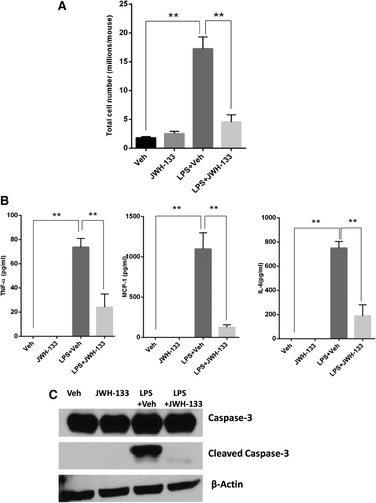
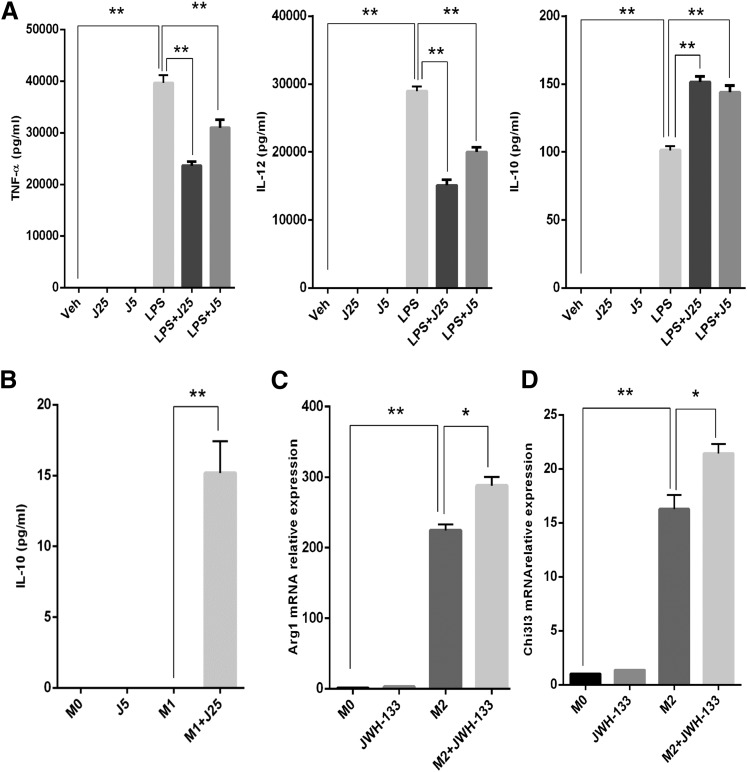
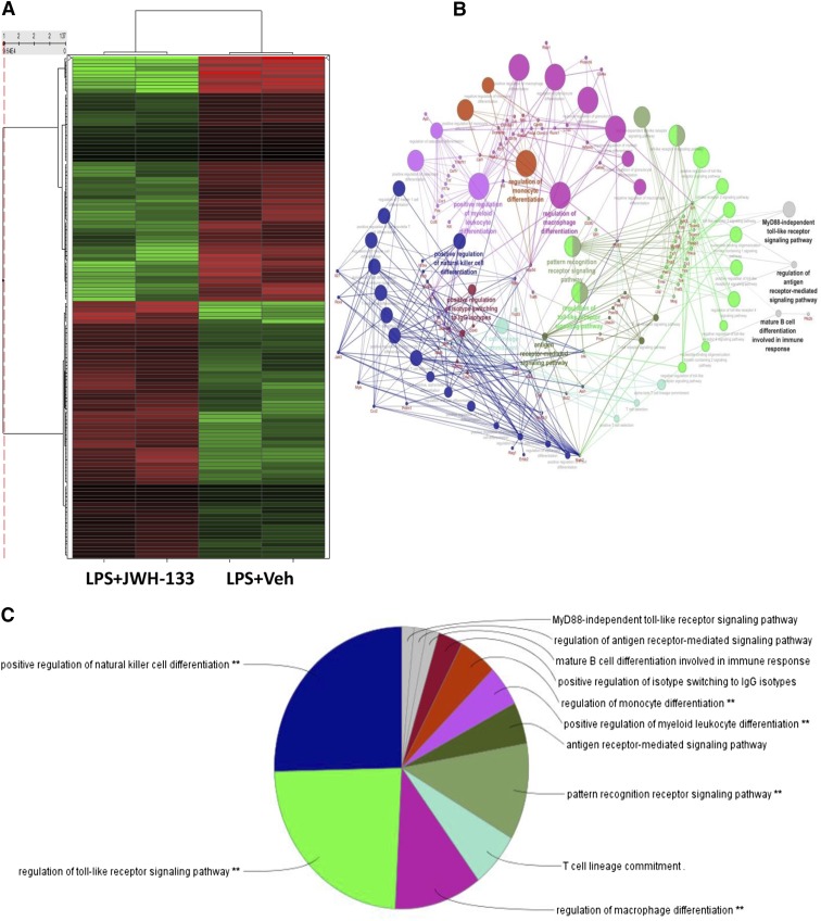
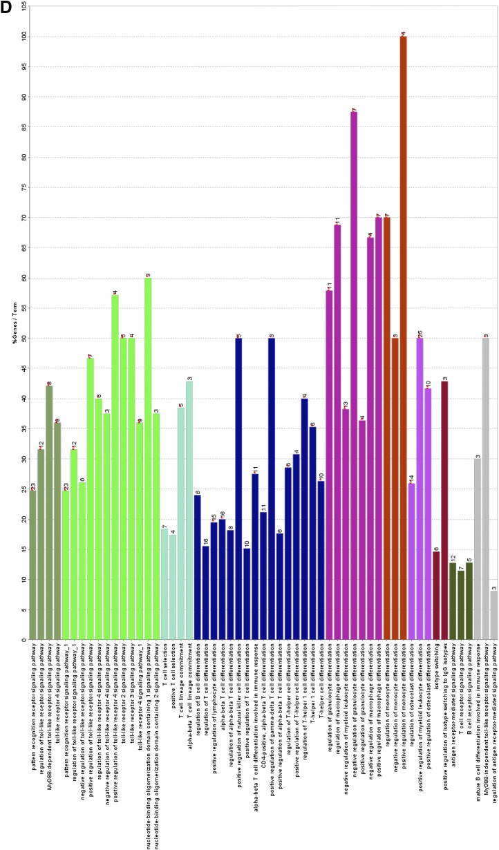
Acute liver failure (ALF) is a potentially life-threatening disorder without any effective treatment strategies. d-Galactosamine (GalN)/lipopolysaccharide (LPS)-induced ALF is a widely used animal model to identify novel hepato-protective agents. In the present study, we investigated the potential of a cannabinoid receptor 2 (CB2) agonist, JWH-133 [(6aR,10aR)-3-(1,1-dimethylbutyl)-6a,7,10,10a-tetrahydro-6,6,9-trimethyl-6H-dibenzo[b,d]pyran], in the amelioration of GalN/LPS-induced ALF. JWH-133 treatment protected the mice from ALF-associated mortality, mitigated alanine transaminase and proinflammatory cytokines, suppressed histopathological and apoptotic liver damage, and reduced liver infiltration of mononuclear cells (MNCs). Furthermore, JWH-133 pretreatment of M1/M2-polarized macrophages significantly increased the secretion of anti-inflammatory cytokine interleukin-10 (IL-10) in M1 macrophages and potentiated the expression of M2 markers in M2-polarized macrophages. In vivo, JWH-133 treatment also suppressed ALF-triggered expression of M1 markers in liver MNCs, while increasing the expression of M2 markers such as Arg1 and IL-10. microRNA (miR) microarray analysis revealed that JWH-133 treatment altered the expression of only a few miRs in the liver MNCs. Gene ontology analysis of the targets of miRs suggested that Toll-like receptor (TLR) signaling was among the most significantly targeted cellular pathways. Among the altered miRs, miR-145 was found to be the most significantly decreased. This finding correlated with concurrent upregulated expression of its predicted target gene, interleukin-1 receptor-associated kinase 3, a negative regulator of TLR4 signaling. Together, these data are the first to demonstrate that CB2 activation attenuates GalN/LPS-induced ALF by inducing an M1 to M2 shift in macrophages and by regulating the expression of unique miRs that target key molecules involved in the TLR4 pathway.


Figures

Fig. 1.
Fig. 1.
JWH-133 treatment attenuates LPS-induced ALF. (A) Percent survival of mice at different time (hours) intervals after coadministration of GalN/LPS+vehicle (depicted as LPS+Veh) or GalN/LPS+JWH-133 (LPS+JWH-133), as described in Materials and Methods. (B) Sera collected at 12 hours post-LPS treatment were analyzed for ALT levels. (C) Histologic examination of livers from mice following H&E staining. *P < 0.05; **P < 0.01.
Fig. 2.
Fig. 2.
JWH-133 treatment decreases liver infiltration, apoptosis, and production of systemic proinflammatory cytokines. (A) Liver-infiltrating MNCs were isolated as described in Materials and Methods. The cells were counted to get absolute numbers of infiltrating cells in liver. Blood was collected from mice at 12 hours after GalN/LPS+vehicle (veh) injection and (B) sera were analyzed for cytokines using sandwich enzyme-linked immunosorbent assay for TNF-α, MCP-1, and IL-6. (C) Protein samples were isolated from liver tissues harvested 12 hours after GalN/LPS+vehicle injection and run on SDS-PAGE for immunoblotting to probe for caspase-3 (both uncleaved and cleaved) and β-actin as a control. **P < 0.01.
Fig. 3.
Fig. 3.
CB2 stimulation by JWH-133 decreases M1 while increasing M2 phenotype activation of macrophages in vitro. (A) Thioglycollate-induced peritoneal macrophages were treated with JWH-133 (25 and 5 µM, shown as J25 and J5), and 1 hour later, cells were stimulated with LPS (100 ng/ml). Conditioned media from macrophage cultures were analyzed for proinflammatory cytokines (TNF-α, IL-12, and anti-inflammatory cytokine IL-10) by enzyme-linked immunosorbent assay. Peritoneal macrophages were treated with JWH-133 (5 µM) for 1 hour and then stimulated toward M1 or M2 phenotypes. (B) IL-10 secretion was quantified in conditioned medium from M1-polarized macrophages using enzyme-linked immunosorbent assay. RNA isolated from the macrophage cultures was used for quantitative PCR to quantify relative expression of genes: Arg-1 (C) and Chi3l3 (D). *P < 0.05; ** P < 0.01. Veh, vehicle.
Fig. 4.
Fig. 4.
JWH-133 treatment induces in vivo M2 polarization of macrophages in mice with LPS-induced ALF. Liver MNCs were isolated for the different groups of animals treated with vehicle (Veh) alone, JWH-133 alone, LPS+Veh, or LPS+JWH-133 at 12 hours after GalN/LPS injection. Relative expression of typical M1 markers, TNF-α and Nos2 (A), and typical M2 markers, Arg-1 and IL-10 (B), in liver MNCs as quantified by qRT-PCR is shown. *P < 0.05; **P < 0.01.
Fig. 5.
Fig. 5.
JWH-133 treatment causes a differential expression of a number of miRNAs in liver MNCs that can potentially regulate TLR signaling pathway. Mice were treated with LPS+vehicle or LPS+JWH-133 as described in Fig. 1, and liver MNCs were isolated for each group as described in Materials and Methods. (A) Heat map for the expression of 2023 miRNAs, with the first two lanes showing LPS+JWH-133 treatment and the latter two lanes showing LPS+vehicle treatment. (B) Gene ontology analysis of the predicted target genes of miRNAs identifies the major cellular pathways altered in the JWH-133+LPS group as compared with the LPS+vehicle group. A pie chart (C) and bar graph analysis (D) of the major cellular pathways altered in the JWH-133+LPS group when compared with the LPS+vehicle based on miRNAs altered by more than 1.5-fold.
Fig. 5.
Fig. 5.
JWH-133 treatment causes a differential expression of a number of miRNAs in liver MNCs that can potentially regulate TLR signaling pathway. Mice were treated with LPS+vehicle or LPS+JWH-133 as described in Fig. 1, and liver MNCs were isolated for each group as described in Materials and Methods. (A) Heat map for the expression of 2023 miRNAs, with the first two lanes showing LPS+JWH-133 treatment and the latter two lanes showing LPS+vehicle treatment. (B) Gene ontology analysis of the predicted target genes of miRNAs identifies the major cellular pathways altered in the JWH-133+LPS group as compared with the LPS+vehicle group. A pie chart (C) and bar graph analysis (D) of the major cellular pathways altered in the JWH-133+LPS group when compared with the LPS+vehicle based on miRNAs altered by more than 1.5-fold.
Fig. 6.
Fig. 6.
JWH-133 treatment suppresses TLR signaling in liver MNCs by decreasing the expression of miR-145 to regulate IRAK3. (A) Table of miRNAs significantly increased or decreased by more than 1.5-fold change in LPS+JWH-133–treated mice when compared with LPS+vehicle (Veh)-treated mice. (B) Toll-like receptor pathway showing the predicted interaction between miR-145, the most significantly altered miRNAs in the LPS+JWH-133group with its predicted target IRAK3 (shown in blue), and how they relate to the rest of the members of the pathway. (C) miRanda algorithm generated miR-145 and mRNA alignment at the 3′-untranslated region of IRAK3 mRNA. mirSVR and PhastCons conservation scores are also shown. miR-145 (D) and its predicted target IRAK3 in liver MNCs (E), as quantified by qRT-PCR at 12 hours after GalN/LPS injection. *P < 0.05; **P < 0.01. CYLD, cylindromatosis (turban tumor syndrome); IKK, inhibitor of κ light polypeptide gene enhancer in B cells, kinase β; MAPK, mitogen-activated protein kinase; NF-κB, nuclear factor κB; PPARA, peroxisome proliferator-activated receptor α; TIRAP, Toll-interleukin 1 receptor (TIR) domain containing adaptor protein.

References

    1. Adhikary S, Kocieda VP, Yen JH, Tuma RF, Ganea D. (2012) Signaling through cannabinoid receptor 2 suppresses murine dendritic cell migration by inhibiting matrix metalloproteinase 9 expression. Blood 120:3741–3749. - PMC - PubMed
    1. Antoniades CG, Berry PA, Wendon JA, Vergani D. (2008) The importance of immune dysfunction in determining outcome in acute liver failure. J Hepatol 49:845–861. - PubMed
    1. Bátkai S, Osei-Hyiaman D, Pan H, El-Assal O, Rajesh M, Mukhopadhyay P, Hong F, Harvey-White J, Jafri A, Haskó G, et al. (2007) Cannabinoid-2 receptor mediates protection against hepatic ischemia/reperfusion injury. FASEB J 21:1788–1800. - PMC - PubMed
    1. Bernal W, Auzinger G, Dhawan A, Wendon J. (2010) Acute liver failure. Lancet 376:190–201. - PubMed
    1. Bindea G, Mlecnik B, Hackl H, Charoentong P, Tosolini M, Kirilovsky A, Fridman WH, Pagès F, Trajanoski Z, Galon J. (2009) ClueGO: a Cytoscape plug-in to decipher functionally grouped gene ontology and pathway annotation networks. Bioinformatics 25:1091–1093. - PMC - PubMed

Publication types

MeSH terms