. 2015 Jul;13(3):367-81.
doi: 10.1007/s12021-015-9264-7.

Segmentation of the Cerebellar Peduncles Using a Random Forest Classifier and a Multi-object Geometric Deformable Model: Application to Spinocerebellar Ataxia Type 6

Affiliations

Segmentation of the Cerebellar Peduncles Using a Random Forest Classifier and a Multi-object Geometric Deformable Model: Application to Spinocerebellar Ataxia Type 6

Chuyang Ye et al. Neuroinformatics. 2015 Jul.

Abstract

The cerebellar peduncles, comprising the superior cerebellar peduncles (SCPs), the middle cerebellar peduncle (MCP), and the inferior cerebellar peduncles (ICPs), are white matter tracts that connect the cerebellum to other parts of the central nervous system. Methods for automatic segmentation and quantification of the cerebellar peduncles are needed for objectively and efficiently studying their structure and function. Diffusion tensor imaging (DTI) provides key information to support this goal, but it remains challenging because the tensors change dramatically in the decussation of the SCPs (dSCP), the region where the SCPs cross. This paper presents an automatic method for segmenting the cerebellar peduncles, including the dSCP. The method uses volumetric segmentation concepts based on extracted DTI features. The dSCP and noncrossing portions of the peduncles are modeled as separate objects, and are initially classified using a random forest classifier together with the DTI features. To obtain geometrically correct results, a multi-object geometric deformable model is used to refine the random forest classification. The method was evaluated using a leave-one-out cross-validation on five control subjects and four patients with spinocerebellar ataxia type 6 (SCA6). It was then used to evaluate group differences in the peduncles in a population of 32 controls and 11 SCA6 patients. In the SCA6 group, we have observed significant decreases in the volumes of the dSCP and the ICPs and significant increases in the mean diffusivity in the noncrossing SCPs, the MCP, and the ICPs. These results are consistent with a degeneration of the cerebellar peduncles in SCA6 patients.


Figures

Fig. 1
Fig. 1
A manually drawn schematic of the cerebellar peduncles. Blue: left SCP; green: right SCP; red: MCP; orange: left ICP; yellow: right ICP. Shown together with the cerebellum (gray) and the brainstem (purple)
Fig. 2
Fig. 2
The SCPs (blue and green) shown with the red nuclei (red) and the dentate nuclei (yellow): (a) typical incorrect SCPs obtained from DTI and (b) segmentation of the SCPs including the decussation in the proposed method. Note that our SCPs do not extend through the dentate nuclei, which leads to a different appearance of the dentate nuclei due to transparency
Fig. 3
Fig. 3
Diffusion properties and cerebellar peduncles on two representative slices (Row 1 and 2 in each subfigure): (a) the 5D Knutsson vector, (b) the cerebellar peduncles for reference, and (c) the Westin indices. Within each subfigure, Row 1 shows an axial slice cutting through the brainstem where the SCPs decussate, and Row 2 shows an axial slice cutting through the body of the MCP
Fig. 4
Fig. 4
Manual delineations of the cerebellar peduncles overlaid on the PEV edge map (left) and the Cl map (right) on two representative slices (Row 1 and 2) in correspondence with Fig. 3. Row 1 shows an axial slice cutting through the brainstem where the SCPs decussate, and Row 2 shows an axial slice cutting through the body of the MCP
Fig. 5
Fig. 5
Means and standard deviations of the variable importance in the cross-validation test. The order of the variables is the same as in the feature vector f = (v, C, ϕ, x)
Fig. 6
Fig. 6
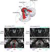
A segmentation result. (a) A 3D rendering (oblique view) of the cerebellar peduncles segmented by the proposed method in the cross-validation test, shown together with the cerebellum (gray). Axial cross sections of (b) the manual delineations and (c) the proposed segmentation contours overlaid on the FA map. Slice 1: a cut through the brainstem where the SCPs decussate. Slice 2: a cut through the cerebellum where all the cerebellar peduncles are visible. In all figures here, the left and right noncrossing SCPs are combined respectively with the dSCP to obtain the complete left and right SCPs. Blue: left complete SCP; green: right complete SCP; red: MCP; orange: lICP; yellow: rICP
Fig. 7
Fig. 7
Box plots of (a) tract volumes, (b) average FAs, and (c) average MDs of the segmented cerebellar peduncles. The numbers are compared between the control and the SCA6 group. Asterisks (*) indicate that statistically significant difference (p < 0.05) is observed in both the Student’s t-test and the Wilcoxon rank-sum test

Similar articles

Cited by

References

    1. Asman AJ, Landman BA. Formulating spatially varying performance in the statistical fusion framework. IEEE Transactions on Medical Imaging. 2012;31(6):1326–1336. - PMC - PubMed
    1. Avants BB, Epstein CL, Grossman M, Gee JC. Symmetric diffeomorphic image registration with cross-correlation: evaluating automated labeling of elderly and neurodegenerative brain. Medical Image Analysis. 2008;12(1):26–41. - PMC - PubMed
    1. Awate SP, Zhang H, Gee JC. A fuzzy, nonparametric segmentation framework for DTI and MRI analysis: With applications to DTI-tract extraction. IEEE Transactions on Medical Imaging. 2007;26(11):1525–1536. - PubMed
    1. Bazin PL, Ye C, Bogovic JA, Shiee N, Reich DS, Prince JL, Pham DL. Direct segmentation of the major white matter tracts in diffusion tensor images. NeuroImage. 2011;58(2):458–468. doi: 10.1016/j.neuroimage.2011.06.020. - DOI - PMC - PubMed
    1. Behrens TEJ, Berg HJ, Jbabdi S, Rushworth MFS, Woolrich MW. Probabilistic diffusion tractography with multiple fibre orientations: What can we gain? NeuroImage. 2007;34(1):144–155. - PMC - PubMed

Publication types

MeSH terms

LinkOut - more resources