Review
. 2015;78(5-6):307-320.
doi: 10.1007/s10337-014-2813-7. Epub 2014 Dec 9.

Clinical Glycomics Employing Graphitized Carbon Liquid Chromatography-Mass Spectrometry

Affiliations
Review

Clinical Glycomics Employing Graphitized Carbon Liquid Chromatography-Mass Spectrometry

Kathrin Stavenhagen et al. Chromatographia. 2015.

Abstract

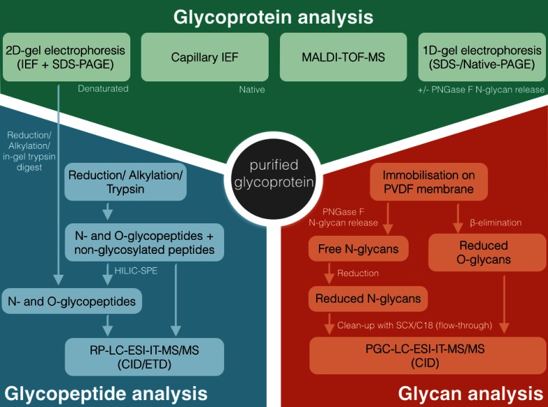
Glycoconjugates and free glycan are involved in a variety of biological processes such as cell-cell interaction and cell trafficking. Alterations in the complex glycosylation machinery have been correlated with various pathological processes including cancer progression and metastasis. Mass Spectrometry (MS) has evolved as one of the most powerful tools in glycomics and glycoproteomics and in combination with porous graphitized carbon-liquid chromatography (PGC-LC) it is a versatile and sensitive technique for the analysis of glycans and to some extent also glycopeptides. PGC-LC-ESI-MS analysis is characterized by a high isomer separation power enabling a specific glycan compound analysis on the level of individual structures. This allows the investigation of the biological relevance of particular glycan structures and glycan features. Consequently, this strategy is a very powerful technique suitable for clinical research, such as cancer biomarker discovery, as well as in-depth analysis of recombinant glycoproteins. In this review, we will focus on how PGC in conjunction with MS detection can deliver specific structural information for clinical research on protein-bound N-glycans and mucin-type O-glycans. In addition, we will briefly review PGC analysis approaches for glycopeptides, glycosaminoglycans (GAGs) and human milk oligosaccharides (HMOs). The presented applications cover systems that vary vastly with regard to complexity such as purified glycoproteins, cells, tissue or body fluids revealing specific glycosylation changes associated with various biological processes including cancer and inflammation.

Keywords: Clinical glycomics; Glycopeptides; Mass spectrometry; N-glycans; O-glycans; Porous graphitized carbon.


Conflict of interest statement

The authors declare no conflict of interest.

Figures

Fig. 1
Fig. 1
PGC–LC–ESI–IT–MS EICs of m/z 913.84, showing the different elution times of three N-glycan isomers with the composition Hexose4N-acetylhexosamine4Fucose1, derived from human colon tissue of an ulcerative colitis patient. Separation of the isobaric structures allows separate MS/MS analyses for in-depth structural characterization of the respective N-glycans. The EIC illustrates the different elution of structures with different glycan features, as the N-glycans containing a bisecting GlcNAc elute earlier than structural isomers with an additional antenna
Fig. 2
Fig. 2
PGC–LC–ESI–IT–MS EICs of monosialylated hybrid N-glycans in a the non-cancerous epithelial cells (HOSE 6.3) and b ovarian cancer cell line (SKOV 3). The authors found a set of different N-glycan structures containing α(2,6)-linked sialic acid exclusively in ovarian cancer cell lines but not in non-cancerous cell lines as represented for HOSE 6.3 and SKOV 3. The EICs further illustrate the different retention behaviors of linkage isomers with α(2,6)-linked and α(2,3)-linked sialic acid, as α(2,3)-linked sialic acid containing glycans are stronger retained and elutes later in the gradient [61]. © 2014 American Society for Biochemistry and Molecular Biology
Fig. 3
Fig. 3
PGC–LC–ESI–IT–MS EICs of sulfated core 1 O-glycan isomers (m/z 464.1) from acidic glycoproteins of synovial fluid from a patient with ReA (a) and RA (b). ReA patients showed a more diverse isomer pattern as it contains one structure with the sulfate linked to the GalNAc (RT 15.4 min) and two structures with a Gal-linked sulfate (RT 16.6 and 17.2 min). In contrast RA patients carry just a single Gal-linked sulfate structure on their synovial acidic glycoproteins (RT 15.6 min) [96]. © 2014 American Society for Biochemistry and Molecular Biology
Fig. 4
Fig. 4
Flow cytometry-based assay to quantify the adhesion of Candida albicans to BEC. a5 shows the adhesion of C. albicans to BEC normalized to 100 % and a4 the corresponding autofluorescence of BEC only. The inhibition of interaction was analyzed after incubation with 0.5 mL of whole saliva a3, N- and O-glycans released from 0.5 mL saliva a2 and N- and O-glycans released from 5 mL saliva a1. Salivary glycans as well as whole saliva inhibits binding of C. albicans to BEC. The graph contains the mean ± standard error of three independent biological replicates and their technical triplicates. (b) shows relative intensities of glycan epitopes from O-glycans of salivary and BEC membrane proteins, which express similar glycan epitopes in different relative intensities. Relative quantification was performed on MS ion intensities of all glycans carrying these epitopes. The graph contains the mean ± standard error of three technical replicates of both saliva and BEC collected from an individual of blood group A secretor status [44]. © Oxford University Press
Fig. 5
Fig. 5
A representative workflow for a multi-experimental comprehensive characterization of a purified glycoprotein that shows the suitability of PGC–LC–ESI–MS/MS implementation into an analytical workflow. The glycoprotein is analyzed on three different levels, including the analysis of the intact (glyco)protein in a top-down approach (upper part). Besides that glycans are characterized in a bottom-up approach by blotting the protein on a PDVF membrane and subsequent release of N- and O-glycans, which are then analyzed on PGC–LC–ESI–MS/MS (lower right part). This information is then be used for peptide and glycopeptide characterization in a bottom-up approach to elucidate the full micro- and macroheterogeneity of the glycoprotein (lower left part). Modified from Sumer-Bayraktar et al. [106]

References

    1. Varki A, Cummings RD, Esko JD, et al. Essentials of glycobiology. 2. NY: Cold Spring Harbor Laboratory Press; 2009. - PubMed
    1. Haslam SM, Julien S, Burchell JM, et al. Characterizing the glycome of the mammalian immune system. Immunol Cell Biol. 2008;86:564–573. - PubMed
    1. Van Kooyk Y, Rabinovich GA. Protein-glycan interactions in the control of innate and adaptive immune responses. Nat Immunol. 2008;9:593–601. - PubMed
    1. Hakomori S. Glycosylation defining cancer malignancy: new wine in an old bottle. Proc Natl Acad Sci USA. 2002;99:10231–10233. - PMC - PubMed
    1. Drake PM, Cho W, Li B, et al. Sweetening the pot: adding glycosylation to the biomarker discovery equation. Clin Chem. 2010;56:223–236. - PMC - PubMed

LinkOut - more resources