

Sports-related changes of the synovial membrane
- PMID: 25750907
- PMCID: PMC4334218
Sports-related changes of the synovial membrane
Abstract
Purpose: the aim of this study is to differentiate the behavior of the synovial membrane in the presence of various stimuli in patients who practice sports.
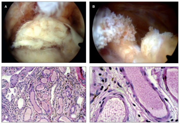
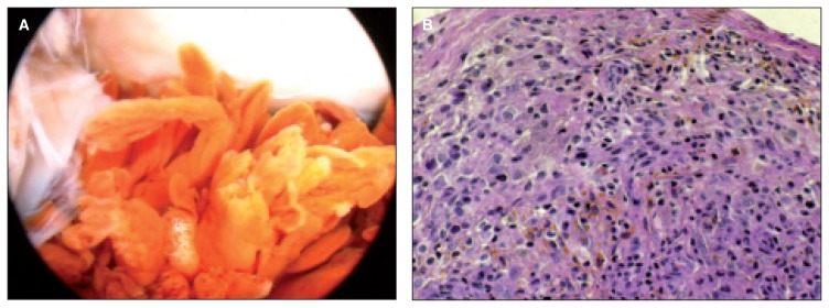
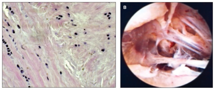
Methods: fifty one patients (30 males and 21 females, mean age 48 years, range 31-59 years) who actively practiced non-competitive sports underwent a biopsy of the synovial membrane during arthroscopic surgery performed for joint effusion secondary to meniscal lesion (24 cases), anterior cruciate ligament injury (ACL) (17 cases), postoperative knee joint stiffness (2 cases), aseptic loosening or dislocation of the polyethylene component of uni-compartmental knee arthroplasty (5 cases), and anterior fibrous impingement of the ankle (3 cases). Synovial tissue samples were obtained during surgery from all the patients and processed for light microscopy, transmission electron microscopy and scanning electron microscopy observation.
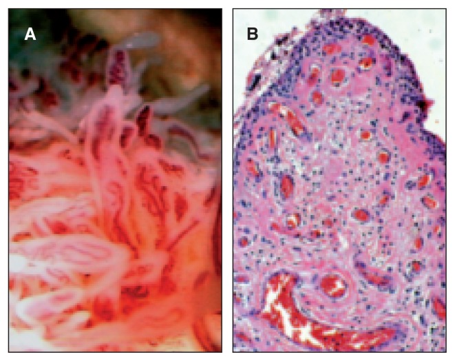
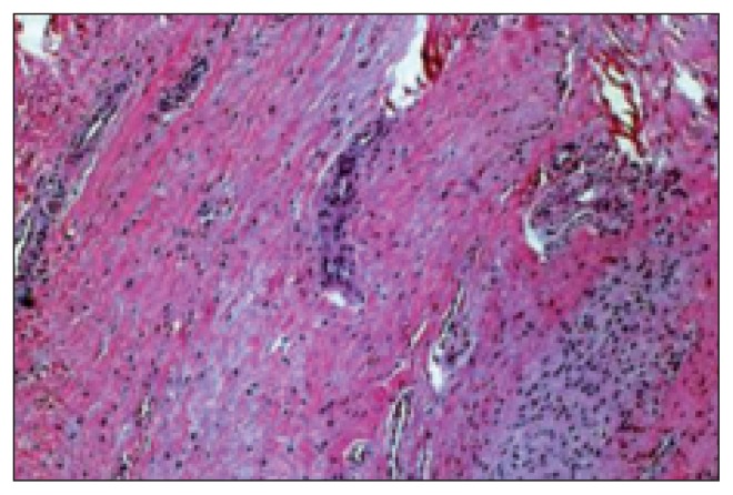
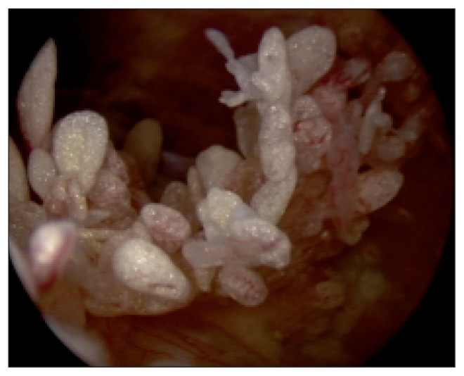
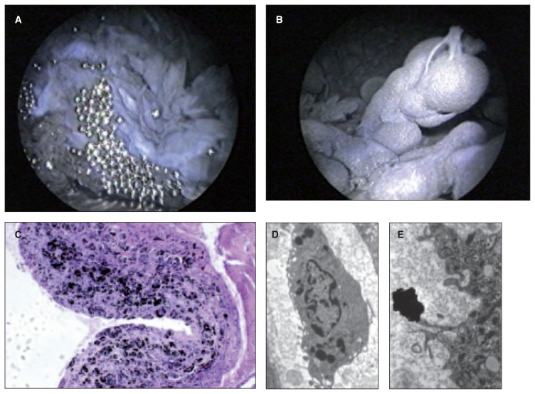
Results: circulatory phenomena were observed in acute inflammatory processes, characterized by hyperemia and vasodilation. Exudative and infiltrative phenomena were observed in the presence of foreign bodies and were characterized by leukocytic exudation (presence of polynuclear neutrophils), accompanied by lymphomonocytic infiltration. Proliferative phenomena were observed in post-traumatic forms of synovitis (ACL and meniscal injuries), characterized by hypertrophy and proliferation of villous formations. Degenerative and regressive phenomena were observed in cases of fibrous reaction (ankle impingement and joint stiffness) and were characterized by formation of dense fibrous connective tissue with hyaline patches, evolving towards sclerosis.
Conclusions: the activation of inflammatory processes in patients who expose their joints to excessive stress may lead to the formation of hyperplastic tissue. Ultramicroscopic debris is usually capable of transforming the structural organization of the synovial tissue.
Level of evidence: Level IV, observational case series.
Keywords: athlete; sports; synovial membrane; synovitis; ultrastructure.
Figures
References
-
- Berumen-Nafarrate E, Leal-Berumen I, Luevano E, et al. Synovial tissue and synovial fluid. J Knee Surg. 2002;15:46–48. - PubMed
-
- Soeur R. The synovial membrane of the knee in pathological conditions. J Bone Joint Surg Am. 1949;31A:317–340. - PubMed
-
- Dabby D, Dekel S. Synovial knee pain arising from chronic inflammatory disorders of the knee. J Knee Surg. 2002;15:53–56. - PubMed
-
- Frassica FJ, Combs JJ, Jr, Sim FH. Synovial proliferative disorders: differential diagnosis. Arthroscopy. 1985;1:183–189. - PubMed
LinkOut - more resources
Full Text Sources
Research Materials