. 2015 Apr;125(4):1713-25.
doi: 10.1172/JCI78578. Epub 2015 Mar 9.

CXCR3 blockade protects against Listeria monocytogenes infection-induced fetal wastage

CXCR3 blockade protects against Listeria monocytogenes infection-induced fetal wastage

Vandana Chaturvedi et al. J Clin Invest. 2015 Apr.

Abstract

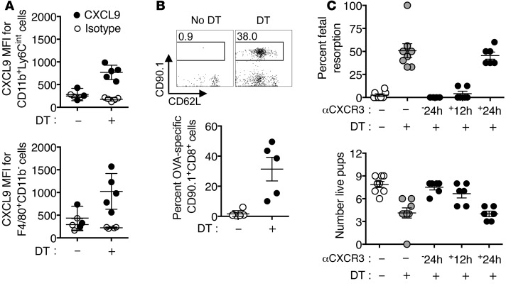
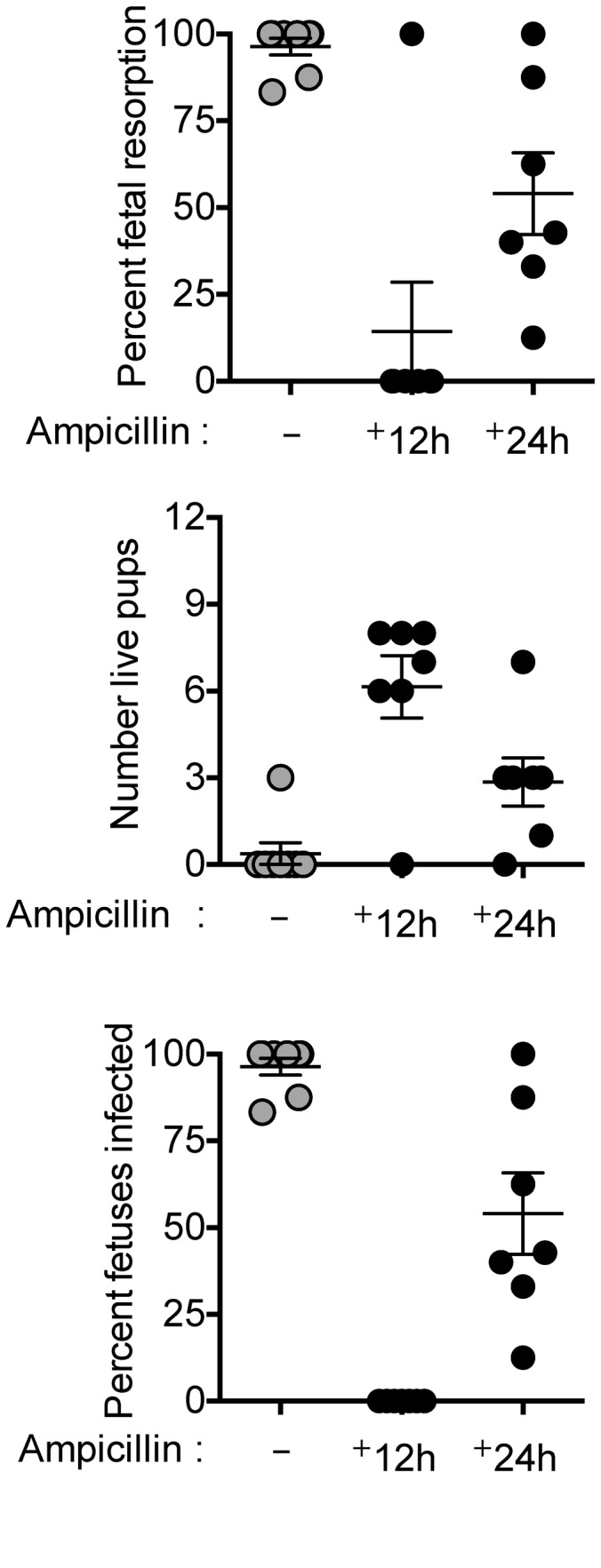
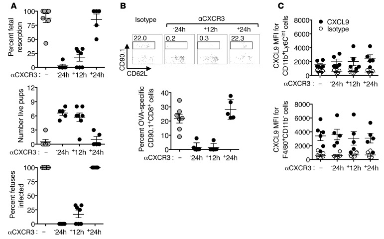
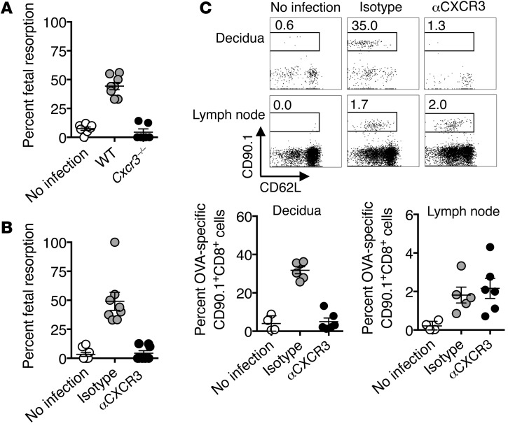
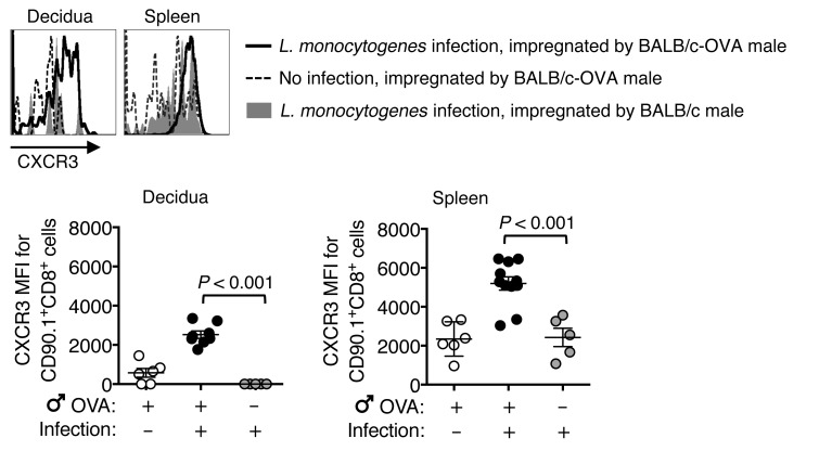
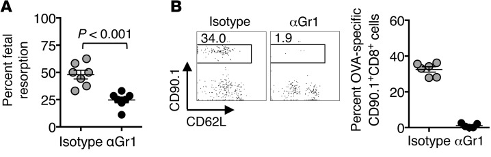
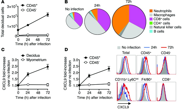
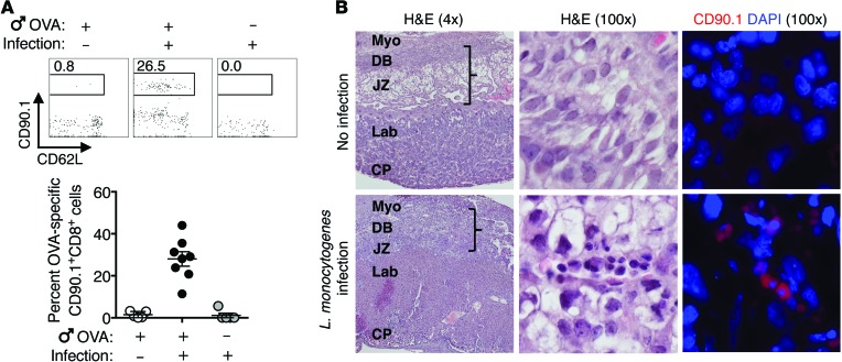
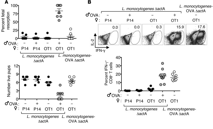
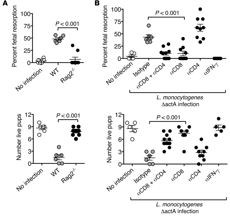
Mammalian pregnancy requires protection against immunological rejection of the developing fetus bearing discordant paternal antigens. Immune evasion in this developmental context entails silenced expression of chemoattractant proteins (chemokines), thereby preventing harmful immune cells from penetrating the maternal-fetal interface. Here, we demonstrate that fetal wastage triggered by prenatal Listeria monocytogenes infection is driven by placental recruitment of CXCL9-producing inflammatory neutrophils and macrophages that promote infiltration of fetal-specific T cells into the decidua. Maternal CD8+ T cells with fetal specificity upregulated expression of the chemokine receptor CXCR3 and, together with neutrophils and macrophages, were essential for L. monocytogenes-induced fetal resorption. Conversely, decidual accumulation of maternal T cells with fetal specificity and fetal wastage were extinguished by CXCR3 blockade or in CXCR3-deficient mice. Remarkably, protection against fetal wastage and in utero L. monocytogenes invasion was maintained even when CXCR3 neutralization was initiated after infection, and this protective effect extended to fetal resorption triggered by partial ablation of immune-suppressive maternal Tregs, which expand during pregnancy to sustain fetal tolerance. Together, our results indicate that functionally overriding chemokine silencing at the maternal-fetal interface promotes the pathogenesis of prenatal infection and suggest that therapeutically reinforcing this pathway represents a universal approach for mitigating immune-mediated pregnancy complications.


Figures

Figure 10
Figure 10. CXCR3 blockade protects against fetal wastage and decidual fetal-specific CD8+ T cell accumulation triggered by partial depletion of maternal FOXP3+ Tregs.
(A) Mean fluorescent intensity after staining with anti-CXCL9 compared with isotype control antibody among neutrophils (CD11b+ Ly6Cint) and macrophages (F4/80+ CD11b) recovered from the decidua 3 days after initiating DT treatment to E11.5 Foxp3DTR/WT female mice on the C57BL/6 background bearing allogeneic pregnancies after mating with BALB/c-OVA males. (B) Representative FACS plots and composite data showing the percentage of fetal-OVA257–264-specific cells (CD90.1+) among CD8+ T cells recovered from the decidua 3 days after initiating DT treatment compared with mice with no DT treatment described in A. (C) Percentage of resorbed fetuses and number of live pups for Foxp3DTR/WT female mice on the C57BL/6 background bearing allogeneic pregnancies after mating with BALB/c males that were administered anti-CXCR3 antibody (500 μg per mouse) 24 hours before or 12 or 24 hours after initiating sustained daily DT treatment (E11.5), compared with controls with no DT or anti-CXCR3 antibody treatment. Each symbol indicates the data from a single mouse, and these results, containing 3 to 8 mice per group, are representative of 3 independent experiments, each with similar results. Error bars represent mean ± 1 SEM.
Figure 9
Figure 9. Ampicillin administration early after virulent L. monocytogenes prenatal infection protects against fetal wastage.
Percentage of resorbed fetuses, number of live pups, and frequency of L. monocytogenes recovery from each concepti 5 days after virulent L. monocytogenes (104 CFU) infection initiated midgestation (E11.5) among female C57BL/6 mice bearing allogeneic pregnancies after mating with BALB/c males that were administered ampicillin in the drinking water beginning 12 or 24 hours after infection compared with controls maintained on autoclaved drinking water without supplementation. Each symbol indicates the data from a single mouse, and these results, containing 7–8 mice per group, are representative of 3 independent experiments, each with similar results. Error bars represent mean ± 1 SEM.
Figure 8
Figure 8. CXCR3 blockade initiated before or shortly after virulent L. monocytogenes prenatal infection protects against fetal wastage and mitigates decidual fetal-specific CD8+ T cell accumulation.
(A) Percentage of resorbed fetuses, number of live pups, and frequency of L. monocytogenes recovery from each concepti 5 days after virulent L. monocytogenes (104 CFU) infection initiated midgestation (E11.5) among C57BL/6 female mice bearing allogeneic pregnancies after mating with BALB/c males that were administered anti-CXCR3 antibody (500 μg per mouse) 24 hours before or 12 or 24 hours after infection compared to controls without antibody treatment. (B) Representative FACS plots and composite data showing the percentage of fetal-OVA257–264-specific cells (CD90.1+) among CD8+ T cells recovered from the decidua 3 days after virulent L. monocytogenes (104 CFU) infection initiated midgestation (E11.5) among C57BL/6 female mice bearing allogeneic pregnancies after mating with BALB/c-OVA males that were administered anti-CXCR3 antibody (500 μg per mouse) 24 hours before or 12 or 24 hours after infection compared to controls without antibody treatment. (C) Mean fluorescent intensity after staining with anti-CXCL9 compared with isotype control antibody among neutrophils (CD11b+Ly6Cint) and macrophages (F4/80+ CD11b) recovered from the decidua 3 days after virulent L. monocytogenes (104 CFU) infection for mice described in B. Each symbol indicates the data from a single mouse, and these results, containing 5–7 mice per group, are representative of 3 independent experiments, each with similar results. Error bars represent mean ± 1 SEM.
Figure 7
Figure 7. CXCR3 deprivation protects against prenatal L. monocytogenes infection–induced fetal wastage.
(A) Percentage of resorbed fetuses among C57BL/6 mice compared with isogenic CXCR3-deficient female mice 5 days after L. monocytogenes ΔactA (107 CFU) infection initiated midgestation (E11.5) during allogeneic pregnancies after mating with BALB/c males and controls without infection. (B) Percentage of resorbed fetuses among C57BL/6 female mice 5 days after L. monocytogenes ΔactA (107 CFU) infection initiated midgestation (E11.5) among C57BL/6 female mice during allogeneic pregnancies after mating with BALB/c males that were administered anti-CXCR3 compared with isotype control antibody (500 μg per mouse) 1 day prior to infection and controls without infection. (C) Representative FACS plots and composite data showing the percentage of fetal-OVA257–264-specific cells (CD90.1+) among CD8+ T cells recovered from the decidua or paraaortic lymph node 3 days after L. monocytogenes ΔactA (107 CFU) infection initiated midgestation (E11.5) for C57BL/6 female mice during allogeneic pregnancies after mating with by BALB/c-OVA males. Each symbol indicates the data from a single mouse, and these results, containing 4–8 mice per group, are representative of 3 independent experiments, each with similar results. Error bars represent mean ± 1 SEM.
Figure 6
Figure 6. Prenatal L. monocytogenes infection selectively primes CXCR3 expression by maternal CD8+ T cells with fetal specificity.
Representative plots and composite analysis showing relative expression of CXCR3 by OVA257–264-specific (CD90.1+) CD8+ T cells recovered from the decidua or spleen among C57BL/6 female mice bearing allogeneic pregnancies after mating with BALB/c-OVA mice compared with BALB/c males 3 days after L. monocytogenes ΔactA (107 CFU) infection initiated midgestation (E11.5) and controls without infection. Each symbol reflects the data from a single mouse, and these results, containing 5–11 mice per group, are representative of 3 independent experiments, each with similar results. Error bars represent mean ± 1 SEM. Differences between the indicated groups were evaluated using the 1-way ANOVA statistical test. MFI, mean fluorescent intensity.
Figure 5
Figure 5. Depletion of CXCL9-producing neutrophils and macrophages protects against L. monocytogenes infection–induced fetal wastage.
(A) Percentage of resorbed fetuses 5 days after L. monocytogenes ΔactA (107 CFU) infection initiated midgestation (E11.5) among C57BL/6 female mice bearing allogeneic pregnancies after mating with BALB/c males that were administered anti-Gr1 compared with isotype control antibody (500 μg per mouse) 1 day prior to infection. (B) Representative FACS plots and composite data showing the percentage of fetal-OVA257–264-specific cells (CD90.1+) among CD8+ T cells recovered from the decidua 3 days after L. monocytogenes ΔactA (107 CFU) infection for C57BL/6 female mice bearing allogeneic pregnancies after mating with BALB/c-OVA males that were administered anti-Gr1 compared with isotype control antibody (500 μg per mouse) 1 day prior to infection. Each symbol reflects the data from a single mouse, and these results, containing 5–7 mice per group, are representative of 3 independent experiments, each with similar results. Error bars represent mean ± 1 SEM. Differences between the indicated groups were evaluated using a 2-tailed unpaired Student’s t test.
Figure 4
Figure 4. CXCL9-producing inflammatory cells accumulate in the decidua after prenatal L. monocytogenes infection.
(A) Number of CD45+ leukocyte and CD45 nonleukocyte stromal cells recovered from the decidua at each time point after L. monocytogenes ΔactA (107 CFU) infection initiated midgestation (E11.5) among C57BL/6 female mice during allogeneic pregnancies after mating with BALB/c males. (B) Pie chart illustrating quantitative accumulation and quantitative shifts in each CD45+ leukocyte subset recovered from the decidua for mice described in A. Individual leukocyte subsets were delineated after gating on CD45+ cells and identified as neutrophils (CD11b+Ly6Cint), macrophages (F4/80+CD11b), natural killer cells (NK1.1+CD4CD8), B cells (B220+CD4CD8), CD4 cells (CD4+CD8), and CD8 cells (CD8+CD4). (C) Relative CXCL9 expression among cells recovered from the decidua compared with adjacent myometrium after L. monocytogenes ΔactA (107 CFU) infection for mice described in A. (D) Relative CXCL9 expression among CD45+ compared with CD45 decidual cells and representative histogram plots showing CXCL9 expression by each cell type before (gray shaded) and 24 (blue line) or 72 (red line) hours after L. monocytogenes ΔactA infection. These data showing average results from 5 to 10 mice per group per time point are representative of 3 independent experiments, each with similar results. Error bars represent mean ± 1 SEM.
Figure 3
Figure 3. Placental accumulation of maternal CD8+ T cells with fetal specificity triggered by prenatal L. monocytogenes infection.
(A) Representative FACS plots and composite data showing the percentage of fetal-OVA257–264-specific (CD90.1+) cells among CD8+ T cells recovered from the decidua 3 days after L. monocytogenes ΔactA (107 CFU) infection initiated midgestation (E11.5) among C57BL/6 female mice during allogeneic pregnancies after mating with BALB/c-OVA males compared with nontransgenic BALB/c males and controls without infection. Each symbol indicates the data from a single mouse, and these results, containing 5–8 mice per group, are representative of 3 independent experiments, each with similar results. (B) Representative histological analysis of the placentas recovered from mice described in A showing no infection control compared with L. monocytogenes ΔactA (107 CFU) infection among OVA+ concepti after H&E staining, along with anti-CD90.1 (red) and DAPI nuclear immunofluorescence staining. High-magnification fields (original magnification, ×100) show placental tissue intersecting the decidua basalis (DB) and junctional zone (JZ). Brackets in the low-magnification fields (original magnification, ×4) indicate the source of decidual tissue harvested for analysis by flow cytometry. These images are representative of 10 individual placentas analyzed per group from 3 separate litters. Myo, myometrium; Lab, labyrinth; CP, chorionic plate. Error bars represent mean ± 1 SEM.
Figure 2
Figure 2. Prenatal infection-induced fetal resorption requires maternal CD8+ T cells with fetal specificity.
(A) Percentage of resorbed fetuses and number of live pups 5 days after L. monocytogenes ΔactA or L. monocytogenes ΔactA OVA infection (each 107 CFU) initiated midgestation (E11.5) among P14 or OT-1 TCR transgenic mice during allogeneic pregnancies after mating with BALB/c or BALB/c-OVA males. Ten days before mating, P14 and OT-1 TCR transgenic mice maintained on a RAG2-deficient background were reconstituted with polyclonal CD4+ T and B cells from splenocytes of CD8α-deficient mice. (B) Representative FACS plots and composite data showing the percentage IFN-γ production after PMA/ionomycin stimulation among maternal CD8+ splenocytes recovered 5 days after L. monocytogenes ΔactA or L. monocytogenes ΔactA OVA infection (each 107 CFU) for the mice described in A. Each symbol indicates the data from a single mouse, and these results, containing 6–8 mice per group, are representative of 3 independent experiments, each with similar results. Error bars represent mean ± 1 SEM.
Figure 1
Figure 1. Maternal CD8+ T cells are essential for prenatal L. monocytogenes infection–induced fetal wastage.
(A) Percentage of resorbed fetuses and number of live pups 5 days after L. monocytogenes ΔactA (107 CFU) infection initiated midgestation (E11.5) among C57BL/6 wild-type mice compared with RAG2-deficient female mice bearing allogeneic pregnancies after mating with BALB/c males and controls without infection. (B) Percentage of resorbed fetuses and number of live pups 5 days after L. monocytogenes ΔactA (107 CFU) infection initiated midgestation (E11.5) among C57BL/6 female mice bearing allogeneic pregnancies after mating with BALB/c males that were treated with anti-CD4 and/or anti-CD8 or anti–IFN-γ antibody compared with rat IgG control antibody (500 μg per mouse) 1 day prior to infection and controls without infection. Each symbol indicates the data from a single mouse, and these results, containing 5–9 mice per group, are representative of 3 independent experiments, each with similar results. Error bars represent mean ± 1 SEM. Differences between the indicated groups were evaluated using the 1-way ANOVA statistical test.

Comment in

References

    1. Kelley M. Counting stillbirths: women’s health and reproductive rights. Lancet. 2011;377(9778):1636–1637. doi: 10.1016/S0140-6736(11)60279-1. - DOI - PubMed
    1. Goldenberg RL, Thompson C. The infectious origins of stillbirth. Am J Obstet Gynecol. 2003;189(3):861–873. doi: 10.1067/S0002-9378(03)00470-8. - DOI - PubMed
    1. Gibbs RS. The origins of stillbirth: infectious diseases. Semin Perinatol. 2002;26(1):75–78. doi: 10.1053/sper.2002.29839. - DOI - PubMed
    1. Goldenberg RL, McClure EM, Saleem S, Reddy UM. Infection-related stillbirths. Lancet. 2010;375(9724):1482–1490. doi: 10.1016/S0140-6736(09)61712-8. - DOI - PMC - PubMed
    1. Robbins JR, Bakardjiev AI. Pathogens and the placental fortress. Curr Opin Microbiol. 2012;15(1):36–43. doi: 10.1016/j.mib.2011.11.006. - DOI - PMC - PubMed

Publication types

MeSH terms