. 2015 Apr;125(4):1603-19.
doi: 10.1172/JCI79743. Epub 2015 Mar 9.

A versatile modular vector system for rapid combinatorial mammalian genetics

A versatile modular vector system for rapid combinatorial mammalian genetics

Joachim Albers et al. J Clin Invest. 2015 Apr.

Abstract

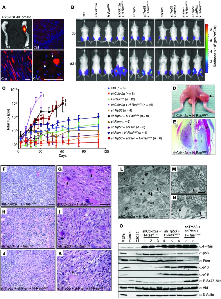
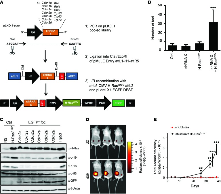
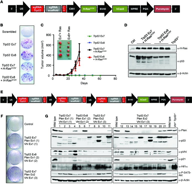
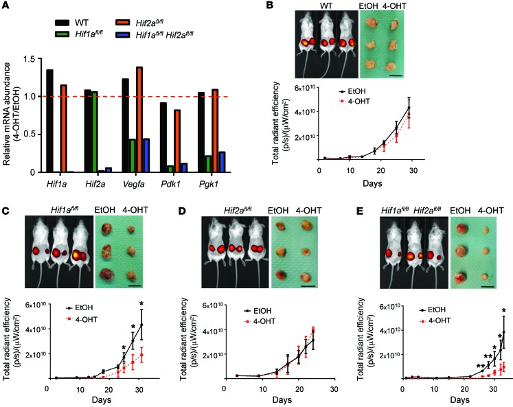
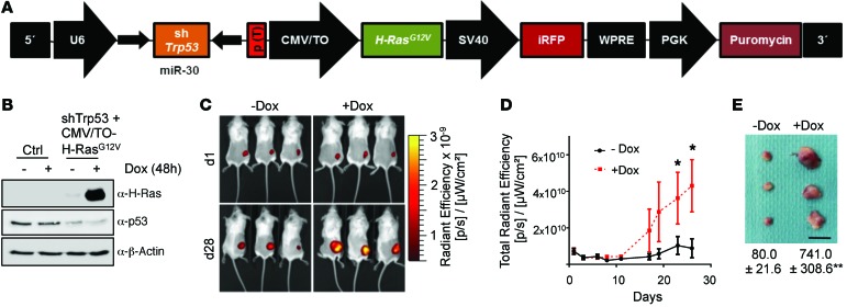
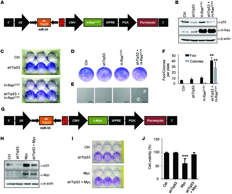
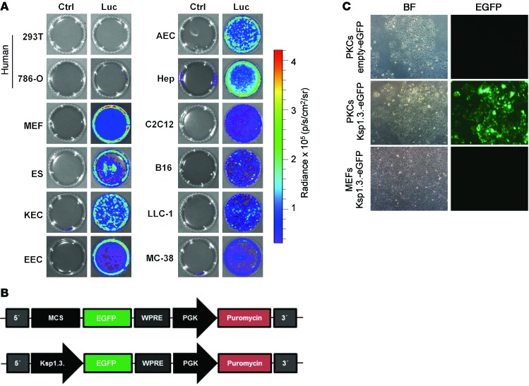
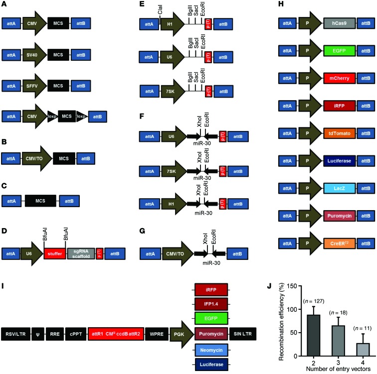
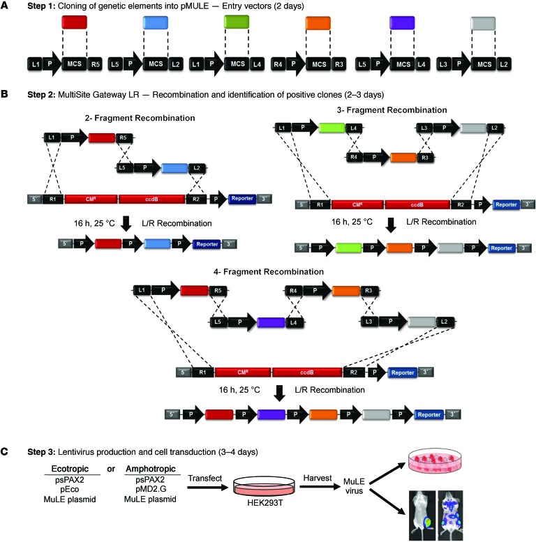
Here, we describe the multiple lentiviral expression (MuLE) system that allows multiple genetic alterations to be introduced simultaneously into mammalian cells. We created a toolbox of MuLE vectors that constitute a flexible, modular system for the rapid engineering of complex polycistronic lentiviruses, allowing combinatorial gene overexpression, gene knockdown, Cre-mediated gene deletion, or CRISPR/Cas9-mediated (where CRISPR indicates clustered regularly interspaced short palindromic repeats) gene mutation, together with expression of fluorescent or enzymatic reporters for cellular assays and animal imaging. Examples of tumor engineering were used to illustrate the speed and versatility of performing combinatorial genetics using the MuLE system. By transducing cultured primary mouse cells with single MuLE lentiviruses, we engineered tumors containing up to 5 different genetic alterations, identified genetic dependencies of molecularly defined tumors, conducted genetic interaction screens, and induced the simultaneous CRISPR/Cas9-mediated knockout of 3 tumor-suppressor genes. Intramuscular injection of MuLE viruses expressing oncogenic H-RasG12V together with combinations of knockdowns of the tumor suppressors cyclin-dependent kinase inhibitor 2A (Cdkn2a), transformation-related protein 53 (Trp53), and phosphatase and tensin homolog (Pten) allowed the generation of 3 murine sarcoma models, demonstrating that genetically defined autochthonous tumors can be rapidly generated and quantitatively monitored via direct injection of polycistronic MuLE lentiviruses into mouse tissues. Together, our results demonstrate that the MuLE system provides genetic power for the systematic investigation of the molecular mechanisms that underlie human diseases.


Figures

Figure 10
Figure 10. Generation of 3 autochthonous mouse models of undifferentiated sarcoma using MuLE vectors.
(A) Intramuscular injection of ROSA26-lox-STOP-lox-tdTomato mice with control or Cre-expressing virus. Bottom left panel shows infected myofibers, and small cells adjacent to myofibers are seen at higher magnification (arrowheads, bottom right panel). (B) Bioluminescence imaging 3 and 31 days after injection of 3 × 10⁵ functional viral particles into each gastrocnemius muscle of 18-day-old SCID/beige mice with MuLE-luciferase viruses expressing combinations of shRNA against Cdkn2a, Trp53, and Pten with or without expression of H-RasG12V. (C) Quantification (mean ± SD) of luminescent signal intensities over time after injection. Sacrifice of all mice in these cohorts by this time point. (D) A tumor (arrow) in a mouse injected with the shCdkn2a plus H-RasG12V MuLE virus only in the right gastrocnemius muscle. (E) Histological image of the tumor (T) from D surrounded by muscle tissue (M). (FK) Representative histology of tumors derived from injection of shCdkn2a plus H-RasG12V (F and G), shTrp53 plus H-RasG12V (H and I), and shTrp53 plus shPTEN plus H-RasG12V (J and K) viruses. Arrowheads in G, I, and K highlight pleomorphic rhabdoid cells. (L) EM showing an example of a tumor cell with sarcomere formation; M and N show higher magnification of the regions in L marked with an arrowhead and arrows, respectively, showing Z-bands or irregular masses of Z-band material with converging filaments. (O) Western blot analysis of independent cell lines (lanes 1–8) derived from independent tumors of the indicated genotypes. MEFs, muscle tissue, and C2C12 myoblast cells served as controls. Scale bars: 50 μm (A and FK); 10 μm (L); 500 nm (M and N).
Figure 9
Figure 9. Combinatorial genetic screening using MuLE vectors.
(A) Schematic representation of the workflow to generate a MuLE Entry vector shRNA library targeting the listed genes (shRNA X) and the final tricistronic lentiviral expression vector library that was used to screen for shRNAs that cause cell transformation in cooperation with oncogenic H-RasG12V overexpression. (B) Quantification of foci that were formed when WT MEFs were transduced with the indicted lentiviruses (MOI = 0.1). (C) Western blot analysis of EGFP-expressing cell clones derived from foci harboring shRNA against the indicated gene. (D) In vivo fluorescence images of mice that were subcutaneously injected with WT MEFs that had been infected with a MuLE virus expressing the identified shRNA against Cdkn2a alone plus iRFP or in combination with H-RasG12V and iRFP. (E) Tumor growth in the same mice monitored by longitudinal in vivo fluorescence imaging. All graphs depict mean ± SD. Student’s t test, n = 3. **P < 0.01; ***P < 0.001.
Figure 8
Figure 8. Combinatorial genetics using the CRISPR/Cas9 system in MuLE vectors.
(A) Schematic of MuLE vector expressing sgRNA against Trp53 and expressing H-RasG12V, hCas9, and puromycin resistance. (B) MEFs were infected with the indicated viruses expressing sgRNAs targeting Trp53 exon 7 (Ex7) or exon 8 with or without H-RasG12V expression, plated at low density 6 days after transduction, and stained with crystal violet 14 days after plating. (C) Growth of cells as tumor xenografts and images of tumors derived from the combination of Trp53 exon 7 or exon 8 sgRNAs with H-RasG12V overexpression. (D) Western blot analysis of tumor cells that were isolated from 3 separate tumors of each genotype 5 weeks after cell injection. (E) Schematic of MuLE vector simultaneously expressing sgRNAs against Trp53, Pten, and Vhl and expressing hCas9 and puromycin resistance. (F) MEFs infected with 3 independent combinations of different sgRNAs formed colonies when plated at low density 10 days after viral transduction. (G) Western blotting of cell lines (lanes 1–21) derived from colonies that formed after infection with viruses expressing the indicated combinations of sgRNAs.
Figure 7
Figure 7. Hif1a but not Hif2a is necessary for efficient growth of Pten/Rb1-deficient, H-RasG12V–expressing tumors.
(A) mRNA expression analysis of the indicated genes in WT, Hif1afl/fl, Hif2afl/fl, and Hif1afl/fl Hif2afl/fl MEFs that were transduced with lentivirus generated from the vector shown in Figure 6A 96 hours after induction of CreERT2 with 300 nm 4-OHT. Shown are ratios of 4-OHT treated to EtOH treated. (BE) In vivo fluorescence imaging at the day of euthanasia (top left panels), excised tumors (top right panels), and longitudinal tumor growth (bottom panels) of mice injected with cells described in A that had been treated with EtOH (left flank) or 4-OHT (right flank) prior to subcutaneous injection. Color intensity in BE is the same as in Figure 6C. Scale bars: 1 cm. All graphs depict mean ± SD. Student’s t test, n = 3–6. *P < 0.05; **P < 0.01.
Figure 6
Figure 6. Generation of genetically complex tumors with multicistronic MuLE vectors.
(A) Pentacistronic vector to simultaneously knock down Rb1 and Pten and to express CreERT2, oncogenic H-RasG12V, and puromycin resistance. (B) Western blot analysis of puromycin-selected Vhlfl/fl Trp53fl/fl primary MEFs that were transduced with lentivirus shown in A, virus containing 4 empty inserts (Ctrl), or 3 empty inserts plus CreERT2, and treated with 300 nM 4-OHT or ethanol (EtOH) for 4 days. (C) In vivo fluorescence images of mice taken 2 and 21 days after subcutaneous injection of Vhlfl/fl Trp53fl/fl MEFs transduced with lentivirus generated from the vector shown in A that had been treated with EtOH (left flank) or with 4-OHT (right flank) prior to injection. (D) Western blot analysis of tumor cells that were isolated from 3 separate tumors of each genotype 3 weeks after cell injection.
Figure 5
Figure 5. Combinatorial genetics using inducible MuLE vectors.
(A) Multicistronic lentiviral vector to simultaneously knock down Trp53, overexpress inducible H-RasG12V, and overexpress iRFP. (B) Western blot analysis demonstrating H-RasG12V overexpression in TetR-expressing MEFs transduced with the lentivirus shown in A upon addition of 1 μM DOX to the culture medium. (C) In vivo fluorescence imaging of mice that were subcutaneously injected with shRNA-Trp53/CMV/TO-H-RasG12V MuLE virus–infected MEFs and fed drinking water without or with DOX. (D) Longitudinal tumor growth quantified by iRFP fluorescence imaging. (E) Image of the isolated tumors and tumor weight after 28 days. Scale bar: 1 cm. Student’s t test, n = 3. *P < 0.05; **P < 0.01.
Figure 4
Figure 4. Combinatorial genetics using MuLE vectors.
(A) Lentiviral vector to simultaneously knock down Trp53 and overexpress oncogenic H-RasG12V. (B) Western blot analysis of primary MEFs transduced with the indicated lentiviruses. (C) Crystal violet staining of the same cells 14 days after seeding at low density and (D) 14 days after seeding in a focus formation assay. (E) Representative images of the same cells seeded in a soft agar colony assay after 3 weeks of growth. Scale bars: 200 μm. (F) Quantification of the foci and colonies growing in assays from D and E. (G) Lentiviral vector generated to simultaneously knock down Trp53 and overexpress Myc. (H) Western blot analysis of primary MEFs transduced with the indicated lentiviruses. (I) Crystal violet staining of the same cells 14 days after seeding at low density. (J) Quantification of viable cells 3 days after transduction with the indicated lentiviruses. All graphs depict mean ± SD. Student’s t test, n = 3. **P < 0.01; ***P < 0.001.
Figure 3
Figure 3. Broad cellular tropism and cell-type–specific expression of ecotropic MuLE vectors.
(A) Luminescent imaging of various human and mouse cultured cells after infection with ecotropic MuLE viruses expressing an empty cassette (Ctrl) or luciferase (Luc). The human kidney cell lines 293T and 786-0 were not infected by these viruses, but various primary mouse cells and cell lines were infected, including MEFs, embryonic stem cells (ES), kidney epithelial cells (KEC), endometrial epithelial cells (EEC), aortic endothelial cells (AEC), hepatocytes (Hep), myoblasts (C2C12), melanoma cells (B16), lung carcinoma cells (LLC-1), and colorectal carcinoma cells (MC-38). (B) Schematic of MuLE vectors with or without the kidney epithelium–specific Ksp1.3 promoter cloned upstream of a cDNA encoding EGFP. (C) Representative bright field (BF) and green fluorescence (EGFP) images of primary mouse kidney epithelial cells (PKCs) and primary MEFs transduced with the lentiviral vectors shown in B. Original magnification, ×1 (A); ×10 (C).
Figure 2
Figure 2. The MuLE vector toolbox.
(A and B) MuLE Entry vectors for pol II promoter–driven constitutive expression (A) or DOX-inducible expression (B). (C) Promoterless MuLE Entry vectors. (D) MuLE Entry vectors for U6-driven expression of sgRNAs. (E and F) MuLE Entry vectors for shRNA-based (E) and shRNA–miR-30–based (F) gene knockdown using pol III promoters and (G) a DOX-inducible miR-30–based shRNA expression vector. Restriction enzyme sites for cloning are shown. (H) Schematic representation of MuLE Entry vectors for expression of hCas9, fluorescent proteins (EGFP, mCherry, iRFP, td­Tomato), firefly-luciferase, β-galactosidase (LacZ), puromycin resistance, or Cre-ERT2. P, various promoters. In all panels, attA and attB denote that multiple combinations of MultiSite Gateway attL-attR sites are available for these vectors. (I) Schematic representation of Destination vectors modified from the pLenti X1 series to contain the different expression cassettes shown. (J) Quantification (mean ± SD) of recombination efficiencies of n independent MultiSite Gateway attL-attR recombinations using 2, 3, or 4 MuLE Entry vectors.
Figure 1
Figure 1. Overview of genetic engineering experiments using MuLE vectors.
(A) Restriction enzyme cloning is used to generate MuLE Entry vectors with a desired genetic insert cloned downstream of a desired promoter (P), with the entire promoter-insert element being surrounded by appropriate attL-attR sites. (B) Schematic overview of the MultiSite Gateway–based recombination cloning of 2, 3, or 4 MuLE Entry vectors into lentiviral destination vectors to generate multicistronic MuLE lentiviral expression vectors. The specific attL-attR sites that mediate each recombination are depicted. CMR, chloramphenicol resistance gene; ccdB, ccdB toxin gene. (C) Transfection of 293T cells with a MuLE expression vector plasmid together with a lentiviral packaging vector (psPAX2) and a vector encoding either amphotropic (MD2G) or ecotropic (pEco) envelope proteins generates MuLE lentiviruses for transduction of cultured cells or for in vivo injection into mouse tissues.

References

    1. Ciriello G, Miller ML, Aksoy BA, Senbabaoglu Y, Schultz N, Sander C. Emerging landscape of oncogenic signatures across human cancers. Nat Genet. 2013;45(10):1127–1133. doi: 10.1038/ng.2762. - DOI - PMC - PubMed
    1. Lawrence MS, et al. Discovery and saturation analysis of cancer genes across 21 tumour types. Nature. 2014;505(7484):495–501. doi: 10.1038/nature12912. - DOI - PMC - PubMed
    1. Dow LE, Lowe SW. Life in the fast lane: mammalian disease models in the genomics era. Cell. 2012;148(6):1099–1109. doi: 10.1016/j.cell.2012.02.023. - DOI - PMC - PubMed
    1. Katzen F. Gateway recombinational cloning: a biological operating system. Expert Opin Drug Discov. 2007;2(4):571–589. doi: 10.1517/17460441.2.4.571. - DOI - PubMed
    1. Paddison PJ, et al. Cloning of short hairpin RNAs for gene knockdown in mammalian cells. Nat Methods. 2004;1(2):163–167. doi: 10.1038/nmeth1104-163. - DOI - PubMed

Publication types

MeSH terms