. 2015 Mar 9;10(3):e0119539.
doi: 10.1371/journal.pone.0119539. eCollection 2015.

Integrin α3β1 signaling through MEK/ERK determines alternative polyadenylation of the MMP-9 mRNA transcript in immortalized mouse keratinocytes

Affiliations

Integrin α3β1 signaling through MEK/ERK determines alternative polyadenylation of the MMP-9 mRNA transcript in immortalized mouse keratinocytes

Dara S Missan et al. PLoS One. .

Abstract

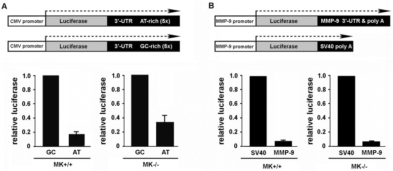
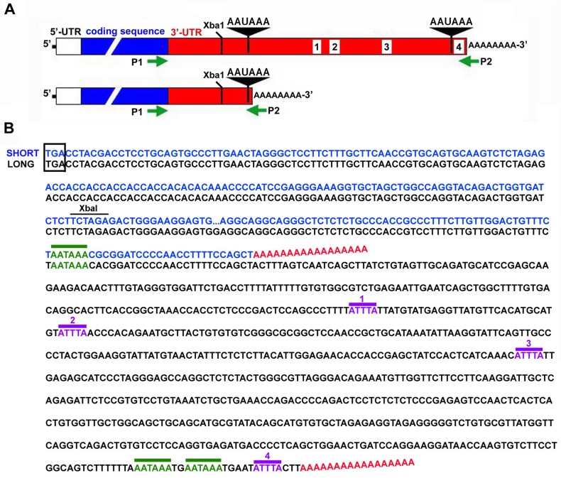
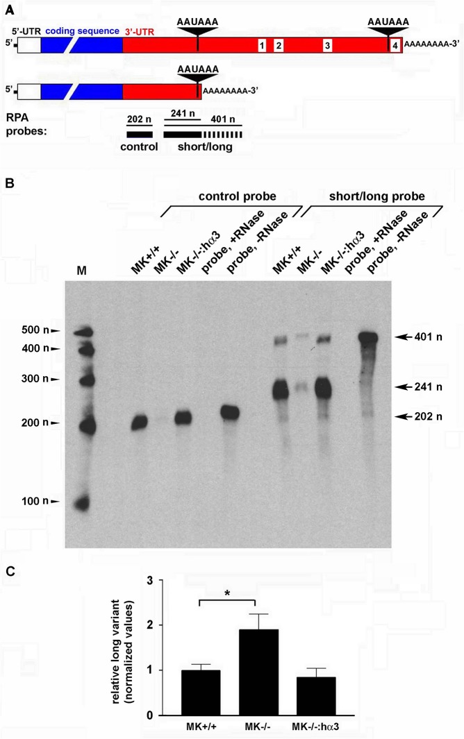
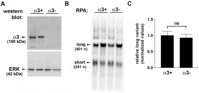
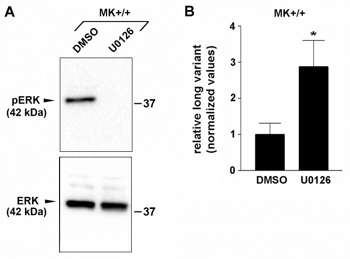
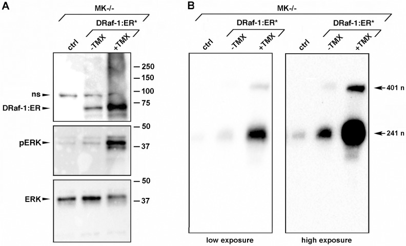
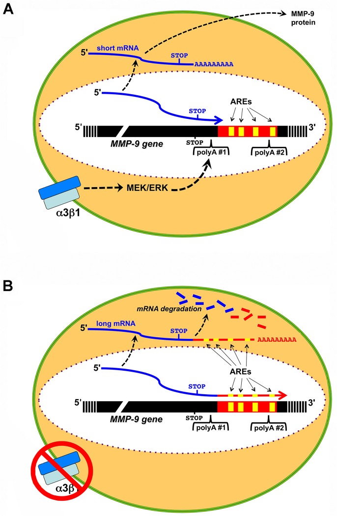
Integrin α3β1 is highly expressed in both normal and tumorigenic epidermal keratinocytes where it regulates genes that control cellular function and extracellular matrix remodeling during normal and pathological tissue remodeling processes, including wound healing and development of squamous cell carcinoma (SCC). Previous studies identified a role for α3β1 in immortalized and transformed keratinocytes in the regulation of genes that promote tumorigenesis, invasion, and pro-angiogenic crosstalk to endothelial cells. One such gene, matrix metalloproteinase-9 (MMP-9), is induced by α3β1 through a post-transcriptional mechanism of enhanced mRNA stability. In the current study, we sought to investigate the mechanism through which α3β1 controls MMP-9 mRNA stability. First, we utilized a luciferase reporter assay to show that AU-rich elements (AREs) residing within the 3'-untranslated region (3'-UTR) of the MMP-9 mRNA renders the transcript unstable in a manner that is independent of α3β1. Next, we cloned a truncated variant of the MMP-9 mRNA which is generated through usage of an alternative, upstream polyadenylation signal and lacks the 3'-UTR region containing the destabilizing AREs. Using an RNase protection assay to distinguish "long" (full-length 3'-UTR) and "short" (truncated 3'-UTR) MMP-9 mRNA variants, we demonstrated that the shorter, more stable mRNA that lacks 3'-UTR AREs was preferentially generated in α3β1-expressing keratinocytes compared with α3β1-deficient (i.e., α3-null) keratinocytes. Moreover, we determined that α3β1-dependent alternative polyadenylation was acquired by immortalized keratinocytes, as primary neonatal keratinocytes did not display α3β1-dependent differences in the long and short transcripts. Finally, pharmacological inhibition of the extracellular signal-regulated kinase (ERK)/mitogen-activated protein kinase (MAPK) pathway in α3β1-expressing keratinocytes caused a shift towards long variant expression, while Raf-1-mediated activation of ERK in α3-null keratinocytes dramatically enhanced short variant expression, indicating a role for ERK/MAPK signaling in α3β1-mediated selection of the proximal polyadenylation site. These findings identify a novel mode of integrin α3β1-mediated gene regulation through alternative polyadenylation.


Conflict of interest statement

Competing Interests: The authors have declared that no competing interests exist.

Figures

Fig 1
Fig 1. Influence of 3’-UTR AREs on luciferase reporter expression in MK cells that express or lack integrin α3β1.
(A) Schematic of CMV promoter-driven luciferase reporter genes containing pentamers that encode either consensus AU-rich ARE sequences or control GC-rich sequences within the 3’-UTR of the mRNA. Reporter plasmids were transfected into α3-expressing MK+/+ cells or α3-null MK−/− cells, and experimental luciferase signals were normalized to that for a co-transfected Renilla luciferase control plasmid (pRLTK). Graph shows luciferase activity from the AU-rich reporter relative to that from the GC-rich reporter. (B) Schematic of MMP-9 promoter-driven luciferase reporter genes containing either the MMP-9 3’-UTR or the SV40 poly(A) signal downstream of luciferase coding sequences. MK+/+ cells or MK−/− cells were co-transfected with reporter and control plasmids as in (A). Graph shows luciferase activity from the MMP-9 3’-UTR reporter relative to that from the SV40 poly(A) reporter. For (A) and (B), MK cells were seeded onto LN-332-rich ECM and transfected for 24 hours, then luciferase expression was assayed as described in the Materials and Methods. Data are the mean of three independent experiments +/- s.e.m.
Fig 2
Fig 2. Keratinocytes express a truncated MMP-9 mRNA variant that lacks 3’-UTR AREs.
(A) Schematic of MMP-9 mRNA 3’-UTR variants. Sequences corresponding to alternative poly(A) signals (black triangles) flank ARE motifs within the long variant (white boxes, 1–4). 5’-UTR (white) and protein coding regions (blue) are also shown (not to scale). An Xba1 site (for reference) and approximate positions of P1 and P2 cloning primers (not to scale) are indicated. (B) 3’-RACE was used to PCR-amplify cDNAs corresponding to the 3’-UTR of the MMP-9 mRNA. The cDNA sequence of a “short” 3’-UTR variant that was amplified from MK+/+ cells (blue) is aligned against the known cDNA sequence of the “long” 3’-UTR variant of murine MMP-9 mRNA (black) (GenBank: BC046991.1). The stop codon (TGA) adjacent to the 3’-UTR is boxed; the position of polyadenylation for each variant is indicated by the red poly(A); poly(A) signals (AATAAA) are overlined in green (two AATAAA motifs occur near the end of the long variant); four canonical AREs are present only in the long variant and are overlined in purple and numbered 1–4.
Fig 3
Fig 3. Integrin α3β1 expression enhances MMP-9 mRNA and promotes use of the proximal poly(A) signal.
(A) Schematic of RPA probes and their alignment with protected regions of long and short variants of the MMP-9 mRNA. The control RPA probe that corresponds to common 3’-UTR sequences upstream of the proximal poly(A) signal is fully protected by either mRNA variant, while the short/long RPA probe that spans the proximal poly(A) signal is not fully protected by the short mRNA variant (indicated by the dashed portion). Lengths of protected regions are indicated in nucleotides (n). (B) RPA was performed as described in the Materials and Methods using the control or short/long probes to assess relative expression of MMP-9 mRNA variants in MK+/+ cells, MK−/− cells, or MK−/−: hα3 cells. Control reactions included probe only in the presence (probe, +RNase) or absence of RNase (probe,-RNase). M, size markers in nucleotides (n); lengths of protected probes are indicated to the right. (C) Signal quantification from three independent RPA experiments (see Materials and Methods) reveals that the MMP-9 mRNA long variant constitutes a higher proportion of the total MMP-9 mRNA expressed in MK−/− cells, compared with α3-expressing MK+/+ cells or MK−/−: hα3 cells. Data are mean ± s.e.m. (n = 3); all values were normalized through dividing by the average for the control (MK+/+). *P<0.05, one-way ANOVA, post-test Tukeys multiple comparison.
Fig 4
Fig 4. MMP-9 mRNA variant expression is not α3β1-dependent in non-immortalized, primary keratinocytes.
(A) Western blot of lysates from representative α3-expressing (α3+) and α3-null (α3−) primary keratinocytes with antiserum against the integrin α3 subunit, or ERK as a loading control; molecular weights are indicated (kDa). (B) Corresponding RPA of primary keratinocytes using the short/long probe to distinguish MMP-9 mRNA variants. Results shown in (A) and (B) are for two representative mice of each genotype. (C) RPA quantification (performed as in Fig. 3) reveals no significant difference in relative expression of the MMP-9 mRNA long variant between α3-expressing and α3-null primary keratinocytes. Data are mean ± s.e.m. (α3+ keratinocytes, n = 10; α3− keratinocytes, n = 17); all values were normalized through dividing by the average for the control (α3+). Not significant (ns), Student’s two-tailed t-test.
Fig 5
Fig 5. Inhibition of MEK/ERK signaling in α3β1-expressing MK cells increases relative expression of the long MMP-9 mRNA variant.
(A) Representative western blot showing inhibition of phospho-ERK (pERK) with U0126 compound in MK+/+ cells, compared with DMSO control; ERK, total ERK; positions of molecular weight markers are indicated at right (kDa). (B) Quantification of RPA (performed as in Fig. 3) showing the effects of ERK inhibition in MK+/+ cells on the relative amount of the long MMP-9 mRNA variant. Data are mean ± s.e.m. (n = 6); all values were normalized through dividing by the average for the control (DMSO). *P<0.05, Student’s two-tailed t-test.
Fig 6
Fig 6. Raf-1-mediated activation of MEK/ERK in α3-null MK cells induces expression of the short MMP-9 mRNA variant.
MK−/− cells were left uninfected (ctrl) or infected with adenovirus expressing a tamoxifen-inducible form of Raf-1 (DRaf-1:ER*) in the absence (−TMX) or presence (+TMX) of 1 μM tamoxifen. (A) Western blot showing that tamoxifen induction of DRaf-1:ER* leads to enhanced ERK phosphorylation (pERK); the latter blot was stripped and reprobed for total ERK (ERK); ns, non-specific band; positions of molecular weight markers are indicated at right (kDa). (B) RPA showing the effects of basal or tamoxifen-induced Raf-1 on expression of MMP-9 mRNA variants. Two different exposures of the same membrane are shown. Lengths of protected probe are indicated as in Fig. 3. Representative results are shown, n = 3.
Fig 7
Fig 7. Model depicting α3β1-dependent alternative polyadenylation of the MMP-9 mRNA.
(A) Integrin α3β1 activates a MEK/ERK signaling pathway that promotes selection of the proximal polyadenylation site (polyA #1) within the MMP-9 gene, thereby generating the short, more stable mRNA transcript and subsequent synthesis of MMP-9 protein. (B) In the absence of α3β1, MEK/ERK signaling is diminished and polyadenylation defaults to the distal polyadenylation site (polyA #2), thereby generating the long, ARE-containing transcript that is subject to mRNA degradation. Positions of the stop codon, AREs (yellow boxes), poly(A) signals, and poly(A+) tails are indicated.

Similar articles

Cited by

References

    1. Kreidberg JA. Functions of α3β1 integrin. Curr Opin Cell Biol. 2000;12: 548–553. - PubMed
    1. DiPersio CM, Hodivala-Dilke KM, Jaenisch R, Kreidberg JA, Hynes RO. α3β1 integrin is required for normal development of the epidermal basement membrane. J Cell Biol. 1997;137: 729–742. - PMC - PubMed
    1. Has C, Spartà G, Kiritsi D, Weibel L, Moeller A, Vega-Warner V, et al. Integrin α3 mutations with kidney, lung, and skin disease. N Engl J Med. 2012;366: 1508–1514. 10.1056/NEJMoa1110813 - DOI - PMC - PubMed
    1. Longmate WM, Monichan R, Chu ML, Tsuda T, Mahoney MG, DiPersio CM. Reduced fibulin-2 contributes to loss of basement membrane integrity and skin blistering in mice lacking integrin α3β1 in the epidermis. J Invest Dermatol. 2014;134: 1609–1617. 10.1038/jid.2014.10 - DOI - PMC - PubMed
    1. Subbaram S, DiPersio CM. Integrin α3β1 as a breast cancer target. Expert Opin Ther Targets. 2011;15: 1197–1210. 10.1517/14728222.2011.609557 - DOI - PMC - PubMed

Publication types