. 2015 Apr;18(4):545-52.
doi: 10.1038/nn.3972. Epub 2015 Mar 9.

NPY signaling inhibits extended amygdala CRF neurons to suppress binge alcohol drinking

Affiliations

NPY signaling inhibits extended amygdala CRF neurons to suppress binge alcohol drinking

Kristen E Pleil et al. Nat Neurosci. 2015 Apr.

Abstract

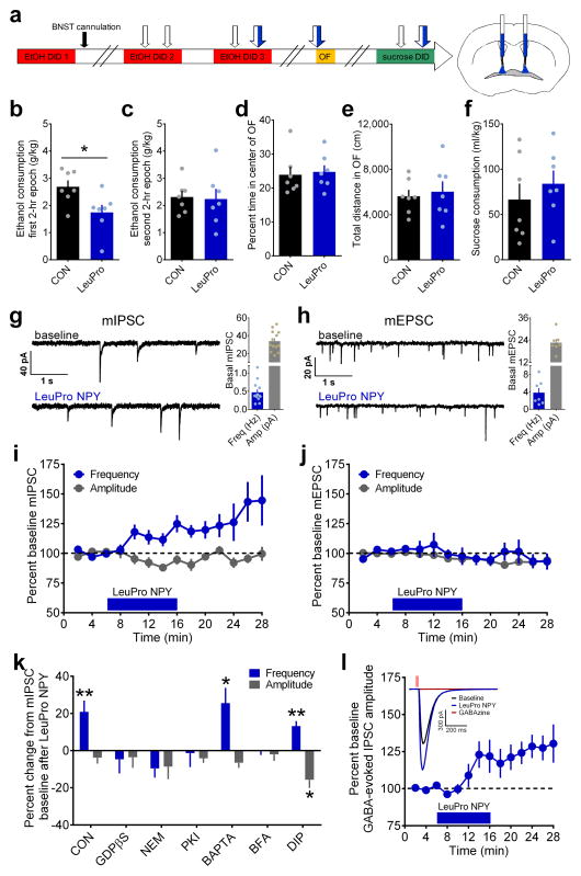
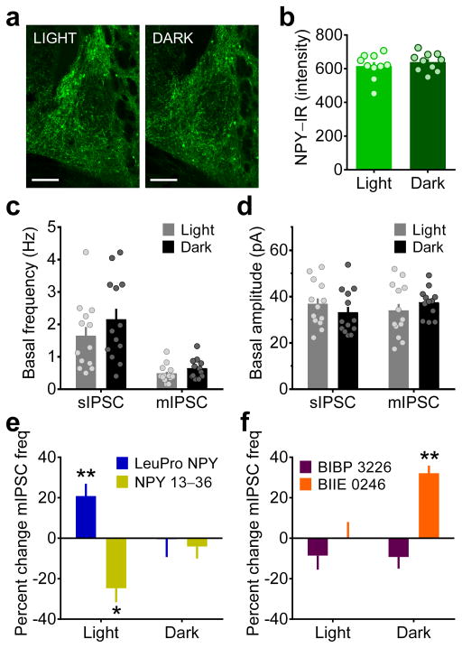
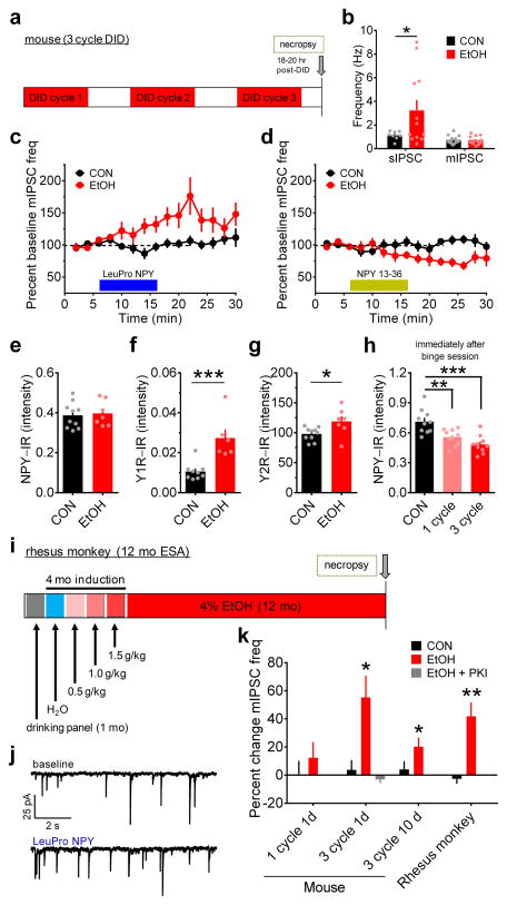
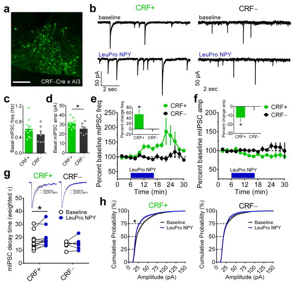
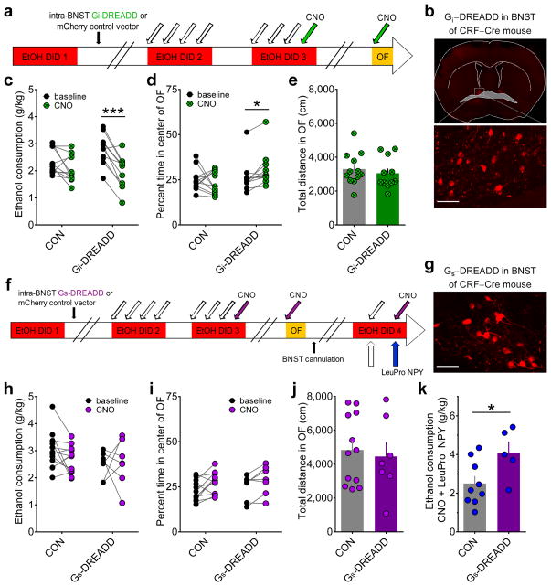
Binge alcohol drinking is a tremendous public health problem because it leads to the development of numerous pathologies, including alcohol abuse and anxiety. It is thought to do so by hijacking brain systems that regulate stress and reward, including neuropeptide Y (NPY) and corticotropin-releasing factor (CRF). The central actions of NPY and CRF have opposing functions in the regulation of emotional and reward-seeking behaviors; thus, dysfunctional interactions between these peptidergic systems could be involved in the development of these pathologies. We used converging physiological, pharmacological and chemogenetic approaches to identify a precise neural mechanism in the bed nucleus of the stria terminalis (BNST), a limbic brain region involved in pathological reward and anxiety behaviors, underlying the interactions between NPY and CRF in the regulation of binge alcohol drinking in both mice and monkeys. We found that NPY Y1 receptor (Y1R) activation in the BNST suppressed binge alcohol drinking by enhancing inhibitory synaptic transmission specifically in CRF neurons via a previously unknown Gi-mediated, PKA-dependent postsynaptic mechanism. Furthermore, chronic alcohol drinking led to persistent alterations in Y1R function in the BNST of both mice and monkeys, highlighting the enduring, conserved nature of this effect across mammalian species. Together, these data provide both a cellular locus and signaling framework for the development of new therapeutics for treatment of neuropsychiatric diseases, including alcohol use disorders.


Figures

Figure 1
Figure 1
Activation of Gi-coupled Y1R in the BNST reduces binge ethanol drinking and enhances GABAergic transmission via a PKA-dependent, postsynaptic insertion of GABAA receptors. (a) Experimental timeline for examination of bilateral intra-BNST infusions of the Y1R agonist LeuPro NPY (99 pmol/200 nL/side, N = 7 mice) or control vehicle (CON; 200 nL/side, N = 7 mice) on EtOH DID, open field test (OF), and sucrose DID. (bf) All statistical comparisons were made using unpaired t-tests. Mice that received infusions of LeuPro NPY consumed significantly less ethanol than CON mice during the first 2–hr epoch (b; t(12) = 2.59, *p = 0.024) but not the second 2–hr epoch (c; p > 0.85) of ethanol DID, but LeuPro NPY infusion did not alter measures of anxiety and locomotion in the open field test (OF), including the percent time spent in the center (d) and total distance traveled (e) of the OF, or sucrose consumption in sucrose DID (f; p’s > 0.45). (gh) Representative traces of miniature inhibitory postsynaptic currents (mIPSCs; g) and excitatory postsynaptic currents (mEPSCs; h) in BNST neurons before and after 10 min bath application of LeuPro NPY (300 nM), and basal frequency and amplitude for neurons used in mPSC experiments. (i) LeuPro NPY increased mIPSC frequency (20.9 ± 6.1%; paired t-test baseline vs. washout: t(12) = 3.42, p = 0.005; n = 13 neurons, N = 11 mice) but did not alter mIPSC amplitude (p > 0.20). (j) Conversely, LeuPro NPY produced a small decrease in the amplitude of mEPSCs (−6.9 ± 2.3%; paired t-test: t(4) = 2.99, p = 0.041; n = 5, N = 3) but did not alter mEPSC frequency (p > 0.60). (k) Inclusion of GDPβS (1 mM; n = 7, N = 4) to block postsynaptic GPCR activity, NEM (50 μM; n = 5, N = 3) to block Gi signaling, PKI (20 μM; n = 6, N = 3) to block PKA signaling, and BFA (200 nM; n = 5, N = 4) to prevent receptor trafficking to the membrane all blocked the ability of bath-applied LeuPro NPY to increase mIPSC frequency (paired t-tests baseline vs. washout: p’s > 0.10), while BAPTA (10 μM; t(5) = 3.12, *p = 0.026; n = 6, N = 5) to block calcium and DIP (2mM; t(5) = 4.76, **p = 0.005; n = 6, N = 3) to block receptor endocytosis did not. LeuPro NPY decreased mIPSC amplitude only when DIP was in the intracellular solution (DIP: paired t-test: t(5) = 3.60, *p = 0.016; others: p’s > 0.05). (l) LeuPro NPY (300 nM) increased the amplitude of the GABAAR-mediated current in response to picospritzed GABA (100 μM) by 25.3 ± 7.0%; paired t-test baseline vs. washout: (t(8) = 3.59, p = 0.007; n = 9, N = 7); inset: representative traces illustrating the effect of LeuPro NPY and complete ablation of the postsynaptic current by the GABAAR blocker GABAzine (20 μM). All data in bl are presented as mean ± SEM.
Figure 2
Figure 2
Circadian regulation of receptor-specific NPY modulation of GABAergic transmission in the BNST. (a) Sample images representing mean NPY–IR in the BNST of mice sacrificed three hr into the light or dark phase of the light cycle (scale bars = 150 μM). Images shown were replicated for each individual data point shown in b. (b) Mean NPY–IR per mouse from five coronal slices (as in panel a) was not different between the light and dark phases of the light cycle (unpaired t-test: p > 0.40; N’s = 10/group). (cd) Basal sIPSC and mIPSC frequency (c) and amplitude (d) were not different across the light cycle (unpaired t-tests: p’s > 0.15; CON n = 13, N = 7, EtOH n = 13, N = 7). (e) Bath application of LeuPro NPY (300 nM) increased, while the Y2R agonist NPY 13–36 (300 nM) decreased, mIPSC frequency during the light phase (LeuPro NPY: as shown in Fig. 1i, **p = 0.005; NPY 13–36: paired t-test baseline vs. washout: t(5) = 3.57, *p = 0.016, n = 6, N = 5) but not the dark phase (p’s > 0.15; LeuPro n = 5, N = 4, NPY 13–36 n = 6, N = 4), of the light cycle. (f) Bath application of the Y1R antagonist BIBP 3226 (1 μM; n = 11, N = 10) or the Y2R antagonist BIIE 0246 (1 μM; n = 4, N = 3) did not alter mIPSC frequency during the light phase (paired t-tests baseline vs. washout: p’s > 0.20); BIIE 0246 increased mIPSC frequency during the dark phase (t(3) = 8.45, **p = 0.004; n = 4, N = 3), while BIBP 3226 did not (p > 0.15; n = 5, N = 4). All data in bl are presented as mean ± SEM.
Figure 3
Figure 3
Chronic binge alcohol drinking alters receptor-specific NPY modulation of GABAergic transmission in the BNST of mice and monkeys. (a) Experimental timeline for 3-cycle DID in mice. (b) BNST neurons from mice that drank ethanol (EtOH) had higher sIPSC frequency than water-drinking controls (CONs; unpaired t-test with Welch’s correction: t(11) = 2.32, *p = 0.040, CON n = 8, N = 5, EtOH n = 12, N = 5), but mIPSC frequency did not differ between groups (unpaired t-test: p > 0.95; CON n = 15, N= 7, EtOH n = 12, N = 6). (c) LeuPro NPY (300 nM) significantly increased mIPSC frequency in EtOH mice (paired t-test baseline vs. washout: t(5) = 3.58, p = 0.016; n = 6, N = 6) but not CONs (p > 0.60, n = 7, N = 7). (d) NPY 13–36 (300 nM) decreased mIPSC frequency in ethanol-drinking mice (t(5) = 2.97, p = 0.031; n = 6, N = 5) but not controls (p > 0.85, n = 7, N = 6). (eg) Mean NPY-IR (average IR from 3–5 slices per mouse) was similar between groups (e; unpaired t-test: p > 0.75; CON N = 10, EtOH N = 7), but Y1R–IR (f; t(13) = 4.23, ***p = 0.001; CON N = 9, EtOH N = 6) and Y2R-IR (g; t(15) = 2.50, *p = 0.025; CON N = 10, EtOH N = 7) were higher in the BNST of EtOH mice than CONs. (h) NPY-IR was significantly decreased in the BNST of EtOH mice compared to water-drinking CONs immediately after the last binge ethanol drinking exposure in 1-cycle and 3-cycle DID (N’s = 10/group), but was not different between one and 3-cycle DID (one-way ANOVA: F(2,27) = 14.25, p < 0.0001; post-hoc Sidak’s multiple comparisons test: CON vs. 1-cycle: t(18) = 3.58,** p = 0.004; CON vs. 3-cycle DID: t(18) = 5.22, ***p < 0.001; 1-cycle DID vs. 3-cycle DID: p > 0.25), suggesting that NPY was similarly recruited acutely during each binge ethanol session across each cycle. (i) Experimental timeline for voluntary ethanol self-administration (ESA; access to 4% ethanol for 22 h/d, 7 d/wk for 12 mo) in adult male rhesus monkeys. (j) Representative traces of mIPSCs from ethanol self-administering rhesus monkey BNST neurons before and after bath application of LeuPro NPY (300 nM). (k) mIPSC frequency was unaltered by LeuPro NPY following one cycle of DID in EtOH mice (n = 8, N = 4) and water-drinking CONs (n = 5, N = 3; paired t-tests baseline vs. washout: p’s > 0.30), but it was increased in EtOH, but not CON, mice 1 d after the final binge session of 3-cycle EtOH DID, as shown in c, which could be blocked by intracellular inclusion of PKI (20 μM; p > 0.35; n = 3, N = 2). The adaptation in LeuPro NPY modulation of mIPSC frequency was still present 10 d after the final binge ethanol session in EtOH mice (t(5) = 3.09, *p = 0.027; n = 6, N = 3) but not CONs (p > 0.50, n = 5, N = 3) and was also observed in rhesus monkeys after 12 mo of continuous access to ethanol (t(8) = 4.21, **p = 0.003; n = 9, N = 5) but not control solution (p > 0.50; n = 4, N = 3). All data in bh and k are presented as mean ± SEM.
Figure 4
Figure 4
Y1R-mediated effects on inhibition in the BNST are specific to CRF neurons. (a) Sample image of the dorsal BNST in a naïve CRF–Cre x Ai3 reporter mouse taken from a coronal brain slice depicting CRF–Cre-positive neurons in green (scale bar = 150 μM). Fluorescence expression shown is typical in this mouse line and was confirmed for all mice used in experiments for panels bh. (b) Representative traces of mIPSCs from CRF–Cre-positive (CRF+) and CRF–Cre-negative (CRF−) neurons in the BNST before and after bath application of LeuPro NPY (300 nM). (cd) Basal mIPSC frequency did not differ in CRF+ and CRF− BNST neurons (unpaired t-test: p > 0.20; CRF+ n = 11, N = 6, CRF− n = 7, N = 4 for all panels), but basal mIPSC amplitude was greater in CRF+ than CRF− neurons (t(16) = 2.25,*p = 0.039). (e) Bath application of LeuPro NPY increased mIPSC frequency in CRF+ neurons in the BNST by 54.7 ± 19.3% (paired t-test baseline vs. washout: t(10) = 2.83, *p = 0.018) but did not alter mIPSC frequency in CRF− neurons (−1.3 ± 7.8%; p > 0.85; magnitude of average percent change in frequency depicted in inset bar graph). (f) LeuPro NPY slightly but significantly decreased mIPSC amplitude in CRF+ neurons (−12.4 ± 5.2%; t(10) = 2.40, *p = 0.038) but did not alter mIPSC amplitude in CRF− neurons (−0.1 ± 4.2%; p > 0.95; magnitude of effects depicted in inset bar graph). (g) Representative averaged traces (top) and weighted tau values (bottom) of CRF+ and CRF− neurons in the BNST before and after bath application of LeuPro NPY showing that LeuPro NPY increased the decay magnitude of mIPSCs in CRF+ neurons (paired t-test: t(10) = 2.40, *p = 0.037) but did not alter the decay of mIPSC events in CRF− neurons (p > 0.50); rise time of mIPSCs was not altered in either group (p’s > 0.10, data not shown). (h) Cumulative probability distributions of mIPSC amplitude showing that there was a leftward shift in the distribution by the 75th percentile of events in CRF+ neurons (paired t-test: t(10) = 2.34, *p = 0.041), indicating that application of LeuPro NPY led to a greater number of mIPSC with smaller amplitude, while there was no effect on the distribution of mIPSC amplitude in CRF− neurons (p’s > 0.45). All data in ch are presented as mean ± SEM.
Figure 5
Figure 5
Direct in vivo chemogenetic activation of Gi signaling in BNST CRF neurons recapitulates, while chemogenetic activation of Gs signaling blocks, the effect of Y1R activation on binge ethanol consumption. (a) Experimental timeline for examination of behavioral effects of in vivo DREADD activation of Gi signaling in BNST CRF neurons in CRF-Cre mice (vehicle = white arrows, CNO (3 mg/kg) = green arrows). (b) Representative image of a coronal slice containing BNST from a CRF-Cre mouse following microinjection of the AAV8-hSyn-DIO-hM4D-mCherry (Gi-DREADD) into the BNST and behavioral testing described in panel a (scale bar = 50 μm). Similar expression was confirmed for all mice in ce. (c) CNO administration produced a decrease in binge ethanol consumption in DID compared to baseline in CRF-Cre mice with the Gi-DREADD (N = 11 for panels be) but not the control vector AAV8-hSyn-DIO-mCherry (CON; N = 12 for panels be), in the BNST (2×2 repeated measures ANOVA: main effect of CNO (F(1,21) = 29.8, p < 0.0001) and significant interaction between Gi-DREADD condition and CNO (F(1,21) = 7.27, p = 0.014); post-hoc t-tests: significant difference between baseline and CNO for the Gi-DREADD mice (t(10) = 6.24, ***p < 0.0001) but not the CON vector mice (p > 0.10)). (d) CNO administration produced an increase in the percent time spent in the center of the OF in CRF-Cre mice with the Gi-DREADD, but not the control vector, in the BNST (2×2 repeated measures ANOVA: significant interaction between Gi-DREADD condition and CNO (F(1,21) = 4.48, p = 0.047); post-hoc t-test: significant difference between baseline and CNO for the Gi-DREADD mice (t(10) = 3.15, *p = 0.010) but not the CON vector mice (p > 0.65)). (e) Gi-DREADD and CON mice did not differ in their locomotor behavior (unpaired t-test: p > 0.50). (f) Experimental timeline for examination of behavioral effects of in vivo DREADD activation of Gs signaling in BNST CRF neurons in CRF-Cre mice and interaction between Gs-DREADD and LeuPro NPY (vehicle = white arrows, CNO (3 mg/kg) = purple arrows, LeuPro NPY (99 pmol/200 nL/side) = blue arrows). (g) Representative image showing expression of the AAV8-hSyn-DIO-rM3D-mCherry (Gs-DREADD) in the BNST of a CRF-Cre mouse that underwent behavioral testing (scale bar = 50 μm). Similar expression was confirmed for all mice in hk. (h) CNO administration did not alter binge ethanol consumption in either the CON (N = 12) or Gs-DREADD (N = 7) groups (2 x 2 repeated measures ANOVA: p’s > 0.15). ij) While there was a main effect of CNO in the percent time spent in the center of the OF with a 2 x 2 repeated measures ANOVA (i; F(1,17) = 9.92, p = 0.006), there was no main effect of DREADD group or interaction (p’s > 0.50), and there was no difference between CON and Gs-DREADD mice in locomotor behavior (j; unpaired t-test: p > 0.70). (k) Activation of the Gs-DREADD blocked the Y1R-mediated suppression of drinking (unpaired t-test: t(12) = 2.36, *p = 0.036; CON N = 9, Gs-DREADD N = 5), suggesting that activation of PKA signaling is sufficient to prevent the behavioral effect of Y1R-mediated inhibition of PKA signaling. Data in e, j, and k are presented as mean ± SEM.

Comment in

Similar articles

Cited by

References

    1. Lindell SG, et al. Functional NPY variation as a factor in stress resilience and alcohol consumption in rhesus macaques. Arch Gen Psychiatry. 2010;67:423–431. - PMC - PubMed
    1. Barr CS, et al. CRH haplotype as a factor influencing cerebrospinal fluid levels of corticotropin-releasing hormone, hypothalamic-pituitary-adrenal axis activity, temperament, and alcohol consumption in rhesus macaques. Arch Gen Psychiatry. 2008;65:934–944. - PMC - PubMed
    1. Ilveskoski E, et al. Association of neuropeptide y polymorphism with the occurrence of type 1 and type 2 alcoholism. Alcohol Clin Exp Res. 2001;25:1420–1422. - PubMed
    1. Chen AC, et al. Single-nucleotide polymorphisms in corticotropin releasing hormone receptor 1 gene (CRHR1) are associated with quantitative trait of event-related potential and alcohol dependence. Alcohol Clin Exp Res. 2010;34:988–996. - PMC - PubMed
    1. Koob GF. A role for brain stress systems in addiction. Neuron. 2008;59:11–34. - PMC - PubMed

Publication types

MeSH terms