Review
. 2015 Mar 5;16(3):5076-124.
doi: 10.3390/ijms16035076.

Ceramide-induced apoptosis in renal tubular cells: a role of mitochondria and sphingosine-1-phoshate

Affiliations
Review

Ceramide-induced apoptosis in renal tubular cells: a role of mitochondria and sphingosine-1-phoshate

Norishi Ueda. Int J Mol Sci. .

Abstract

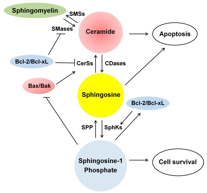
Ceramide is synthesized upon stimuli, and induces apoptosis in renal tubular cells (RTCs). Sphingosine-1 phosphate (S1P) functions as a survival factor. Thus, the balance of ceramide/S1P determines ceramide-induced apoptosis. Mitochondria play a key role for ceramide-induced apoptosis by altered mitochondrial outer membrane permeability (MOMP). Ceramide enhances oligomerization of pro-apoptotic Bcl-2 family proteins, ceramide channel, and reduces anti-apoptotic Bcl-2 proteins in the MOM. This process alters MOMP, resulting in generation of reactive oxygen species (ROS), cytochrome C release into the cytosol, caspase activation, and apoptosis. Ceramide regulates apoptosis through mitogen-activated protein kinases (MAPKs)-dependent and -independent pathways. Conversely, MAPKs alter ceramide generation by regulating the enzymes involving ceramide metabolism, affecting ceramide-induced apoptosis. Crosstalk between Bcl-2 family proteins, ROS, and many signaling pathways regulates ceramide-induced apoptosis. Growth factors rescue ceramide-induced apoptosis by regulating the enzymes involving ceramide metabolism, S1P, and signaling pathways including MAPKs. This article reviews evidence supporting a role of ceramide for apoptosis and discusses a role of mitochondria, including MOMP, Bcl-2 family proteins, ROS, and signaling pathways, and crosstalk between these factors in the regulation of ceramide-induced apoptosis of RTCs. A balancing role between ceramide and S1P and the strategy for preventing ceramide-induced apoptosis by growth factors are also discussed.


Figures

Figure 1
Figure 1
Metabolism of sphingolipids. Ceramide can be generated by three major pathways: (1) the de novo synthesis pathway, which occurs in the endoplasmic reticulum; (2) hydrolysis of sphingomyelin; and (3) the salvage pathway, which occurs in acidic compartment of the late endosomes/lysosomes. A-CDase, acid ceramidase; A-SMase, acid sphingomyelinase; CerSs, ceramide synthases; CK, ceramide kinase; C1P, ceramide-1-phosphate; C1PP, C1P phosphatase; DES, dihydroceramide desaturase; KDS, 3-keto-dihydrosphingosine reductase; SMases, sphingomyelinases; SMSs, sphingomyelin synthases; SphKs, sphingosine kinases; S1P, sphingosine-1-phosphate; SPP, S1P phosphatase; SPT, serine palmitoyl transferase.
Figure 2
Figure 2
Compartmentalization and trafficking of ceramide. Ceramide (Cer) is synthesized de novo on the cytosolic surface of the endoplasmic reticulum (ER), and then transported via ceramide transport protein (CERT) to the Golgi, where it becomes as a substrate for sphingomyelin (SM) synthesis. Cer in the ER can also be transferred to the Golgi by vesicular transport, and subsequently the transport protein, four-phosphate adaptor protein 2 (FAPP2), delivers glucosylceramide (GlcCer) as a precursor for the synthesis of glycosphingolipids (GSL). SM and GSL are then transported to the plasma membrane via vesicular transport. SM and GSL also move from the plasma membrane to the endosomal system and are metabolized via lysosomal degradation. Ceramide is transformed to galactosylceramide (GalCer) in the ER. Ceramide can be exchanged between the ER and mitochondria through mitochondria-associated membranes (MAMs) at the ER. Because of its adequate solubility, sphingosine can leave the lysosome and move between membranes. CDase, ceramidase; dhCer, dihydroceramide; dhSph, dihydrosphingosine; 3kdhSph, 3-keto-dihydrosphingosine; Mito, mitochondria; Nuc, nucleus; Ser, serine; SK, sphingosine kinase; Sph, sphingosine; S1P, sphingosine-1-phosphate.
Figure 3
Figure 3
Mitochondria, Bcl-2 family proteins, and reactive oxygen species (ROS) in ceramide-induced apoptosis of renal tubular cells (RTCs). Once ceramide is synthesized and accumulated upon stimuli, it can induce ROS generation and depletion of redox system by inhibiting mitochondrial respiratory chain complexes. Depletion of ATP can switch the cell death from of apoptosis to necrosis. Ceramide enhances the expression and activity of pro-apoptotic Bcl-2 family proteins (e.g., Bax/Bak) and reduces those of anti-apoptotic Bcl-2 proteins (e.g., Bcl-2/Bcl-xL). Ceramide can regulate at least 4 channel/pore-forming complexes that regulate the MOMP: (1) a proteolipid pore, mitochondrial apoptosis-induced channel (MAC), formed by oligomerization of Bax and Bak mediated by tBid in the MOM; (2) a lipid channel formed by ceramide in the MOM; (3) a voltage-dependent anion channel (VDAC) in the MOM; and (4) MPT pore formed in the MIM in response to apoptotic stimuli. Ceramide can form MAC caused by oligomerization of Bax/Bak. Ceramide also assembles ceramide channel, which is regulated by Bcl-2 family proteins. Ceramide may form pores in the MOM, in concert with VDAC, which is not part of MPT pore, and possibly Bax and Bak. Additionally, ceramide may regulate the function of VDAC, which is regulated by several pro- and anti-apoptotic Bcl-2 family members. These processes lead to MOMP, thereby initiating apoptosis. BcL-2/Bcl-xL has the opposite function that inhibits MOMP. Ceramide can increase MPT pore opening, leading to MOMP. MOMP can enhance ROS generation and the release of apoptogenic proteins such as cytochrome C, which activate a caspase cascade, leading to apoptosis. The MOM is finally ruptured and this in turn contributes to mitochondrial remodeling, fusion and fission, which results in further release of apoptogenic factors, leading to apoptosis. There is an interconnection between the mitochondria and the ER through the regulation of calcium homeostasis via inositol 1,4,5-triphosphate receptor (IP3R) on the ER membrane. Bax/Bak can increase the release of Ca2+ from the ER into the cytosol via pore forming complexes on the ER membrane, whereas Bcl-2/Bcl-xL can prevent this event by directly or indirectly regulating IP3R. This process regulates mitochondrial calcium homeostasis, thereby regulating MOMP and apoptosis. ER, endoplasmic reticulum; Mito, mitochondria; ↑, increase; ┤, suppress the expression and activity.
Figure 4
Figure 4
Ceramide, sphingosine and sphingosine-1-phosphate (S1P) and their regulation by Bcl-2 family proteins in apoptosis. Ceramide and sphingosine function as pro-apoptotic factors, while S1P functions as a survival factor. Bcl-2/Bcl-xL activates sphingosine kinases (SphKs), resulting in accumulation of S1P, thereby reducing apoptosis. Bcl-2/Bcl-xL suppresses CerSs and SMases, reducing ceramide accumulation and apoptosis. In contrast, Bax/Bak increases CerSs activity, thereby enhancing ceramide accumulation and apoptosis. S1P prevents apoptosis by reducing the expression and activity of Bax/Bak and activating those of Bcl-2/Bcl-xL. S1P lyase degrades S1P, reducing S1P levels, thereby enhancing apoptosis. CDases, ceramidases; CerSs, ceramide synthases; SMSs, sphingomyelin synthases; SPP, S1P phosphatase; ↑, increase; ┤, suppress the expression and activity.
Figure 5
Figure 5
Ceramide-induced cell signaling pathways in the regulation of apoptosis of RTCs. Ceramide can regulate various cellular signaling pathways, thereby regulating apoptosis in RTCs. Ceramide activates ERK and p38MAPK, whereas JNK is activated or not activated. A role of MAPKs in the regulation of ceramide-induced apoptosis in RTCs is controversial. Ceramide can also decrease PI3K/Akt. CREB, STAT3, p21/p53, FAK, PKC, and increase PLA2, PKA and calpain, leading to apoptosis in RTCs. A crosstalk existing between MAPKs, PKC and CREB can regulate ceramide-induced apoptosis. S1P may antagonize ceramide-induced alteration of cell signaling molecules involved in apoptotic processes in RTCs. ↑, increase; ↓, decrease; ┤, suppress the expression and activity; Akt, protein kinase B; CREB, cAMP response element binding protein; ERK, extracellular signal-regulated kinase; FAK, focal adhesion kinase; MAPK, mitogen-activated protein kinase; JNK, c-Jun N-terminal kinase; MLKs, mixed lineage kinases; PI3K, phosphatidylinositol-3 kinase; PKA, protein kinase A; PKC, protein kinase C; PLA2, phopspholipase A2; S1P, sphingosine-1-phosphate; STAT3, signal transducer and activator of transcription-3.

References

    1. Hait N.C., Oskeritzian C.A., Paugh S.W., Milstien S., Spiegel S. Sphingosine kinases, sphingosine 1-phosphate, apoptosis and diseases. Biochim. Biophys. Acta. 2006;1758:2016–2026. doi: 10.1016/j.bbamem.2006.08.007. - DOI - PubMed
    1. Bartke N., Hannun Y.A. Bioactive sphingolipids: Metabolism and function. J. Lipid Res. 2009;50:S91–S96. doi: 10.1194/jlr.R800080-JLR200. - DOI - PMC - PubMed
    1. Hannun Y.A., Obeid L.M. Many ceramides. J. Biol. Chem. 2011;286:27855–27862. doi: 10.1074/jbc.R111.254359. - DOI - PMC - PubMed
    1. Siskind L.J. Mitochondrial ceramide and the induction of apoptosis. J. Bioenerg. Biomembr. 2005;37:143–153. doi: 10.1007/s10863-005-6567-7. - DOI - PMC - PubMed
    1. Colombini M. Ceramide channels and their role in mitochondria-mediated apoptosis. Biochim. Biophys. Acta. 2010;1797:1239–1244. doi: 10.1016/j.bbabio.2010.01.021. - DOI - PubMed

Publication types

MeSH terms