. 2015 May 15;593(10):2379-88.
doi: 10.1113/JP270056. Epub 2015 Mar 31.

In vivo evaluation of the dentate gate theory in epilepsy

Affiliations

In vivo evaluation of the dentate gate theory in epilepsy

Esther Krook-Magnuson et al. J Physiol. .

Abstract

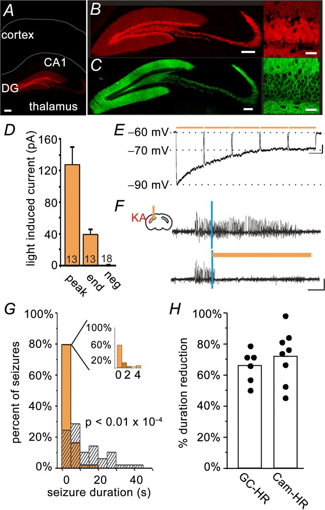
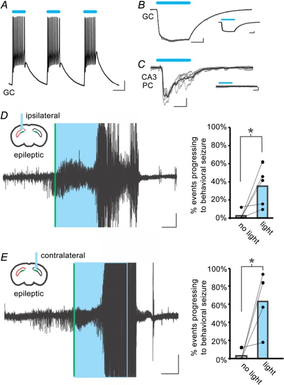
The dentate gyrus is a region subject to intense study in epilepsy because of its posited role as a 'gate', acting to inhibit overexcitation in the hippocampal circuitry through its unique synaptic, cellular and network properties that result in relatively low excitability. Numerous changes predicted to produce dentate hyperexcitability are seen in epileptic patients and animal models. However, recent findings question whether changes are causative or reactive, as well as the pathophysiological relevance of the dentate in epilepsy. Critically, direct in vivo modulation of dentate 'gate' function during spontaneous seizure activity has not been explored. Therefore, using a mouse model of temporal lobe epilepsy with hippocampal sclerosis, a closed-loop system and selective optogenetic manipulation of granule cells during seizures, we directly tested the dentate 'gate' hypothesis in vivo. Consistent with the dentate gate theory, optogenetic gate restoration through granule cell hyperpolarization efficiently stopped spontaneous seizures. By contrast, optogenetic activation of granule cells exacerbated spontaneous seizures. Furthermore, activating granule cells in non-epileptic animals evoked acute seizures of increasing severity. These data indicate that the dentate gyrus is a critical node in the temporal lobe seizure network, and provide the first in vivo support for the dentate 'gate' hypothesis.


Figures

Figure 1
Figure 1
On-demand restoration of the dentate gate inhibits spontaneous temporal lobe seizures Crossing a mouse line expressing Cre in DG GCs (visualized in A and B, by crossing with a tdTomato reporter line; red in the online version) with a mouse line expressing the inhibitory opsin HR in a Cre-dependent fashion, produced mice with HR expressed selectively in DG GCs (GC-HR). Selectivity of opsin expression was maintained in epileptic animals (C, yellow fluorescent protein tagged HR; green in the online version). The edge of the slice and the border between CA1 and the alveus are drawn in A for reference. D and E, whole-cell patch-clamp recordings from epileptic brain slices revealed robust light-induced inhibition of GCs of opsin-expressing but not opsin-negative animals. (Peak, peak-induced currents; End, current measured at the end of 10 s of pulsed light delivery; Neg, opsin-negative controls. The number of cells recorded is indicated in each bar. D, summary voltage clamp data. E, example current clamp recording). F–G, in vivo online detection of spontaneous seizures allowed on-demand light delivery, which rapidly truncated seizures when delivered to the hippocampus ipsilateral to prior KA injection (example animal: vertical blue lines indicate seizure detection; amber bars indicate light delivery; hashed bars indicate events not receiving light; G, inset: expansion of the first 5 s after light delivery; 100 seizures; colour is shown in the online version). H, the inhibition of seizure duration achieved with selective inhibition of GCs is comparable to that achieved with broader inhibition of excitatory cells including CA1 pyramidal cells (Cam-HR; shown for reference in H; each dot represents one animal). Scale bars: A, 200 μm; B and C, left 200 μm, right 30 μm; E, 5 mV, 1 s; F, 0.2 mV, 5 s.
Figure 2
Figure 2
Excitation of GCs worsens spontaneous seizures A, light-induced excitation of a GC in a slice from an epileptic GC-ChR2 animal. B, robust light-induced currents in a GC, which remain in the presence of TTX (inset). Grey traces: individual sweeps; black traces: average. C, optogenetic activation of GCs produces postsynaptic currents in a downstream CA3 pyramidal cell (PC), which are eliminated by the application of TTX (inset). D and E, on-demand light delivery to the dentate gyrus in mice expressing the excitatory opsin ChR2 in GCs (GC-ChR2 mice) ipsilateral (D) or contralateral (E) to previous kainate injection causes electrographic seizures to progress to large behavioural seizures. Right: each dot pair represents one animal. *P < 0.001 (chi-squared). Scale bars: A, 10 mV, 50 ms; B and C, 100 pA, 20 ms; D and E, 0.1 mV, 10 s. Boxes (coloured blue in the online version) denote light delivery. Vertical lines (coloured green in the online version) indicate online seizure detection. Large amplitude signals include a movement artefact and have been truncated.
Figure 3
Figure 3
Excitation of GCs is sufficient to induce seizures in kainate-naïve animals A, example responses to repeated light delivery in a kainate-naïve GC-ChR2 animal. Small grey numbers under traces indicate the number of light delivery. Boxes (coloured blue in the online version) denote 30 s of pulsed light delivery. Large amplitude signals include a movement artefact and have been truncated. Scale bar: 0.1 mV, 20 s. B, seizure duration and severity increases with repeated light delivery. Each symbol represents the seizure duration from a given animal; shading (coloured blue in the online version) indicates the duration of light delivery (left axis). The black line indicates the average behavioural seizure score (right axis). Seizure scoring was based on the extended Racine scale of Pinel & Rover (1978), as detailed in the Methods.

References

    1. Acsady L, Kamondi A, Sik A, Freund T. Buzsaki G. GABAergic cells are the major postsynaptic targets of mossy fibers in the rat hippocampus. J Neurosci. 1998;18:3386–3403. - PMC - PubMed
    1. Ang CW, Carlson GC. Coulter DA. Massive and specific dysregulation of direct cortical input to the hippocampus in temporal lobe epilepsy. J Neurosci. 2006;26:11850–11856. - PMC - PubMed
    1. Armstrong C, Krook-Magnuson E, Oijala M. Soltesz I. Closed-loop optogenetic intervention in mice. Nat Protoc. 2013;8:1475–1493. - PMC - PubMed
    1. Armstrong C, Krook-Magnuson E. Soltesz I. Neurogliaform and Ivy cells: a major family of nNOS expressing GABAergic neurons. Front Neural Circuits. 2012;6:23. - PMC - PubMed
    1. Armstrong C, Szabadics J, Tamas G. Soltesz I. Neurogliaform cells in the molecular layer of the dentate gyrus as feed-forward gamma-aminobutyric acidergic modulators of entorhinal-hippocampal interplay. J Comp Neurol. 2011;519:1476–1491. - PMC - PubMed

Publication types

LinkOut - more resources