. 2015 Apr 7;23(4):677-87.
doi: 10.1016/j.str.2015.01.018. Epub 2015 Mar 5.

Structural basis for VEGF-C binding to neuropilin-2 and sequestration by a soluble splice form

Affiliations

Structural basis for VEGF-C binding to neuropilin-2 and sequestration by a soluble splice form

Matthew W Parker et al. Structure. .

Abstract

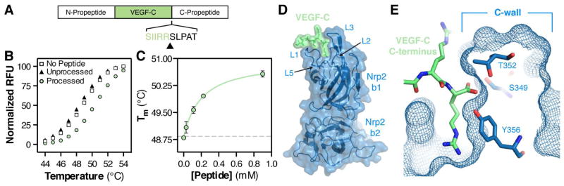
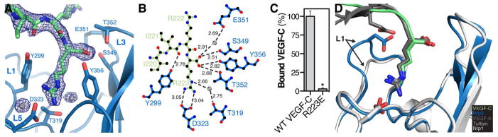
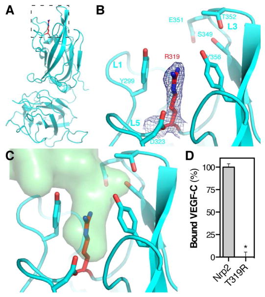
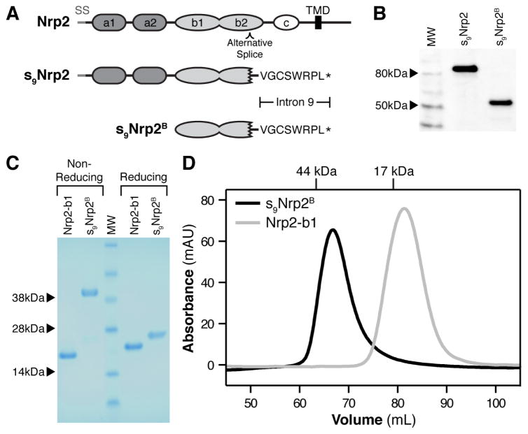
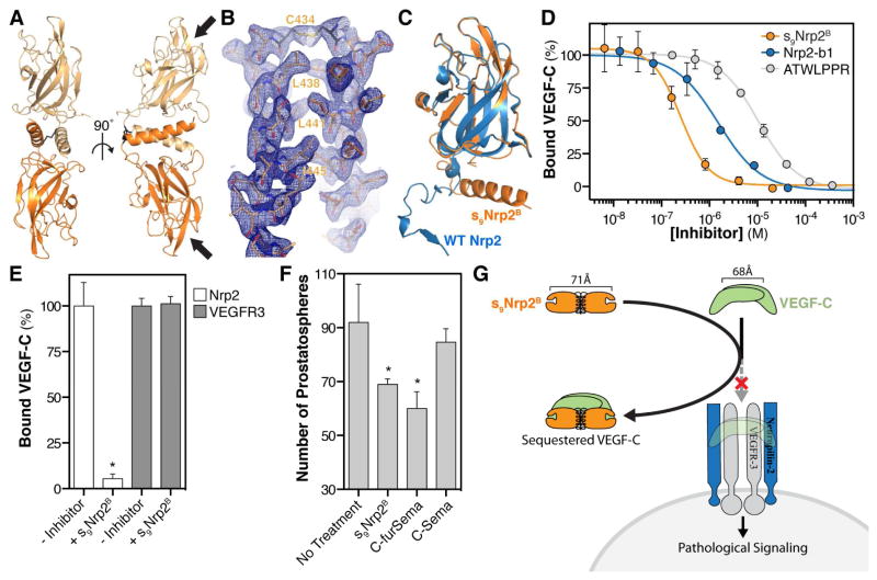
Vascular endothelial growth factor C (VEGF-C) is a potent lymphangiogenic cytokine that signals via the coordinated action of two cell surface receptors, Neuropilin-2 (Nrp2) and VEGFR-3. Diseases associated with both loss and gain of VEGF-C function, lymphedema and cancer, respectively, motivate studies of VEGF-C/Nrp2 binding and inhibition. Here, we demonstrate that VEGF-C binding to Nrp2 is regulated by C-terminal proteolytic maturation. The structure of the VEGF-C C terminus in complex with the ligand binding domains of Nrp2 demonstrates that a cryptic Nrp2 binding motif is released upon proteolysis, allowing specific engagement with the b1 domain of Nrp2. Based on the identified structural requirements for Nrp2 binding to VEGF-C, we hypothesized that the endogenous secreted splice form of Nrp2, s9Nrp2, may function as a selective inhibitor of VEGF-C. We find that s9Nrp2 forms a stable dimer that potently inhibits VEGF-C/Nrp2 binding and cellular signaling. These data provide critical insight into VEGF-C/Nrp2 binding and inhibition.


Conflict of interest statement

The authors declare no conflict of interest.

Figures

Figure 1
Figure 1. Crystal structure of the VEGF-C/Nrp2 complex reveals the basis for proteolytic-dependent binding
(A) Organization of the VEGF-C pro-protein and site of C-terminal processing (black arrow). (B) Peptides corresponding to processed (green circle) and unprocessed (black triangle) VEGF-C were assayed for the ability to bind Nrp2-b1b2 as measured by DSF thermal shift assay. Peptides were added to Nrp2-b1b2 to a final concentration of 0.5 mM and melting was monitored between 20 and 90°C. All samples were measured in triplicate and a representative melting curve is shown for each. (C) Processed VEGF-C dose-dependently enhances the Nrp2-b1b2 Tm. Error bars indicate the standard deviation (SD) of the three measurements. (D) Structure of Nrp2-b1b2 (blue) in complex with the C-terminus of VEGF-C (green). (E) Cross-section of the Nrp2 binding pocket demonstrates that the free carboxy terminus of VEGF-C is buried against the Nrp2 C-wall, which is formed by the third coagulation factor loop.
Figure 2
Figure 2. Mechanism of VEGF-C binding to Nrp2
(A) Zoom of the intermolecular interface between Nrp2 (blue) and VEGF-C (green) with the 2Fo-Fc electron density map for VEGF-C contoured at 1.0σ. Interfacing water is shown as grey spheres. (B) Ligplot+ generated representation of the interaction between VEGF-C (green) and Nrp2 (blue). Bond distances (Å) are labeled in black and water is shown as grey spheres. (C) Nrp2 binding was compared between VEGF-C and VEGF-C R223E. Binding was measured in triplicate and is reported as mean ± SD (*p<0.05). (D) Superimposition of the VEGF-A HBD/Nrp1 complex (PDB=4DEQ) and the tuftsin/Nrp1 complex (PDB=2ORZ) onto the structure of the VEGF-C/Nrp2 complex demonstrates the shared and unique modes of engagement within this ligand/receptor family.
Figure 3
Figure 3. Crystal structure and VEGF-C binding properties of Nrp2-T319R
(A) Structure of Nrp2-T319R with the stick representation for T319R shown in red. (B) Zoom of the Nrp2-T319R binding pocket. The blue mesh illustrates the 2Fo-Fc electron density map for R319 contoured at 1.0σ. (C) Superimposition of VEGF-C (green) onto the structure of Nrp2-T319R demonstrates that the binding pocket normally occupied by VEGF-C is blocked in the mutant. (D) VEGF-C binding was compared between Nrp2-b1b2 and Nrp2-T319R. Binding was measured in triplicate and is reported as mean ± SD (*p<0.05).
Figure 4
Figure 4. s9Nrp2B forms a disulfide-linked dimer
(A) Domain organization of Nrp2 and the protein fragment utilized for our studies, s9Nrp2B. (B) Western blot analysis of Hgh-tagged s9Nrp2 and s9Nrp2B expressed in CHO cells. (C) Non-reducing and reducing SDS-PAGE analysis of Nrp2-b1 and s9Nrp2B. (D) The oligomeric state of s9Nrp2B (black line) was analyzed by size-exclusion chromatography. Nrp2-b1 was run as a reference (grey line).
Figure 5
Figure 5. Crystal structure and inhibitory properties of s9Nrp2B
(A) Crystal structure of the s9Nrp2B dimer (Chain A: light orange; Chain B: dark orange). The intermolecular disulfide is shown in black and the Nrp2-b1 binding pockets are labeled with arrows. (B) Zoom of the dimeric helical bundle with the 2Fo-Fc electron density map contoured at 1σ. (C) The residues of the Nrp2 b1-b2 linker and b2 domain show a dramatic structural reorganization from an extended loop in the b1b2 sequence (blue) to an extended helix in the s9Nrp2B dimer (orange). (D) ATWLPPR (grey), Nrp2-b1 (blue), and s9Nrp2B (orange) were assayed for the ability to inhibit VEGF-C binding to Nrp2. ATWLPPR inhibited binding with an IC50 = 10 μM (log[IC50] = −4.98 ± 0.03), Nrp2-b1 inhibited binding with an IC50 = 1.5 μM (log[IC50] = −5.82 ± 0.09), and s9Nrp2B inhibited binding with an IC50 = 250 nM (log[IC50] = −6.60 ± 0.08). (E) s9Nrp2B was assayed for the ability to alter VEGF-C binding to VEGFR3. Addition of 4μM s9Nrp2B fully inhibited VEGF-C/Nrp2 binding but showed no effect on VEGF-C/VEGFR3 binding. (F) Inhibition of C4-2 cell prostatosphere formation was used to assess the biological activity of s9Nrp2B. Prostatosphere formation was compared in the absence and presence of s9Nrp2B, as well as with C-furSema (positive control) and C-Sema (negative control). (G) Model illustrating the mechanism of action for s9Nrp2B. s9Nrp2B sequesters VEGF-C and prevents activation of the VEGFR3/Nrp2 signaling complex. All inhibition experiments were measured in triplicate and reported as mean ± SD (*p<0.05).

Comment in

References

    1. Adams RH, Lohrum M, Klostermann A, Betz H, Puschel AW. The chemorepulsive activity of secreted semaphorins is regulated by furin-dependent proteolytic processing. EMOB J. 1997;16:6077–6086. - PMC - PubMed
    1. Albuquerque RJ, Hayashi T, Cho WG, Kleinman ME, Dridi S, Takeda A, Baffi JZ, Yamada K, Kaneko H, Green MG, et al. Alternatively spliced vascular endothelial growth factor receptor-2 is an essential endogenous inhibitor of lymphatic vessel growth. Nat Med. 2009;15:1023–1030. - PMC - PubMed
    1. Ambati BK, Nozaki M, Singh N, Takeda A, Jani PD, Suthar T, Albuquerque RJ, Richter E, Sakurai E, Newcomb MT, et al. Corneal avascularity is due to soluble VEGF receptor-1. Nature. 2006;443:993–997. - PMC - PubMed
    1. Appleton BA, Wu P, Maloney J, Yin J, Liang WC, Stawicki S, Mortara K, Bowman KK, Elliott JM, Desmarais W, et al. Structural studies of neuropilin/antibody complexes provide insights into semaphorin and VEGF binding. EMBO J. 2007;26:4902–4912. - PMC - PubMed
    1. Aricescu AR, Lu W, Jones EY. A time- and cost-efficient system for high-level protein production in mammalian cells. Acta Crystallogr D, Biol Crystallogr. 2006;62:1243–1250. - PubMed

Publication types