. 2015 Mar 19;57(6):1124-1132.
doi: 10.1016/j.molcel.2015.01.043. Epub 2015 Mar 5.

Phosphoproteomics reveals distinct modes of Mec1/ATR signaling during DNA replication

Affiliations

Phosphoproteomics reveals distinct modes of Mec1/ATR signaling during DNA replication

Francisco Meirelles Bastos de Oliveira et al. Mol Cell. .

Erratum in

  • Mol Cell. 2015 Apr 2;58(1):194

Abstract

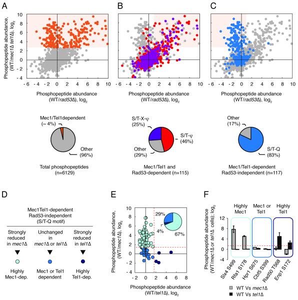
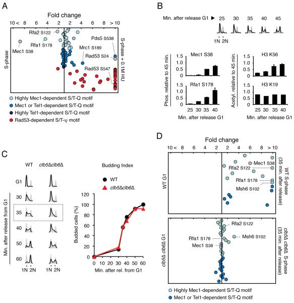
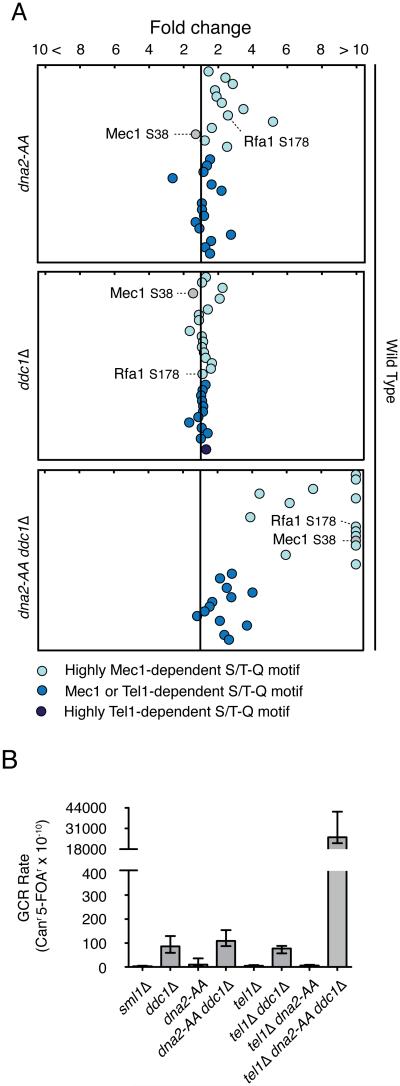
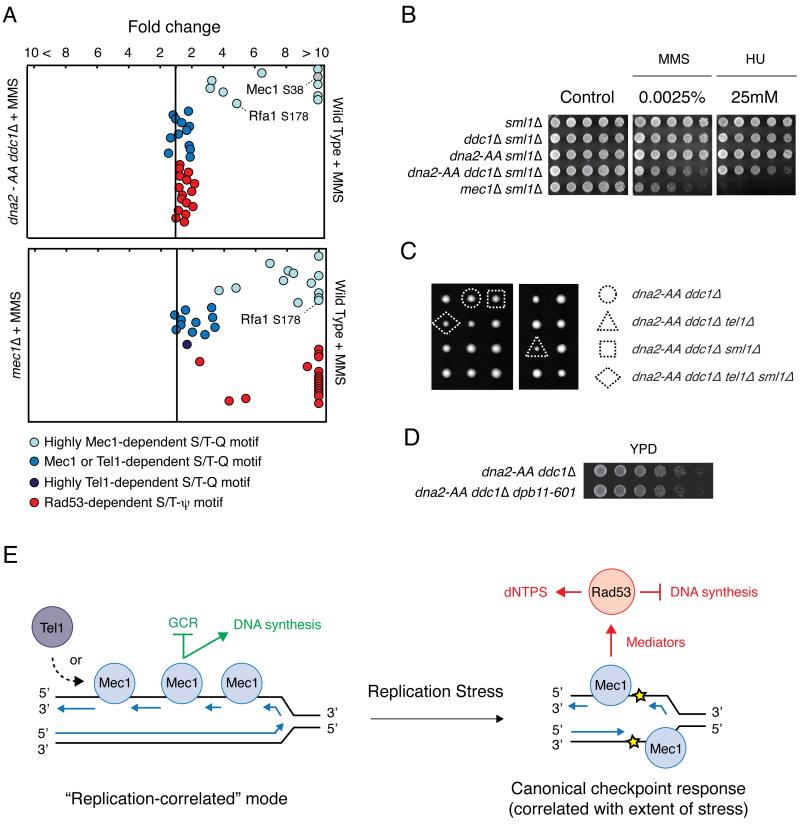
The Mec1/Tel1 kinases (human ATR/ATM) play numerous roles in the DNA replication stress response. Despite the multi-functionality of these kinases, studies of their in vivo action have mostly relied on a few well-established substrates. Here we employed a combined genetic-phosphoproteomic approach to monitor Mec1/Tel1 signaling in a systematic, unbiased, and quantitative manner. Unexpectedly, we find that Mec1 is highly active during normal DNA replication, at levels comparable or higher than Mec1's activation state induced by replication stress. This "replication-correlated" mode of Mec1 action requires the 9-1-1 clamp and the Dna2 lagging-strand factor and is distinguishable from Mec1's action in activating the downstream kinase Rad53. We propose that Mec1/ATR performs key functions during ongoing DNA synthesis that are distinct from their canonical checkpoint role during replication stress.


Figures

Figure 1
Figure 1. Proteome-wide identification of Mec1/Tel1-dependent phosphorylation events using quantitative MS
(A) Identification of Mec1/Tel1-dependent phosphopeptides (cells treated with 0.2M HU or 0.04% MMS). Orange dots correspond to 238 Mec1/Tel1-dependent phosphopeptides. See text for details. (B) Mec1/Tel1 and Rad53-dependent phosphorylation events (light orange shade) are biased towards the S/T-ψ (red) and S/T-X-ψ (purple) motifs. (C) Mec1/Tel1-dependent and Rad53-independent phosphorylation events (light orange shade) are biased towards the S/T-Q motif (blue). (D-E) The phosphoproteome of WT cells was compared to the phosphoproteome of mec1Δ or tel1Δ cells (all cells treated with 0.04% MMS) and phosphopeptides carrying phosphorylation in the S/T-Q motif were categorized according to the observed change in abundance. Dotted red lines represent the established cutoff of 3-fold increase in WT relative to mec1Δ or tel1Δ cells. (F) Examples of phosphopeptides of each of the indicated groups. Data are represented as fold change in phosphopeptide abundance; log2 +/− SEM (n≥2). See also Table S1.
Figure 2
Figure 2. Quantitative analysis of Mec1/Tel1-dependent phosphorylation during normal DNA replication
(A) QMAPS showing the relative abundance of phosphopeptides categorized according to results from Figure 1. Phosphopeptides carrying Mec1 autophosphorylation or Mec1-dependent Rad53 phosphorylation are indicated in grey. α-factor arrested cells were released from arrest in normal SILAC media or SILAC media containing 0.1M HU for 45 minutes. Abscissa indicates fold change in phosphopeptide abundance (linear scale) between S-phase cells treated with 0.1M HU and untreated. (B) Protein extracts were prepared from WT cells at indicated times after release from α-factor-arrest into fresh media. Mec1 (and Mec1-associated Rfa1) was pulled-down and phosphopeptides containing Mec1 autophosphorylation at S38 and Rfa1 phosphorylation at S178 were monitored by quantitative MS analysis. FACS analysis and H3K56 acetylation were used as positive controls for DNA replication progression while acetylation of H3K19 was used as a constitutive control. Data are represented as mean +/− SEM (n≥2). (C) FACS analysis and budding index of WT and clb5Δclb6Δ mutant cells following α-factor arrest and release in drug-free SILAC media. (D) QMAPS showing the relative abundance of phosphopeptides carrying Mec1/Tel1-dependent S/T-Q motifs. Indicated cells were released from α-factor arrest in drug-free SILAC media for 35 minutes. For all the QMAPS in Figure 2, each dot corresponds to a different phosphopeptide identified at least 3 times in 2 independent biological replicates. See also Table S2.
Figure 3
Figure 3. Importance of Dna2 and Ddc1 for replication-correlated mode of Mec1 activation
(A) QMAPS showing the relative abundance of phosphopeptides carrying Mec1/Tel1-dependent S/T Q motifs. WT, dna2-AA, ddc1Δ and dna2-AA ddc1Δ cells were released from α-factor arrest in SILAC media for 45 minutes. See also Table S3. (B) Effects of the dna2-AA mutation on accumulation of gross-chromosomal rearrangements in Ddc1 and Tel1 defective mutants. All strains are sml1Δ. Error bars indicate 95% confidence intervals (CI).
Figure 4
Figure 4. Dna2 and Ddc1 are not essential for Mec1 activation during replication stress.
A) QMAPS analysis comparing WT and indicated mutant cells. Cells were arrested with α-factor and released from arrest in SILAC media containing 0.04% MMS for 45 minutes. See also Table S4. (B) 5-fold serial dilutions of indicated cells with sml1Δ background were plated on YPD plates containing indicated drugs and incubated at 30°C for 48h. (C) Meiotic tetrads from a DNA2/dna-AA DDC1/ddc1Δ TEL1/tel1Δ SML1/sml1Δ diploid strain were dissected on YPD plates and incubated at 30°C for 72h. (D) 4-fold serial dilutions of indicated cells were plated on YPD plates and incubated at 30°C for 36h. (E) Model depicting distinct modes of Mec1 action during DNA replication. See text in the discussion. Blue arrows correspond to newly synthesized DNA strands.

References

    1. Bandhu A, Kang J, Fukunaga K, Goto G, Sugimoto K. Ddc2 mediates Mec1 activation through a Ddc1- or Dpb11-independent mechanism. PLoS genetics. 2014;10 - PMC - PubMed
    1. Branzei D, Foiani M. Maintaining genome stability at the replication fork. Nat Rev Mol Cell Biol. 2010;11:208–219. - PubMed
    1. Brown EJ, Baltimore D. ATR disruption leads to chromosomal fragmentation and early embryonic lethality. Genes Dev. 2000;14:397–402. - PMC - PubMed
    1. Chen SH, Albuquerque CP, Liang J, Suhandynata RT, Zhou H. A proteome-wide analysis of kinase-substrate network in the DNA damage response. J Biol Chem. 2010;285:12803–12812. - PMC - PubMed
    1. Desany BA, Alcasabas AA, Bachant JB, Elledge SJ. Recovery from DNA replicational stress is the essential function of the S-phase checkpoint pathway. Genes Dev. 1998;12:2956–2970. - PMC - PubMed

Publication types

MeSH terms

Substances