. 2015 Apr 2;58(1):95-109.
doi: 10.1016/j.molcel.2015.01.042. Epub 2015 Mar 5.

Assembly and specific recognition of k29- and k33-linked polyubiquitin

Affiliations

Assembly and specific recognition of k29- and k33-linked polyubiquitin

Martin A Michel et al. Mol Cell. .

Abstract

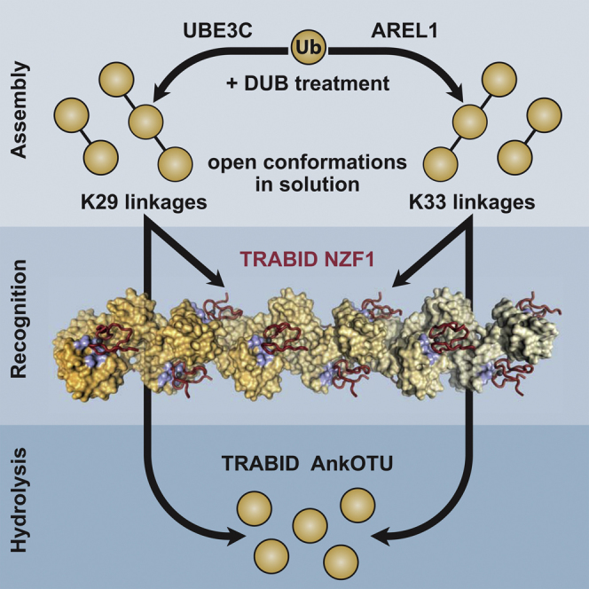
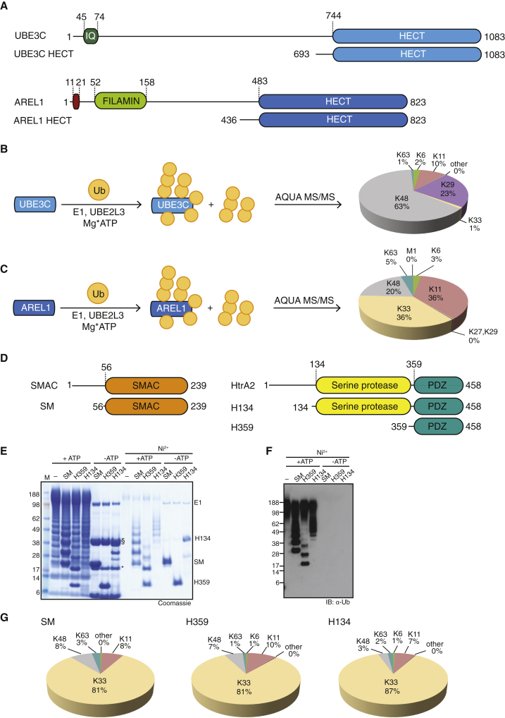
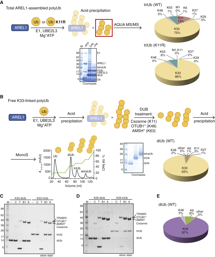
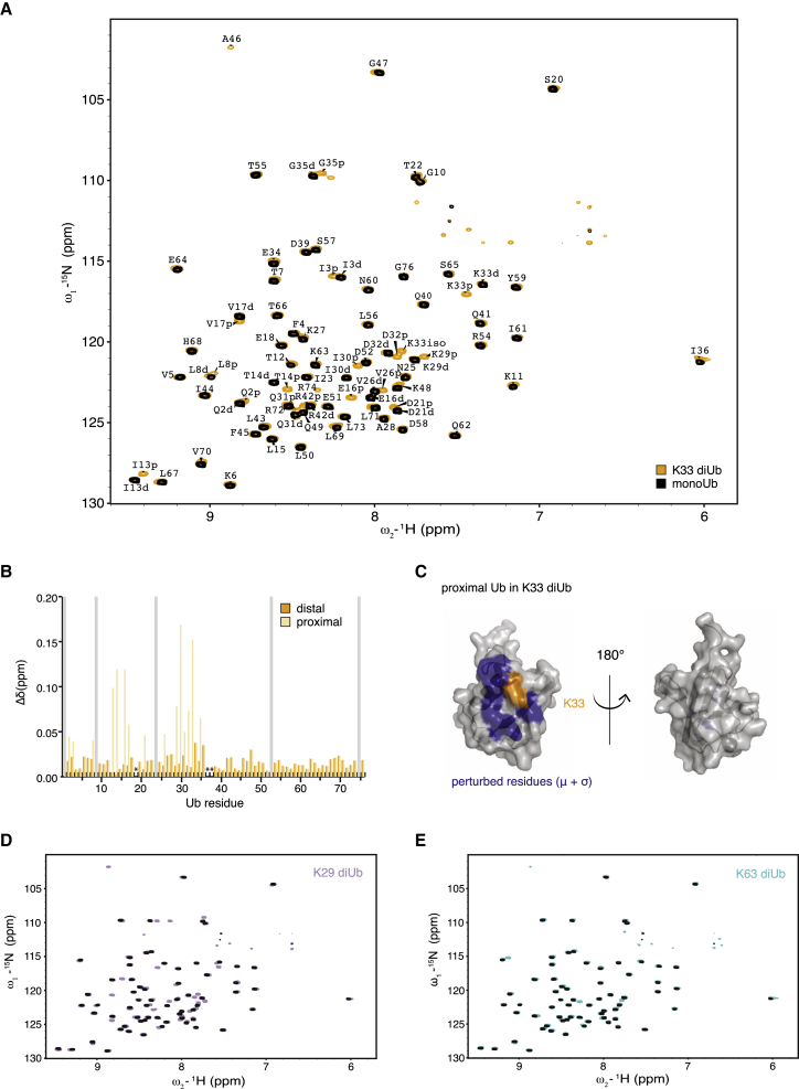
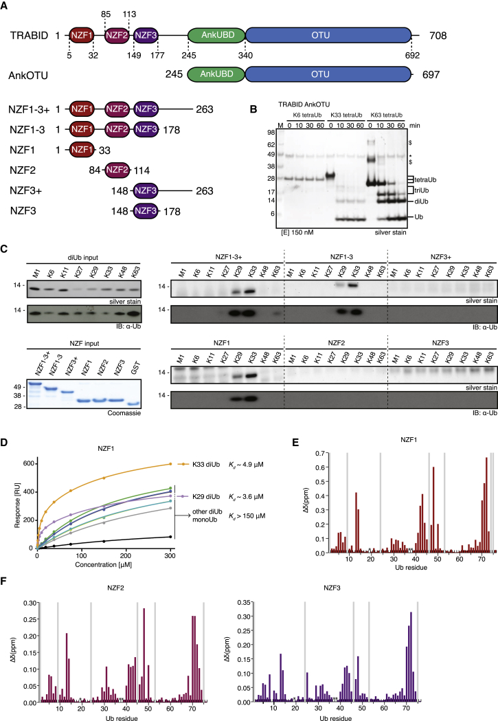
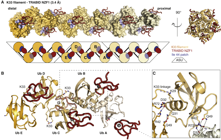
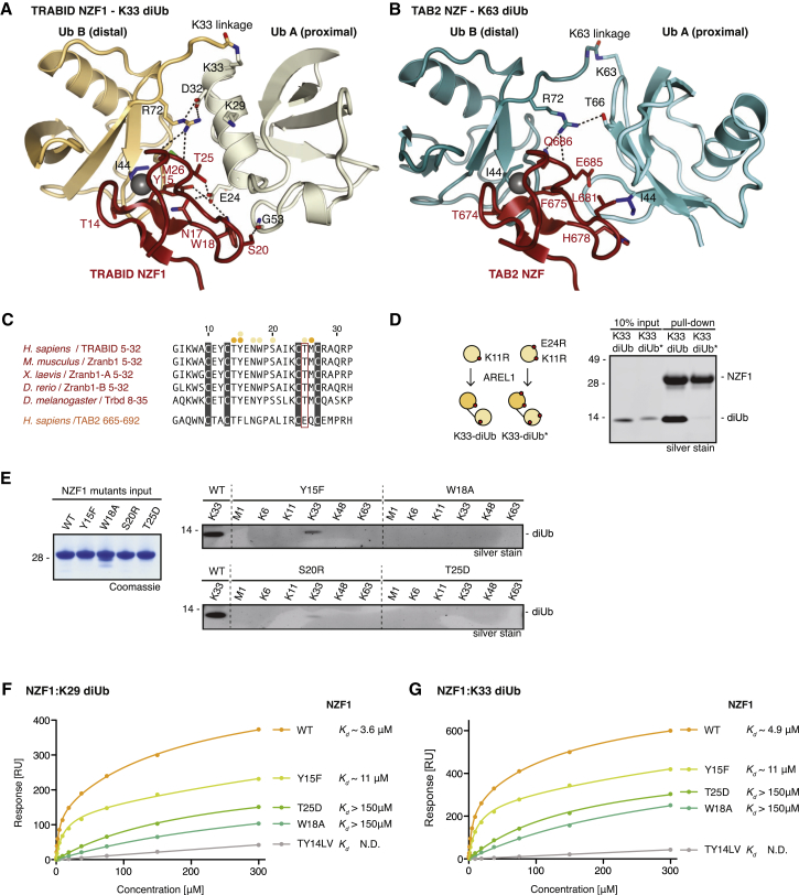
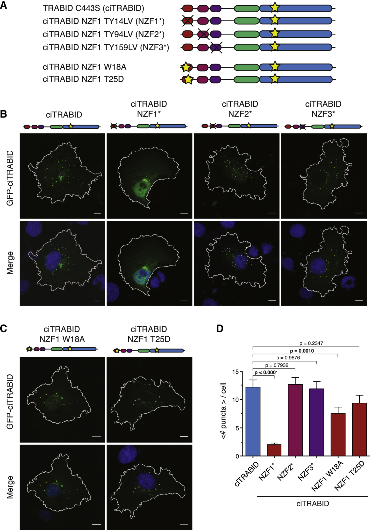
Protein ubiquitination regulates many cellular processes via attachment of structurally and functionally distinct ubiquitin (Ub) chains. Several atypical chain types have remained poorly characterized because the enzymes mediating their assembly and receptors with specific binding properties have been elusive. We found that the human HECT E3 ligases UBE3C and AREL1 assemble K48/K29- and K11/K33-linked Ub chains, respectively, and can be used in combination with DUBs to generate K29- and K33-linked chains for biochemical and structural analyses. Solution studies indicate that both chains adopt open and dynamic conformations. We further show that the N-terminal Npl4-like zinc finger (NZF1) domain of the K29/K33-specific deubiquitinase TRABID specifically binds K29/K33-linked diUb, and a crystal structure of this complex explains TRABID specificity and suggests a model for chain binding by TRABID. Our work uncovers linkage-specific components in the Ub system for atypical K29- and K33-linked Ub chains, providing tools to further understand these unstudied posttranslational modifications.


Figures

None
Graphical abstract
Figure 1
Figure 1
Role of HECT E3 Ligases in Assembling Atypical Ub Chains (A) Domain structures of UBE3C and AREL1 (KIAA0317) (top) and constructs used in this study (bottom). (B) Schematic of an assembly reaction with UBE3C, UBE2L3 (UbcH7), E1, and WT Ub (left). The linkage composition in the reaction mixture was analyzed by AQUA-based MS/MS (right). (C) Reaction as in (B) with AREL1, UBE2L3, E1, and WT Ub. (D) Domain structures of the pro-apoptotic proteins SMAC and HtrA2 (top) and the expressed constructs used in this work (bottom). (E) AREL1 is able to assemble chains onto SMAC and HtrA2 in an in vitro ubiquitination reaction that depends on ATP. Ubiquitinated, His6-tagged substrates are enriched following Ni2+ affinity binding. SM, SMAC (56–239); H359, HtrA2 (359–458); H134, HtrA2 (134–458); §, AREL1; , UBE2L3. (F) Western blot against Ub of the Ni2+-enriched reaction from (E). (G) AQUA MS/MS profiles of the ubiquitinated substrates purified from (E).
Figure 2
Figure 2
Purification of Unanchored K29/K33 PolyUb Chains (A) Schematic of the assembly of K33-linked Ub chains using either WT or K11R Ub (top). AQUA profiles of triUb using either WT (top right) or K11R Ub (bottom right; K6 linkage was excluded from the quantitative analysis because of the K11R substitution). Bottom: corresponding SDS-PAGE gel for assembly of free chains. −ATP, initial reaction without ATP addition; O/N, overnight incubation of the assembly reaction; +PA, perchloric acid treatment of the assembly reaction. (B) Schematic representation of the purification of K33-linked polyUb chains. Following the assembly reaction, perchloric acid treatment removes the ubiquitinated and unmodified forms of E1, E2, and E3. Linkage-selective DUBs are then used to remove undesired Ub linkages. An additional perchloric acid step is required to inactivate the DUBs prior to cation exchange chromatography (bottom), which resolves the homotypic chains based on linkage length. Bottom center: SDS-PAGE of purified K33-linked di-, tri-, and tetraUb. Bottom right: AQUA MS/MS of purified K33-linked diUb. (C) Deubiquitinase assay of purified K33-linked di- and triUb. –, no DUB; C, 200 nM Cezanne (K11-specific); T, 350 nM TRABID (K29/K33-specific); B1, 1 μM OTUB1 (K48-specific); A, 1 μM AMSH (K63-specific). (D) K29-linked polyUb chains can be purified analogous to the schematic shown in (B). Purified K29-linked di- and triUb were treated with DUBs as in (C). (E) AQUA mass spectrometry profile of purified K29 diUb.
Figure 3
Figure 3
NMR Analysis of K29/K33 Chains (A) BEST-TROSY spectra for 15N-K33 diUb (orange) and 15N-monoUb (black). Complete assignment of resonances from the proximal (p) or distal (d) moieties from a 13C, 15N-K33 diUb sample are shown. (B) Chemical shift perturbation of distal (orange) and proximal (beige) resonances with respect to monoUb. Grey bars, exchange-broadened resonances; asterisks, proline residues. (C) Resonances that display a perturbation of more than 1 σ are mapped onto the surface of monoUb (purple) and cluster around the K33 residue (orange). No significant perturbations were observed on the distal Ub moiety, consistent with an open conformation of K33 diUb. (D) BEST-TROSY spectra for 15N K29 diUb (purple) and monoUb (black). (E) BEST-TROSY spectra for 15N K63 diUb (cyan) and monoUb (black).
Figure 4
Figure 4
Characterization of TRABID Specificity (A) Domain structure of human TRABID. The AnkOTU fragment has been characterized in detail in Licchesi et al. (2012). Boundaries of the NZF domain fragments analyzed here are shown. (B) Deubiquitination assay of TRABID AnkOTU against K6-, K33-, and K63-linked tetraUb. See Figure S4A for a reaction at a lower DUB concentration. (C) Pull-down analysis of NZF fragments with a panel of diUb covering all linkage types. Left: the input chains and GST-NZF constructs used. Right: pull-down analysis shown by silver stain and anti-Ub western blot. See Figure S4B for additional controls. (D) SPR binding experiment of NZF1 to monoUb and the eight different diUb species with error bars representing SEs. Kd values derived from two experiments are shown. See Figure S4C for best-fit parameters and values of SEs. (E) NMR analysis of isolated NZF1 binding to 15N-labeled monoUb. The chemical shift perturbation for Ub from binding to 600 μM of NZF1 is shown. Grey bars, exchange-broadened residues; asterisks, proline residues. See Figures S4E–S4G for titration data. (F) NMR analyses as in (E) but for NZF2 and NZF3.
Figure 5
Figure 5
Structure of K33 Filaments Bound to NZF1 (A) Structure of the K33-linked Ub filament as observed in the crystal, showing three adjacent asymmetric units (black outline). Ub molecules are shown as a surface representation with a gradient from orange (distal) to beige (proximal), and Ile44 patches are indicated in blue. NZF1 is shown as a red ribbon with gray Zn2+ atoms. A schematic is shown below. Right: view of the filament down the 5-fold symmetry axis. ASU, asymmetric unit. (B) Content of the asymmetric unit, colored as in (A). K33 isopeptide linkages are shown as stick representations; see Figure S5C for electron density. (C) Close-up view of one Ub in the filament, showing interacting residues as a stick representation. Hydrogen bonds are shown as black dashed lines. The K33 and K29 side chains are also shown.
Figure 6
Figure 6
Explaining the K29/K33 Specificity of TRABID NZF1 (A) Detailed view of the interactions between TRABID NZF1 (red) and K33-linked diUb (orange/beige). Interacting residues are labeled, and hydrogen bonds are shown as black dashed lines. (B) As in (A) for the TAB2 NZF interaction with K63-linked diUb (cyan). (C) Sequence alignment of TRABID NZF1 from a diverse range of species and human TAB2 NZF domains. Interacting residues are indicated with orange (distal Ub) and beige (proximal Ub) dots. Thr25 in TRABID NZF1 is replaced with Glu685 in TAB2 NZF, which would prevent K29/K33 binding in TAB2 NZF. (D) Left: Ub chains were assembled into K33 diUb with AREL1 using K11R or K11R/E24R Ub. Right: pull-down assays with TRABID NZF1 and diUb variants. (E) Pull-down assays as in Figure 4C for TRABID NZF1 mutants. (F and G) SPR binding experiment of NZF1 and its mutants against K29 diUb (F) and K33 diUb (G) with the respective Kd values indicated. SEs from two experiments are shown as error bars. See Figure S6F for values of SEs and best-fit parameters.
Figure 7
Figure 7
Localization of Catalytically Inactive TRABID Mutants in Cells (A) Constructs used in localization experiments for GFP-TRABID fusions. Yellow stars indicate single amino acid substitutions, whereas black crosses denote two amino acid substitutions that abrogate Ub binding in the respective domain. (B) Localization of catalytically inactive full-length GFP-TRABID (ciTRABID) constructs. GFP-ciTRABID localizes to distinct puncta in COS-7 cells. Mutations in this background that abrogate Ub binding of NZF1 (NZF1) lead to a significant decrease in the number of dots, whereas the equivalent mutations in NZF2 (NZF2) or NZF3 (NZF3) do not lead to a change in the number of puncta. Cartoon representations of the constructs are shown as in (A). Scale bars, 10 μm. (C) The same experiment with single amino acid substitutions in the proximal Ub binding site of NZF1. (D) Statistical analysis of experiments in (B) and (C) with an average number of puncta per cell for the different mutants and corresponding SEs. p Values are given in reference to the ciTRABID mutant, and significant values (α < 0.05) are shown in boldface. Error bars represent SEs.

Comment in

References

    1. Al-Hakim A.K., Zagorska A., Chapman L., Deak M., Peggie M., Alessi D.R. Control of AMPK-related kinases by USP9X and atypical Lys(29)/Lys(33)-linked polyubiquitin chains. Biochem. J. 2008;411:249–260. - PubMed
    1. Alam S.L., Sun J., Payne M., Welch B.D., Blake B.K., Davis D.R., Meyer H.H., Emr S.D., Sundquist W.I. Ubiquitin interactions of NZF zinc fingers. EMBO J. 2004;23:1411–1421. - PMC - PubMed
    1. Berndsen C.E., Wolberger C. New insights into ubiquitin E3 ligase mechanism. Nat. Struct. Mol. Biol. 2014;21:301–307. - PubMed
    1. Bremm A., Komander D. Emerging roles for Lys11-linked polyubiquitin in cellular regulation. Trends Biochem. Sci. 2011;36:355–363. - PubMed
    1. Bremm A., Freund S.M.V., Komander D. Lys11-linked ubiquitin chains adopt compact conformations and are preferentially hydrolyzed by the deubiquitinase Cezanne. Nat. Struct. Mol. Biol. 2010;17:939–947. - PMC - PubMed

Publication types

MeSH terms