. 2015 Apr 2;58(1):60-70.
doi: 10.1016/j.molcel.2015.01.028. Epub 2015 Mar 5.

Two distinct DNA binding modes guide dual roles of a CRISPR-Cas protein complex

Affiliations

Two distinct DNA binding modes guide dual roles of a CRISPR-Cas protein complex

Timothy R Blosser et al. Mol Cell. .

Abstract

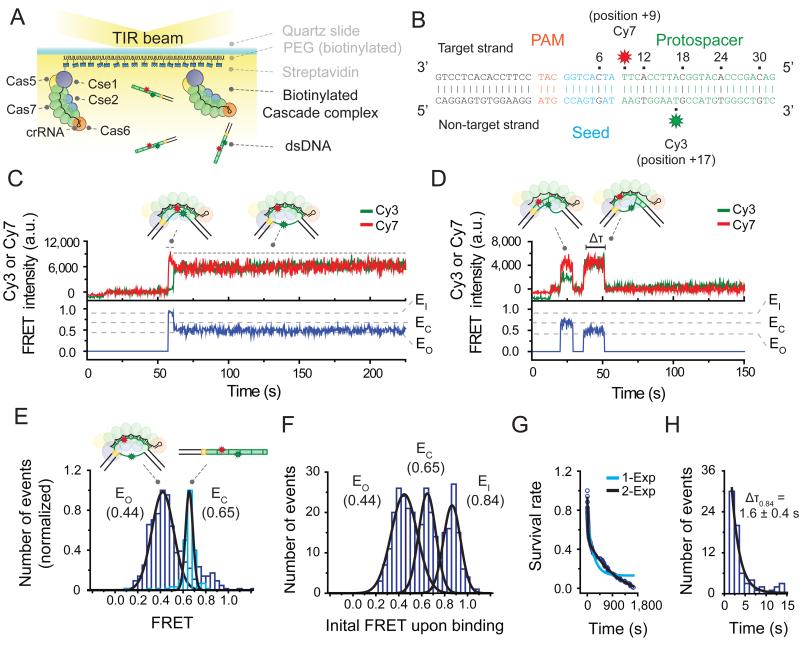
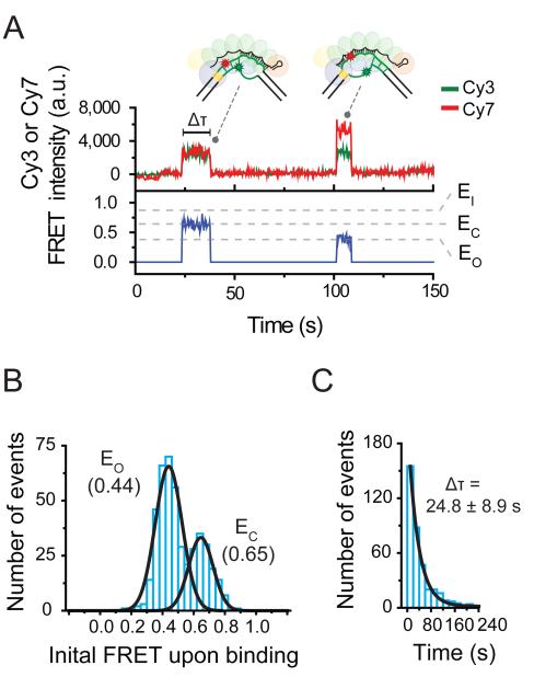
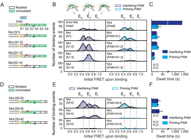
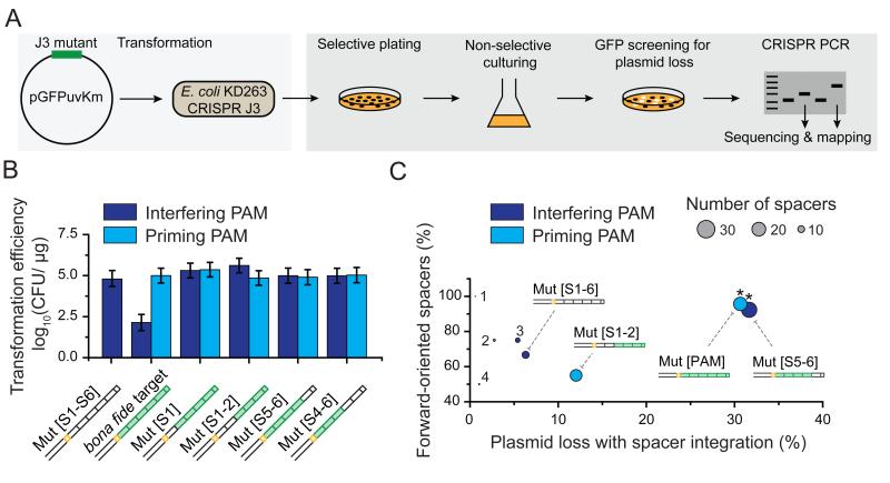
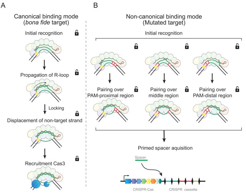
Small RNA-guided protein complexes play an essential role in CRISPR-mediated immunity in prokaryotes. While these complexes initiate interference by flagging cognate invader DNA for destruction, recent evidence has implicated their involvement in new CRISPR memory formation, called priming, against mutated invader sequences. The mechanism by which the target recognition complex mediates these disparate responses-interference and priming-remains poorly understood. Using single-molecule FRET, we visualize how bona fide and mutated targets are differentially probed by E. coli Cascade. We observe that the recognition of bona fide targets is an ordered process that is tightly controlled for high fidelity. Mutated targets are recognized with low fidelity, which is featured by short-lived and PAM- and seed-independent binding by any segment of the crRNA. These dual roles of Cascade in immunity with distinct fidelities underpin CRISPR-Cas robustness, allowing for efficient degradation of bona fide targets and priming of mutated DNA targets.


Figures

Figure 1
Figure 1. Two binding modes of Cascade revealed by a single-molecule FRET assay
(A) Schematic of a single-molecule FRET experiment used to monitor binding of Cascade to target DNA substrates. (B) The bona fide target construct consists of a 15 bp flank (black), a PAM (orange), and a protospacer (green), with its seed highlighted in blue. Cy7 (red star) was attached to position +9 of the target strand and Cy3 (green star) to position +17 of the non-target strand. (C) A representative time trace of donor (Cy3, green) and acceptor (Cy7, red) fluorescence and corresponding FRET (blue) exhibiting the long-lived binding of the bona fide target. High FRET (~0.84, named EI for FRET efficiency of an intermediate state) exhibited upon binding is followed by low FRET (~0.44, named EO for FRET efficiency of an open state). DNA was added at time 10 sec. (D) A representative time trace exhibiting the short-lived binding of the bona fide target exhibits two FRET states (EO ~0.44 and EC ~0.65). EC is for FRET efficiency of a closed state). The duration of each state is measured as the dwell time (Δτ). DNA was added at time 10 sec. (E) The FRET distribution of the bona fide target DNA alone (light blue) or after equilibration with immobilized Cascade (purple) with peaks at EC (0.65) and EO (0.44), respectively (derived from Gaussian fit, black line). Data obtained from 5 fields of view each. (F) A histogram of the initial FRET upon binding (average of first 1.5 sec of each event) of the bona fide target exhibits three peaks at FRET = EO (0.44), EC (0.65), EI (0.84) (derived from Gaussian fit, black line). (G) The survival rate of events that start at EI (0.84) was fitted using a single (light blue color) and a double (black color) exponential curve. The double exponential fit resulted in two characteristic times (25.9 and 1040 sec). (H) The dwell time distribution of EI (0.84) state of bona fide target binding with mean ΔτE0.84 (derived from single exponential fit, black line). Error represent standard deviation (3 individual data sets). See also Figure S1 and Table S1.
Figure 2
Figure 2. Short-binding of Cascade to PAM-mutated targets
(A) A representative time trace exhibiting the short-lived binding of the PAM-mutated target exhibits two FRET states, EO (0.44) and EO (0.65). The duration of each state is measured as the dwell time (Δτ). DNA was added at time 12 sec. (B) A histogram of the initial FRET upon binding (average of first 1.5 sec of each event) of Mut[PAM] exhibits peaks at EO (0.44) and EC (0.65) (derived from Gaussian fit, black line). (C) The dwell time distribution of Mut[PAM] binding events with mean Δτ (derived from single exponential fit, black line). Error represents std (3 individual data sets).
Figure 3
Figure 3. Cascade exhibits non-canonical binding to protospacers with PAM-proximal or PAM-distal segmented mutations
(A) Schematics of DNA targets in the PAM-proximal mutation series illustrating mutated (white) or unmutated (green) segments (S1-S6) of the protospacer. Mut[S1], Mut[S1-2], Mut[S1-3] and Mut[S1-4] have segments 1, 1-2, 1-3, and 1-4 mutated, respectively. (B) Histograms of the initial FRET upon binding (average of first 1.5 sec of each event) of each PAM-proximal mutant from [A] bearing either an interfering (purple bars, left column) or priming (light blue bars, right column) PAM exhibit peaks (Gaussian fits, black lines) positioned similar to that of the bona fide and Mut[PAM] targets (top row, same as Figures 1F and 2B) at EO (0.44), EC (0.65), or EI (0.84) (dashed black lines). The recorded events are from one field-of-view of the detector. (C) Mean binding dwell time of each PAM-proximal mutant from [A] bearing either an interfering (purple bars) or a priming (light blue bars) PAM (derived from dwell time distributions, see Figure S2A). Error represents std (3 individual data sets). The dwell time of the bona fide target could not be measured accurately due to the photobleaching and thus arbitrarily set 1040 sec to represent the longer characteristic time scale in Figure 1G. (D) Schematics of DNA targets in the PAM-distal mutation series illustrated as in [A]. Mut[S5-6], Mut[S4-6], and Mut[S3-6] have segments 5-6, 4-6, and 3-6 mutated, respectively. (E) Histograms of the initial FRET upon binding of each PAM-distal mutant from [D] displayed in a similar fashion to [B]. (F) Mean binding dwell of each PAM-distal mutant from [D] bearing either an interfering (purple bars) or a priming (light blue bars) PAM (derived from dwell time distributions, see Figure S2A). Error represents std (3 individual data sets). N.D. is “Not Determined”. See also Table S2.
Figure 4
Figure 4. Non-canonical binding leads to primed spacer acquisition
(A) Cartoon representation of the in vivo assay used to determine primed plasmid loss and spacer acquisition. (B) Transformation efficiencies of plasmids harboring different target sequences (see schematics) with an interfering (purple bar) or a priming (light blue bar) PAM. CFU is “Colony-Forming Unit.” Error is std of 3 individual measurements. (C) A two-dimensional bubble plot showing the fraction of forward-oriented spacers acquired versus the percentage of plasmid loss for those targets in [B] that exhibited spacer integration. Circle size represents the total number of spacers that were acquired and circle color represents an interfering (purple) or a priming (light blue) PAM. A star (*) indicates a forward directional bias (relative to random) with a P-value < 1× 10⁻⁵ based on binomial statistics. The numbers of 1, 2, 3 and 4 indicate data points from constructs Mut[PAM+S5-6], Mut[PAM+S1], Mut[S4-6] and Mut[PAM+S4-6], respectively. See also Figure S3, Table S1 and Table S3..
Figure 5
Figure 5. Two binding modes of Cascade lead to different functional outcomes
Cascade employs two distinct target-DNA binding modes that trigger (A) interference or (B) priming. (A) In the interference pathway, target recognition initiates from the PAM and PAM-proximal region. R-loop formation then propagates toward the PAM-distal region. When Cascade senses the fully paired structure, it brings this complex into a lower energy state (“locking”) that displaces the non-target strand out of Cascade. This exposed strand is then cleaved by Cas3. (B) In the priming pathway, DNA is probed through brief interactions. PAM recognition facilitates this priming pathway but is not required. The brief interactions may initiate from the PAM-proximal (left), the PAM-distal region (right), or the middle of the protospacer (middle), which becomes stable when paired over 3 or more segments. This non-canonical (“unlocked”) binding mode leads to a unique conformation of the R-loop and signals for primed spacer acquisition.

References

    1. Barrangou R. CRISPR-Cas systems and RNA-guided interference. Wiley interdisciplinary reviews RNA. 2013;4:267–278. - PubMed
    1. Charpentier E, Marraffini LA. Harnessing CRISPR-Cas9 immunity for genetic engineering. Current opinion in microbiology. 2014;19:114–119. - PMC - PubMed
    1. Chen Z, Yang H, Pavletich NP. Mechanism of homologous recombination from the RecA-ssDNA/dsDNA structures. Nature. 2008;453:489–484. - PubMed
    1. Cisse II, Kim H, Ha T. A rule of seven in Watson-Crick base-pairing of mismatched sequences. Nat Struct Mol Biol. 2012;19:623–627. - PMC - PubMed
    1. Datsenko KA, Pougach K, Tikhonov A, Wanner BL, Severinov K, Semenova E. Molecular memory of prior infections activates the CRISPR/Cas adaptive bacterial immunity system. Nature communications. 2012;3:945. - PubMed

Publication types

MeSH terms

LinkOut - more resources