. 2015 Apr 24;290(17):10994-1007.
doi: 10.1074/jbc.M115.636951. Epub 2015 Mar 9.

Variable Substrate Preference among Phospholipase D Toxins from Sicariid Spiders

Affiliations

Variable Substrate Preference among Phospholipase D Toxins from Sicariid Spiders

Daniel M Lajoie et al. J Biol Chem. .

Abstract

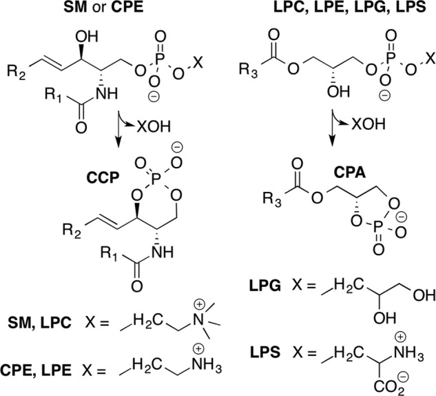
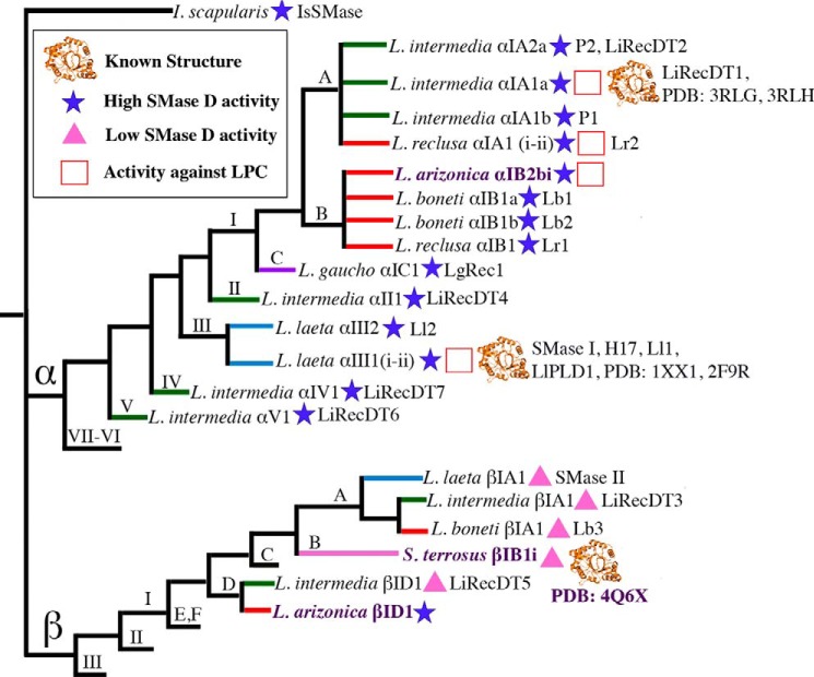
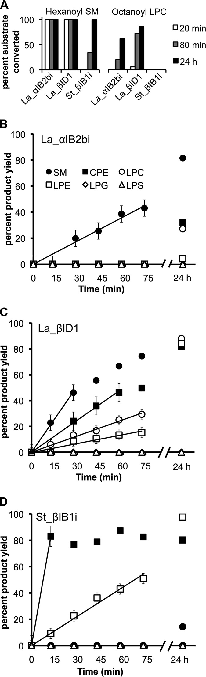
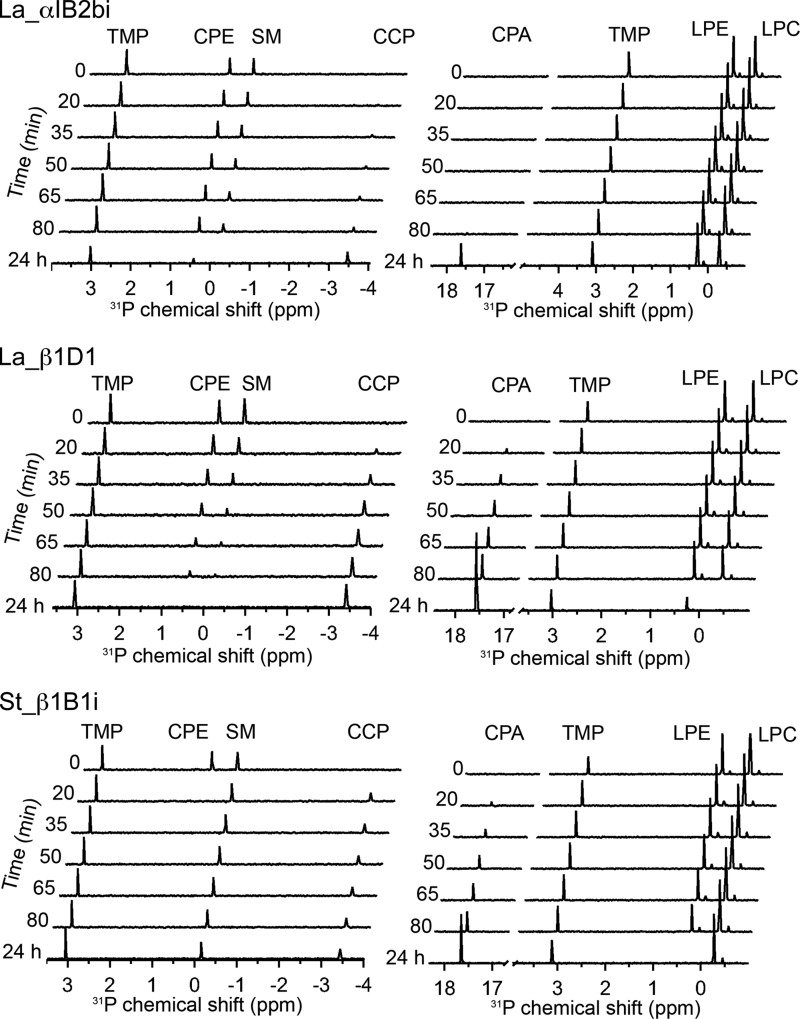
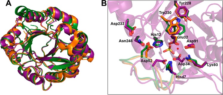
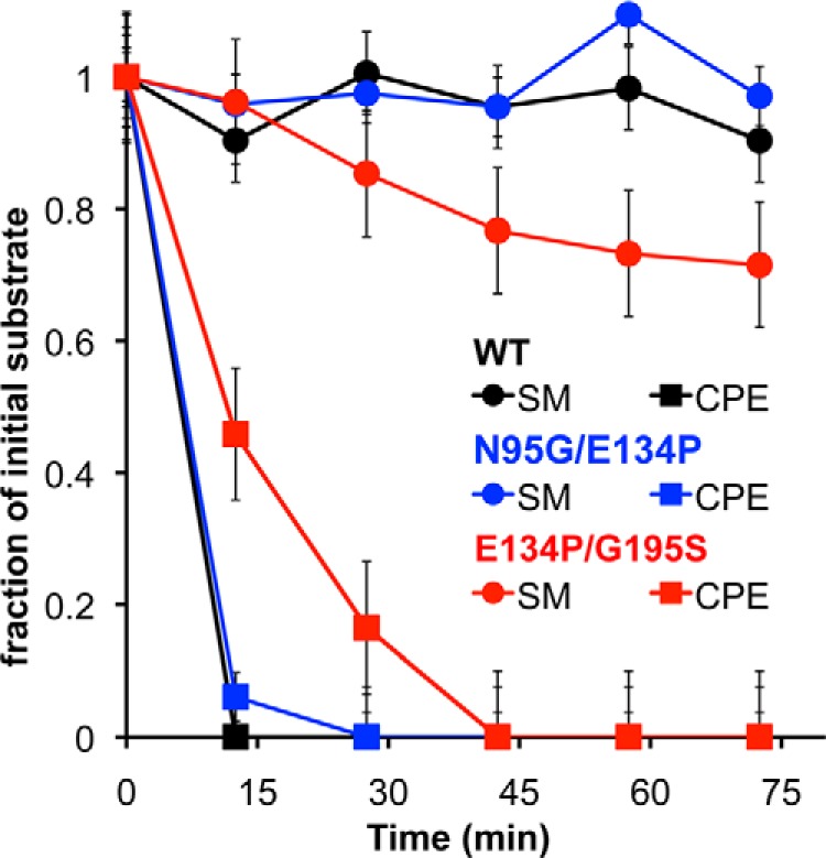
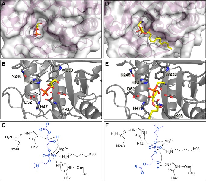
Venoms of the sicariid spiders contain phospholipase D enzyme toxins that can cause severe dermonecrosis and even death in humans. These enzymes convert sphingolipid and lysolipid substrates to cyclic phosphates by activating a hydroxyl nucleophile present in both classes of lipid. The most medically relevant substrates are thought to be sphingomyelin and/or lysophosphatidylcholine. To better understand the substrate preference of these toxins, we used (31)P NMR to compare the activity of three related but phylogenetically diverse sicariid toxins against a diverse panel of sphingolipid and lysolipid substrates. Two of the three showed significantly faster turnover of sphingolipids over lysolipids, and all three showed a strong preference for positively charged (choline and/or ethanolamine) over neutral (glycerol and serine) headgroups. Strikingly, however, the enzymes vary widely in their preference for choline, the headgroup of both sphingomyelin and lysophosphatidylcholine, versus ethanolamine. An enzyme from Sicarius terrosus showed a strong preference for ethanolamine over choline, whereas two paralogous enzymes from Loxosceles arizonica either preferred choline or showed no significant preference. Intrigued by the novel substrate preference of the Sicarius enzyme, we solved its crystal structure at 2.1 Å resolution. The evolution of variable substrate specificity may help explain the reduced dermonecrotic potential of some natural toxin variants, because mammalian sphingolipids use primarily choline as a positively charged headgroup; it may also be relevant for sicariid predatory behavior, because ethanolamine-containing sphingolipids are common in insect prey.

Keywords: Crystal Structure; Enzyme Mechanism; Loxosceles; Loxoscelism; Phospholipase D; Sicarius; Sphingomyelinase; Substrate Specificity; Toxin.


Figures

SCHEME 1.
SCHEME 1.
Relevant substrates and products for our study of SicTox enzymes. The reaction at left shows conversion of sphingolipid substrates to cyclic ceramide phosphate; the reaction at right shows conversion of lysophospholipid substrates to cyclic phosphatidic acid. R1, R2, and R3 represent alkyl chains of varying length. The four different headgroup alcohols (XOH) represented here are choline (SM and LPC), ethanolamine (CPE and LPE), glycerol (LPG), and serine (LPS).
FIGURE 1.
FIGURE 1.
Simplified cladogram of SicTox gene products that have been biochemically characterized. The tree, which is based on a previously reported phylogeny (24), includes all SicTox gene products that have been tested for PLD activity following isolation from whole venom or purification from recombinant sources. The names of toxins investigated in this study are highlighted in purple. The ingroup includes two major clades (α and β). IsSMase from Ixodes scapularis (74) is included as an outgroup. Homologs with >95% amino acid sequence identity are considered isoforms and grouped under the same name. Names follow the SicTox nomenclature (24), but alternative names used in the literature are also given, and PDB codes are given for known protein structures. Branch colors correspond to species groups as previously presented (24): red, L. reclusa; green, Loxosceles spadicea; purple, Loxosceles gaucho; blue, L. laeta, and pink, South America Sicarius species. Branches lacking terminal names represent clades for which no member has been characterized biochemically. Except for toxins characterized in this study, high or low SMase D activity is based on comparisons with the activity of whole venom (at the same protein amount) from the toxin source species. In the case of β1A1 (SMase II) from L. laeta, SMase D activity has been observed but with a 3-fold higher Km value and a reported 2-fold lower reaction rate at saturating substrate concentrations compared with αIII1 (SMase I) from the same species (26). References for structure and activity data are as follows: IsSMase (74); LiRecDT6 (11); LiRecDT7 (14); SMase I (8, 18, 27, 35, 59, 64, 75); Ll2 (8); LiRecDT4 (9); LgRec1 (76); Lr1/Lb1/Lb2 (8, 25); L. arizonica αIB2bi (15, 30); Lr2 (19, 25); P1/P2 (77–79); LiRecDT1 (6, 7, 12, 13, 20, 60); 3RLG (61); 3RLH (28); LiRecDT2 (7); LiRecDT5 (9); Lb3 (8, 25); LiRecDT3 (7, 10); and SMase II (26).
FIGURE 2.
FIGURE 2.
Multiple amino acid sequence alignment of selected SicTox enzymes. Residues 1–285 are shown. Amino acid residues deemed essential for catalytic activity as previously inferred from the structures of Ll_αIII1 and Li_αIA1a (PDB 1XX1 and 3RLH, respectively) are indicate by red boxes. Despite variation in SMase D activity, all 11 of these residues are perfectly conserved in the family. Conserved cysteine residues involved in disulfide bonds are indicated by yellow boxes. Amino acid residues mutated in this study are indicated by blue boxes.
FIGURE 3.
FIGURE 3.
31P NMR assay results of three different SicTox PLD toxins with different phospholipid substrates. A, preliminary assessment of activity against SM and LPC using 10 μg of pure enzyme with either 2 mm hexanoyl sphingomyelin (6:0 SM) and 2 mm Triton X-100, or 80 mm octanoyl lysophosphatidylcholine (8:0 LPC). St_βIB1i shows much lower activity toward the two substrates than La_αIB2bi and La_βID1, (B–D). Panel assays using 10 μg of pure enzyme with ∼2 mm each of six different phospholipid substrates solubilized in CHAPS detergent. Fitted slopes of product accumulation during the linear portion of the reaction were used to extract initial reaction rates. Error bars have been added to points used for slope determination; these are based on statistical analysis of instrument precision and represent standard deviations. In some cases, no product was observed after 75 min but did appear after 24 h. Substrate conversion and product yield were derived from integration of NMR signals relative to a trimethyl phosphate internal standard, as described under “Experimental Procedures.”
FIGURE 4.
FIGURE 4.
Stacked 31P NMR spectra from competition assays of three SicTox PLD enzymes with choline- and ethanolamine-containing substrates. Left panels show SM/CPE competition experiments, and right panels show LPC/LPE competition experiments. The spectra illustrate the preference, or lack thereof, of the different enzymes for choline (SM and LPC) or ethanolamine (CPE and LPE) headgroups. Assays contained 10 μg of each SicTox PLD and ∼2 mm of each substrate. In the SM/CPE assay, cleavage of either substrate yields cyclic ceramide phosphate (CCP), a six-membered ring phosphate with an upfield 31P resonance. In the LPC/LPE assay, cleavage of either substrate yields cyclic phosphatidic acid (CPA), a five-membered ring phosphate with a far downfield 31P resonance.
FIGURE 5.
FIGURE 5.
Structural and amino acid sequence comparison of the SicTox PLD toxins. A, superposition of St_β1B1i from S. terrosus (purple; PDB code 4Q6X) on Li_αIA1a/LiRecDT1 from L. intermedia (gold; PDB code 3RLH), and Ll_αIII1/SMase I from L. laeta (green; PDB code 1XX1). B, active sites of the same three enzymes, showing putatively important amino acid residues for catalysis and/or substrate binding (see also Fig. 2). The essential magnesium ion cofactor from the structure of St_β1B1i is shown as a small red sphere, along with a sulfate ion from the L. laeta SMase I structure. With the exception of Asp-52, in the flexible/variable catalytic loop, all the amino acid residues have a similar location and conformation in St_β1B1i, a β clade member, as in the two α clade representatives. Residue numbering is referenced to Ll_αIII1/SMase I.
FIGURE 6.
FIGURE 6.
In silico docking of LPE and LPC into the active site of St_βIB1i. A, surface rendering of St_βIB1i (tan), with lysophosphatidylcholine (blue) and lysophosphatidylethanolamine (lavender) docked into the active site. The magnesium cofactor is shown as a small green sphere interacting with the phosphate group of each substrate. For both substrates, the headgroup is situated in a binding pocket that has been previously identified as potentially important for substrate specificity (24). B, side chains in the putative headgroup binding pocket of St_βIB1i (tan; PDB code 4Q6X) compared with those of LiRecDT1, an α clade protein from L. intermedia (green; PDB code 3RLH). The two pockets are quite similar but contain amino acid sequence differences at positions 95, 134, and 195 (see also amino acid residues in blue boxes in Fig. 2). Residue numbering is referenced to Ll_αIII1/SMase I.
FIGURE 7.
FIGURE 7.
31P NMR competition assays of St_βIB1i variants against SM and CPE. The data for wild-type St_βIB1i (black) derive from the spectra shown in Fig. 4 and the N95G/E134P (blue) and E134P/G195S (red) variants that were assayed in the same manner. All three variants turn over CPE considerably faster than SM, although E134P/G195S may exhibit a decreased preference for CPE. See “Experimental Procedures” for details of assay conditions. Error bars are based on statistical analysis of instrument precision and represent standard deviations.
FIGURE 8.
FIGURE 8.
Two plausible mechanisms of LPC cyclization by SicTox enzymes, based on two possible substrate binding modes. A, active site pocket of St_βIB1i with glycerol 3-phosphate bound as seen in a GDPD enzyme from O. antarctica (PDB code 3QVQ). B, this binding mode places the nucleophilic 2′-OH of the glycerol moiety adjacent to His-12. The putative leaving group position is not evident, as glycerol 3-phosphate is the product of removal of the leaving group by a GDPD enzyme. However, the phosphate oxygen opposite the nucleophile (see arrow) is likely to represent the leaving group oxygen of substrate. C, mechanism for LPC/LPE cyclization derived from the glycerol 3-phosphate binding mode. His-12 deprotonates the 2′-hydroxyl generating the nucleophile for attack on the phosphate. His-47 acts as a general acid to protonate the leaving group. D, active site pocket of St_βIB1i with LPE docked as in Fig. 6. E, this binding mode inverts the orientation of the glycerol phosphate moiety relative to that shown for glycerol 3-phosphate in A–C, and places the 2′-OH of glycerol adjacent to His-47. F, mechanism for LPC/LPE cyclization based on the LPC/LPE docking. His-47 activates the 2′-OH for in-line attack on the phosphodiester. His-12 protonates the leaving group.

Similar articles

Cited by

References

    1. Tambourgi D. V., Gonçalves-de-Andrade R. M., van den Berg C. W. (2010) Loxoscelism: From basic research to the proposal of new therapies. Toxicon 56, 1113–1119 - PubMed
    1. Gremski L. H., Trevisan-Silva D., Ferrer V. P., Matsubara F. H., Meissner G. O., Wille A. C., Vuitika L., Dias-Lopes C., Ullah A., de Moraes F. R., Chávez-Olortegui C., Barbaro K. C., Murakami M. T., Arni R. K., Senff-Ribeiro A., et al. (2014) Recent advances in the understanding of brown spider venoms: from the biology of spiders to the molecular mechanisms of toxins. Toxicon 83, 91–120 - PubMed
    1. Hogan C. J., Barbaro K. C., Winkel K. (2004) Loxoscelism: old obstacles, new directions. Ann. Emerg. Med. 44, 608–624 - PubMed
    1. da Silva P. H., da Silveira R. B., Appel M. H., Mangili O. C., Gremski W., Veiga S. S. (2004) Brown spiders and loxoscelism. Toxicon 44, 693–709 - PubMed
    1. Swanson D. L., Vetter R. S. (2006) Loxoscelism. Clin. Dermatol. 24, 213–221 - PubMed

Publication types

Associated data

LinkOut - more resources