. 2015 Apr 24;290(17):10729-40.
doi: 10.1074/jbc.M115.648410. Epub 2015 Mar 9.

High structural resolution hydroxyl radical protein footprinting reveals an extended Robo1-heparin binding interface

Affiliations

High structural resolution hydroxyl radical protein footprinting reveals an extended Robo1-heparin binding interface

Zixuan Li et al. J Biol Chem. .

Abstract

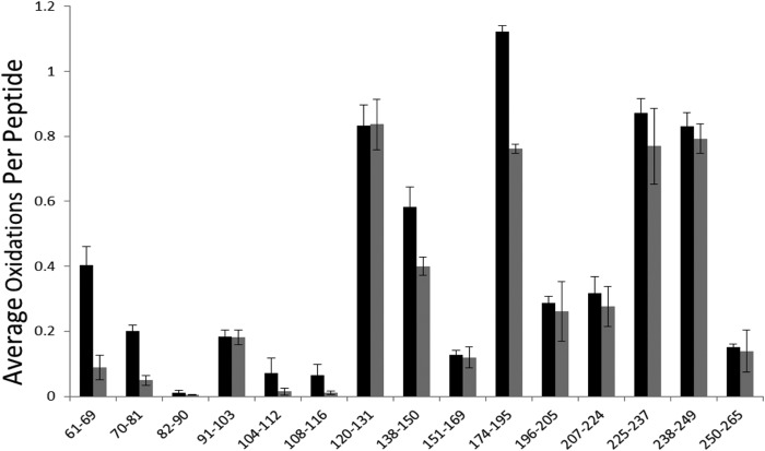
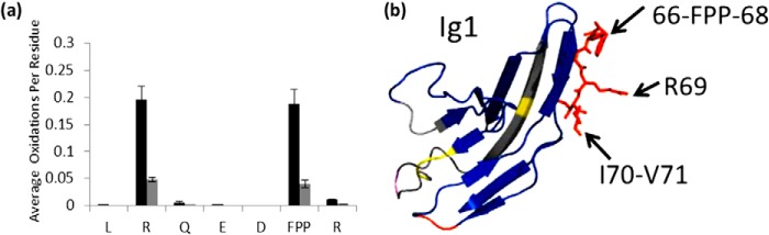
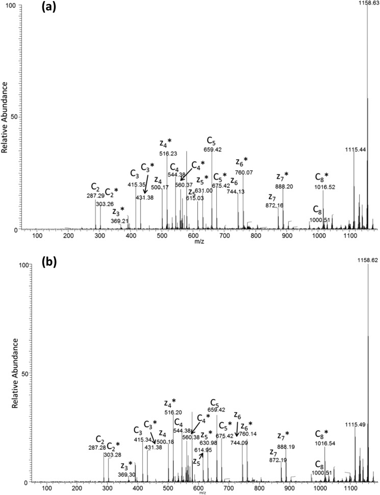
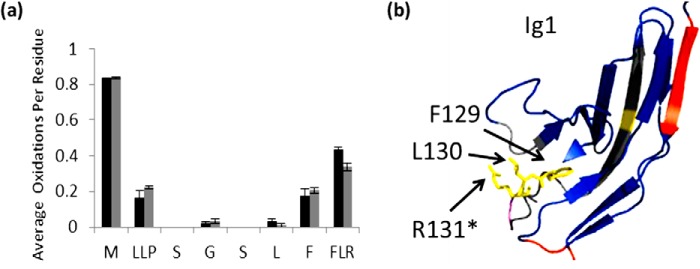
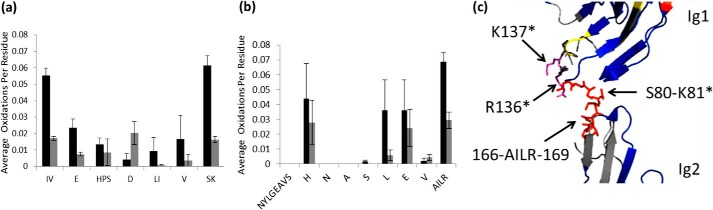
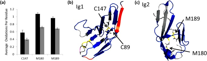
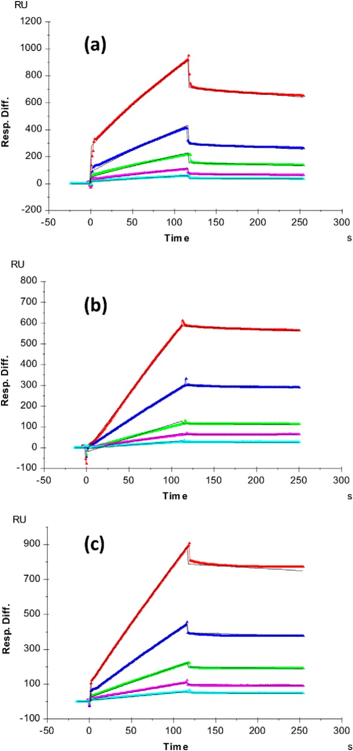
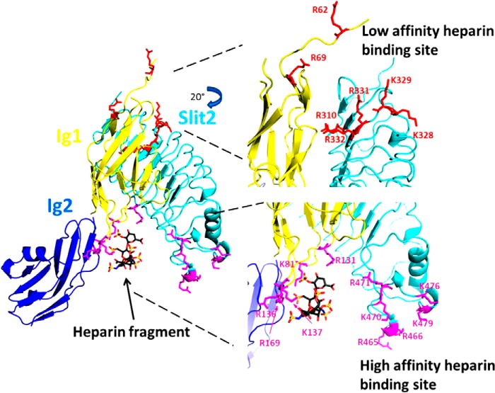
Interaction of transmembrane receptors of the Robo family and the secreted protein Slit provides important signals in the development of the central nervous system and regulation of axonal midline crossing. Heparan sulfate, a sulfated linear polysaccharide modified in a complex variety of ways, serves as an essential co-receptor in Slit-Robo signaling. Previous studies have shown that closely related heparin octasaccharides bind to Drosophila Robo directly, and surface plasmon resonance analysis revealed that Robo1 binds more tightly to full-length unfractionated heparin. For the first time, we utilized electron transfer dissociation-based high spatial resolution hydroxyl radical protein footprinting to identify two separate binding sites for heparin interaction with Robo1: one binding site at the previously identified site for heparin dp8 and a second binding site at the N terminus of Robo1 that is disordered in the x-ray crystal structure. Mutagenesis of the identified N-terminal binding site exhibited a decrease in binding affinity as measured by surface plasmon resonance and heparin affinity chromatography. Footprinting also indicated that heparin binding induces a minor change in the conformation and/or dynamics of the Ig2 domain, but no major conformational changes were detected. These results indicate a second low affinity binding site in the Robo-Slit complex as well as suggesting the role of the Ig2 domain of Robo1 in heparin-mediated signal transduction. This study also marks the first use of electron transfer dissociation-based high spatial resolution hydroxyl radical protein footprinting, which shows great utility for the characterization of protein-carbohydrate complexes.

Keywords: Glycosaminoglycan; Heparin; Heparin-binding Protein; Hydroxyl Radical Protein Footprinting; Mass Spectrometry (MS); Protein-Carbohydrate Interaction; Structural Biology.


Figures

FIGURE 1.
FIGURE 1.
Amino acid sequence of Robo1 Ig1-2 domain. Identified oxidized amino acids are colored red. The underline indicates the N-glycan site. Unoxidized residues in the Ig1 domain are colored green; unoxidized residues in the Ig2 domain are colored blue.
FIGURE 2.
FIGURE 2.
Hydroxyl radical protein footprinting of Robo1-heparin. Extent of oxidation of Robo1 with unfractionated heparin (gray bars) and without unfractionated heparin (black bars) at the peptide level is shown. Error bars, S.D. from a triplicate set of HRPF analyses.
FIGURE 3.
FIGURE 3.
Residues involved in binding with unfractionated heparin. a, extent of oxidation of Robo1 alone (black bars) compared with heparin-bound Robo1 (gray bars) at the residue level. Error bars, S.D. from a triplicate set of experiments. b, protected residues Phe66-Pro67-Pro68, Ile70, and Val71 surround protected basic residue Arg69 in three-dimensional space, suggesting that Arg69 interacts with heparin, which shields neighboring residues. Protected residue Arg62 is not shown in the crystal structure.
FIGURE 4.
FIGURE 4.
Representative ETD spectrum of singly oxidized peptide LRQEDFFPR for Robo1 alone (a) and Robo1 with heparin (b). The asterisks indicate the product ions that are oxidized. The ETD spectrum shows unoxidized and oxidized c ions as pairs from c2 to c8, whereas unoxidized and oxidized c1, c6, and c7 ions are absent in the spectrum. Oxidized z3 and unoxidized and oxidized z4–z7 ions generated by ETD are also shown in the spectrum. The intensities of c ions are preferred to calculate for quantitation because the interference between unoxidized y ions and oxidized z ions prevents accurate quantification in mass spectrometers with lower resolution, as demonstrated by our previous work.
FIGURE 5.
FIGURE 5.
Residues involved in binding with unfractionated heparin. a, extent of oxidation of Robo1 alone (black bars) compared with heparin-bound Robo1 (gray bars) at the residue level. Error bars, S.D. from a triplicate set of experiments. b, structure highlighting Phe129-Leu130-Arg131 are shown in yellow. An asterisk indicates the residue involved in a heparin dp8 binding reported previously (21).
FIGURE 6.
FIGURE 6.
Residues identified as binding sites around basic patch. Error bars, S.D. from a triplicate set of experiments. a, residue level extent of oxidation for peptide 70–81 of Robo1 (black bars) and Robo1-heparin (gray bars). b, residue level extent of oxidation for peptide 151–169 of Robo1 (black bars) and Robo1-heparin (gray bars). c, structure shows the protected residues Ser80-Lys81 and Ala166-Ile167-Leu168-Arg169 (red) are at a basic patch with basic residues in magenta unidentified by HRPF. Asterisks indicate the residue identified as interacting with heparin dp8 previously (21).
FIGURE 7.
FIGURE 7.
Non-binding residues show significant protections. a, residue level extent of oxidation of Robo1 alone (black bars) compared with heparin-bound Robo1 (gray bars) for residues Cys147, Met180, and Met189. Error bars, S.D. from a triplicate set of experiments. b, structural model showing Cys147 (yellow) involved in the disulfide bond with Cys89 in Ig1 domain. c, hydrophobic residues Met180 and Met189 are shown in yellow in the Ig2 domain of Robo1.
FIGURE 8.
FIGURE 8.
Heparin affinity chromatography of Robo1 site-directed mutants. The chromatogram is a representative replicate; the analyses were performed in duplicate, and the elution profiles were reproducible. Orange, WT Robo1; green, Mutant I (R136A/K137A); blue, Mutant II (R62A/R69A). The purple curve shows the conductivity of the elution buffer (millisiemens/cm) due to the increasing NaCl concentration, with the scale shown on the right y axis.
FIGURE 9.
FIGURE 9.
SPR sensorgrams of Robo1-heparin interaction. a, Robo1 WT/heparin; b, Mutant I (R136A/K137A)/heparin; c, Mutant II (R62A/R69A)/heparin. Concentrations of Robo1 (from top to bottom) are as follows: 1000 nm (red), 500 nm (blue), 250 nm (green), 125 nm (magenta), and 63 nm (cyan). The black curves are the fitting curves from a 1:1 Langmuir model from BIAevaluate version 4.0.1. Resp. Diff., response difference.
FIGURE 10.
FIGURE 10.
A model of the mechanism of Slit2-Robo1-heparin interactions. The Slit2-Robo1 complex has two binding sites for heparin: the previously identified high affinity binding site near the Ig1-Ig2 interface of Robo1 (magenta) and a novel low affinity binding site located near the disordered N terminus of Robo1 as well as within adjacent conserved basic residues in Slit2 (cyan). Full-length heparin/HS binds first to the high affinity binding site, which then allows for binding of a separate portion of the heparin/HS chain to the low affinity binding site. The binding to the low affinity binding site prompts conformational changes required for signal transduction. This model was generated using the x-ray crystal structure of the second LRR domain of human Slit2 in complex with the Ig1 domain of human Robo1 (Protein Data Bank code 2V9T). The heparin tetrasaccharide and the Ig2 domain of Drosophila Robo1 were aligned and joined from the x-ray crystal structure of dRobo1 bound to heparin dp8 (Protein Data Bank code 2VRA).

Similar articles

Cited by

References

    1. Garbe D. S., Bashaw G. J. (2004) Axon guidance at the midline: from mutants to mechanisms. Crit. Rev. Biochem. Mol. Biol. 39, 319–341 - PubMed
    1. Dickson B. J., Gilestro G. F. (2006) Regulation of commissural axon pathfinding by slit and its robo receptors. Annu. Rev. Cell Dev. Biol. 22, 651–675 - PubMed
    1. Evans T. A., Bashaw G. J. (2012) Slit/Robo-mediated axon guidance in Tribolium and Drosophila: divergent genetic programs build insect nervous systems. Dev. Biol. 363, 266–278 - PMC - PubMed
    1. Kidd T., Brose K., Mitchell K. J., Fetter R. D., Tessier-Lavigne M., Goodman C. S., Tear G. (1998) Roundabout controls axon crossing of the CNS midline and defines a novel subfamily of evolutionarily conserved guidance receptors. Cell 92, 205–215 - PubMed
    1. Huminiecki L., Gorn M., Suchting S., Poulsom R., Bicknell R. (2002) Magic roundabout is a new member of the roundabout receptor family that is endothelial specific and expressed at sites of active angiogenesis. Genomics 79, 547–552 - PubMed

Publication types

MeSH terms