

Lack of association between epicardial fat volume and extent of coronary artery calcification, severity of coronary artery disease, or presence of myocardial perfusion abnormalities in a diverse, symptomatic patient population: results from the CORE320 multicenter study
- PMID: 25752899
- PMCID: PMC4355954
- DOI: 10.1161/CIRCIMAGING.114.002676
Lack of association between epicardial fat volume and extent of coronary artery calcification, severity of coronary artery disease, or presence of myocardial perfusion abnormalities in a diverse, symptomatic patient population: results from the CORE320 multicenter study
Abstract
Background: Epicardial fat may play a role in the pathogenesis of coronary artery disease (CAD). We explored the relationship of epicardial fat volume (EFV) with the presence and severity of CAD or myocardial perfusion abnormalities in a diverse, symptomatic patient population.
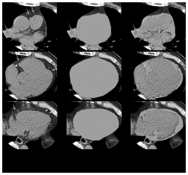
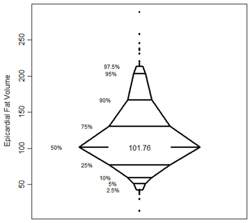
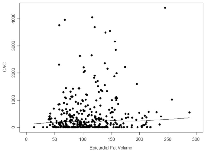
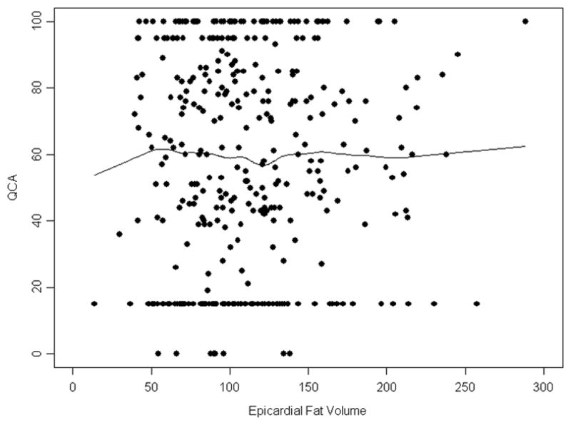
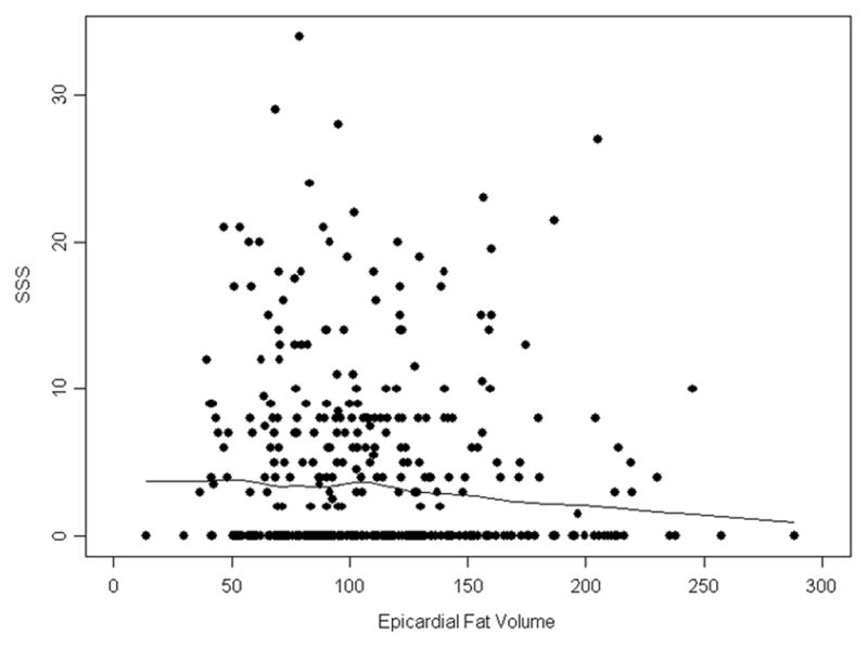
Methods and results: Patients (n=380) with known or suspected CAD who underwent 320-detector row computed tomographic angiography, nuclear stress perfusion imaging, and clinically driven invasive coronary angiography for the CORE320 international study were included. EFV was defined as adipose tissue within the pericardial borders as assessed by computed tomography using semiautomatic software. We used linear and logistic regression models to assess the relationship of EFV with coronary calcium score, stenosis severity by quantitative coronary angiography, and myocardial perfusion abnormalities by single photon emission computed tomography (SPECT). Median EFV among patients (median age, 62.6 years) was 102 cm(3) (interquartile range: 53). A coronary calcium score of ≥1 was present in 83% of patients. Fifty-nine percent of patients had ≥1 coronary artery stenosis of ≥50% by quantitative coronary angiography, and 49% had abnormal myocardial perfusion results by SPECT. There were no significant associations between EFV and coronary artery calcium scanning, presence severity of ≥50% stenosis by quantitative coronary angiography, or abnormal myocardial perfusion by SPECT.
Conclusions: In a diverse population of symptomatic patients referred for invasive coronary angiography, we did not find associations of EFV with the presence and severity of CAD or with myocardial perfusion abnormalities. The clinical significance of quantifying EFV remains uncertain but may relate to the pathophysiology of acute coronary events rather than the presence of atherosclerotic disease.
Keywords: coronary artery disease; coronary stenosis; myocardial ischemia.
© 2015 American Heart Association, Inc.
Figures
Comment in
-
Epicardial adipose tissue: a benign consequence of obesity?Circ Cardiovasc Imaging. 2015 Mar;8(3):10.1161/CIRCIMAGING.115.003156 e003156. doi: 10.1161/CIRCIMAGING.115.003156. Circ Cardiovasc Imaging. 2015. PMID: 25752745 Free PMC article. No abstract available.
References
-
- Rosito GA, Massaro JM, Hoffmann U, Ruberg FL, Mahabadi AA, Vasan RS, O’Donnell CJ, Fox CS. Pericardial fat, visceral abdominal fat, cardiovascular disease risk factors, and vascular calcification in a community-based sample: The Framingham Heart study. Circulation. 2008;117:605–613. - PubMed
-
- Ahmadi N, Nabavi V, Yang E, Hajsadeghi F, Lakis M, Flores F, Zeb I, Bevinal M, Ebrahimi R, Budoff M. Increased epicardial, pericardial, and subcutaneous adipose tissue is associated with the presence and severity of coronary artery calcium. Academic Radiology. 2010;17:1518–1524. - PubMed
-
- De Vos AM, Prokop M, Roos CJ, Meijs MF, van der Schouw YT, Rutten A, Gorter PM, Cramer MJ, Doevendans PA, Rensing BJ, Bartelink ML, Velthuis BK, Mosterd A, Bots ML. Peri-coronary epicardial adipose tissue is related to cardiovascular risk factors and coronary artery calcification in post-menopausal women. European Heart Journal. 2008;29:777–783. - PubMed
-
- Nakanishi R, Rajani R, Cheng VY, Gransar H, Nakazato R, Shmilovich H, Otaki Y, Hayes SW, Thomson LE, Friedman JD, Slomka PJ, Berman DS, Dey D. Increase in epicardial fat volume is associated with greater coronary artery calcification progression in subjects at intermediate risk by coronary calcium score: A serial study using non-contrast cardiac ct. Atherosclerosis. 2011;218:363–368. - PubMed
Publication types
MeSH terms
Grants and funding
LinkOut - more resources
Full Text Sources
Medical
Miscellaneous
