. 2015 Mar 23;32(6):756-64.
doi: 10.1016/j.devcel.2015.01.032. Epub 2015 Mar 5.

A CRISPR/Cas9 vector system for tissue-specific gene disruption in zebrafish

Affiliations

A CRISPR/Cas9 vector system for tissue-specific gene disruption in zebrafish

Julien Ablain et al. Dev Cell. .

Abstract

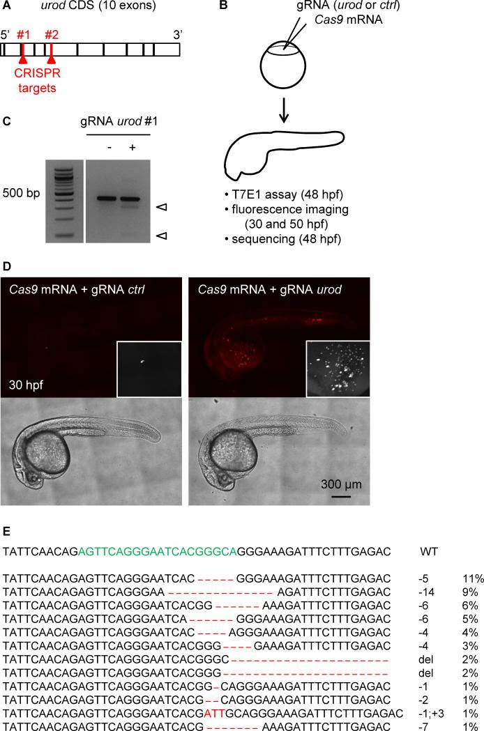
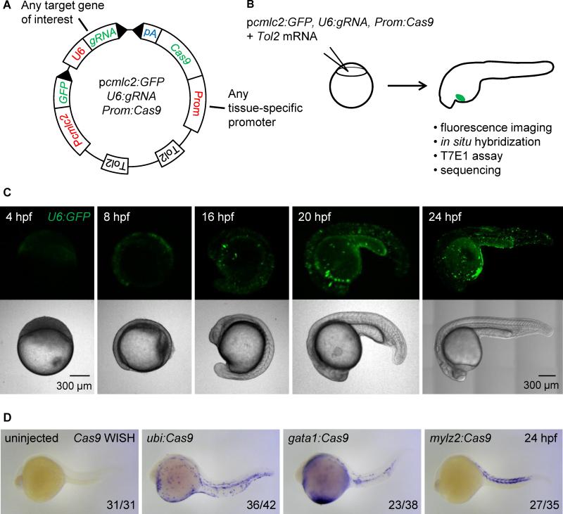
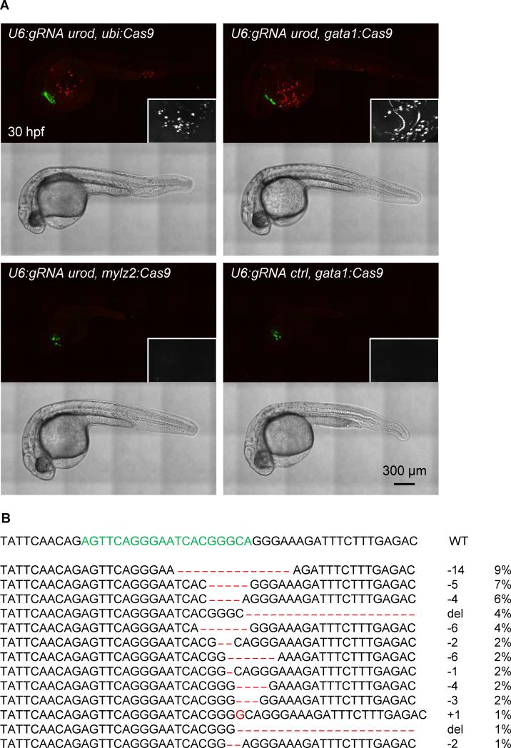
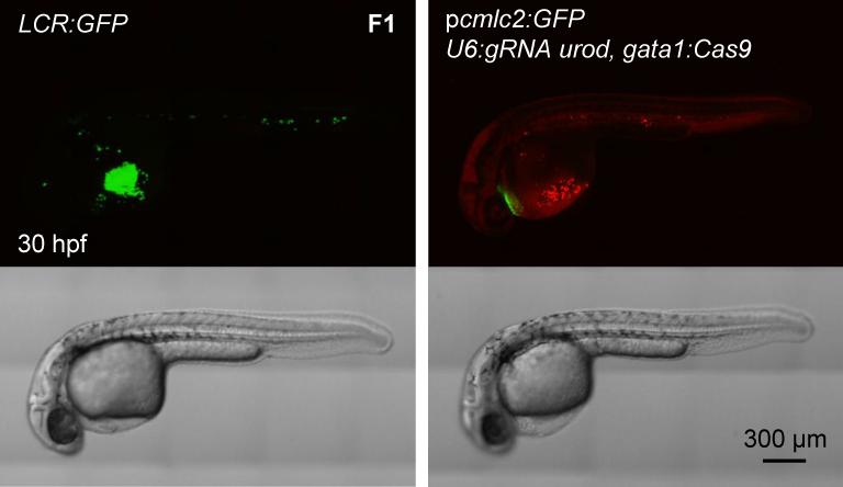
CRISPR/Cas9 technology of genome editing has greatly facilitated the targeted inactivation of genes in vitro and in vivo in a wide range of organisms. In zebrafish, it allows the rapid generation of knockout lines by simply injecting a guide RNA (gRNA) and Cas9 mRNA into one-cell stage embryos. Here, we report a simple and scalable CRISPR-based vector system for tissue-specific gene inactivation in zebrafish. As proof of principle, we used our vector with the gata1 promoter driving Cas9 expression to silence the urod gene, implicated in heme biosynthesis, specifically in the erythrocytic lineage. Urod targeting yielded red fluorescent erythrocytes in zebrafish embryos, recapitulating the phenotype observed in the yquem mutant. While F0 embryos displayed mosaic gene disruption, the phenotype appeared very penetrant in stable F1 fish. This vector system constitutes a unique tool to spatially control gene knockout and greatly broadens the scope of loss-of-function studies in zebrafish.


Figures

Figure 1
Figure 1. Fluorescent phenotype resulting from CRISPR targeting of urod
(A) Schematic representation of urod coding sequence (CDS). The position of the CRISPR target sequences at the beginning of exon three and five are indicated. (B) Outline of the experiment: Cas9 mRNA is injected into one-cell stage embryos along with a gRNA targeting either urod or p53 as a negative control (ctrl). Targeted mutation efficiency is assessed by T7E1 assay and the presence of red fluorescent erythrocytes by confocal microscopy. (C) T7E1 mutagenesis assay at the CRISPR target site in the urod gene. The assay was performed on genomic DNA from 2 dpf embryos injected at the one-cell stage with Cas9 mRNA and either a gRNA against p53 (negative control) or a gRNA against urod. Cleavage bands (arrowheads) indicate the presence of mutations at the target site. See also Figure S1B. (D) Confocal images reveal the presence of fluorescent blood cells in urod mosaic knock-out embryos at 30 hpf. The black and white insets show a 2x magnification of the red fluorescent signal in the yolk region. See also Figure S1E. (E) Most frequent (>1%) mutant urod alleles found by deep-sequencing in whole embryos injected with Cas9 mRNA and urod gRNA. The CRISPR target sequence is in green, mutations in red. The type of mutation and the associated frequency (in % of all mutated alleles) are indicated. del: large deletion.
Figure 2
Figure 2. Integratable CRISPR vector for tissue-specific gene targeting
(A) Schematic representation of the tissue-specific CRISPR vector. Prom denotes any promoter of interest used to drive Cas9 expression in a tissue-restricted manner. GFP expression in the heart of injected embryos is used as a transgenesis marker. Tol2 indicates transposition sites for the Tol2 transposase. pA: SV40 polyA sequence. See Figure S2A for the sequence of U6:gRNA, which comprises two BseRI restriction sites allowing easy cloning of any gene-specific target sequence at the 5’ end of the gRNA. (B) Outline of the experiment: the CRISPR vector is injected into one-cell stage embryos along with Tol2 mRNA. Embryos with GFP-positive hearts are sorted at 24 hpf and further analyzed. Cas9 expression is assessed by in situ hybridization of Cas9 mRNA, targeted mutation efficiency by T7E1 assay, and the presence of fluorescent red blood cells by confocal microscopy. (C) Confocal images of embryos injected with Tol2 mRNA and the pcmlc2:GFP, U6:GFP, gata1:Cas9 vector at various time points. Note early (4 hpf) and ubiquitous (24 hpf) GFP expression. (D) Representative images show whole mount in situ hybridization (WISH) using an anti-sense RNA probe against Cas9 mRNA in 24 hpf embryos injected with Tol2 mRNA and pcmlc2:GFP, U6:gRNA urod vectors expressing Cas9 under the control of the indicated promoters. Cas9 expression pattern is governed by the tissue-specificity of the promoters. See also Figure S2B.
Figure 3
Figure 3. Tissue-specific urod inactivation using the CRISPR vector
(A) Confocal images of 30 hpf embryos injected with Tol2 mRNA and the indicated vectors. The fluorescent blood phenotype associated with urod knockout is seen when using the ubiquitous ubi promoter or the erythrocyte-specific gata1 promoter with the gRNA targeting urod, but not when using the muscle-specific mylz2 promoter or a gRNA against p53. See also Figure S3A and Movie S1. (B) Most frequent (>1%) mutant urod alleles found by deep-sequencing in sorted fluorescent red cells from the blood of embryos injected with the pcmlc2:GFP, U6:gRNA urod, gata1:Cas9 vector. The CRISPR target sequence is in green, mutations in red. The type of mutation and the associated frequency (in % of all mutated alleles) are indicated.
Figure 4
Figure 4. Generation of transgenic, tissue-specific knockout fish
Confocal images of 30 hpf embryos of the LCR:GFP transgenic line and F1 generation of fish stably expressing pcmlc2:GFP, U6:gRNA urod, gata1:Cas9 vector. See also Figure S4A.
Figure 5
Figure 5. Rescue of an anemia phenotype by erythrocyte-specific targeting of p53
(A) Benzidine staining of rps29-/− embryos injected with a pcmlc2:GFP, U6:gRNA, gata1:Cas9 vector targeting urod (ctrl) or p53 at 48 hpf. (B) Quantification of the hemoglobin rescue in rps29-/− embryos. urod (ctrl): n=18; p53: n=20. Pooled results from two independent experiments.

References

    1. Balwani M, Desnick RJ. The porphyrias: advances in diagnosis and treatment. Blood. 2012;120:4496–4504. - PMC - PubMed
    1. Cho SW, Kim S, Kim JM, Kim JS. Targeted genome engineering in human cells with the Cas9 RNA-guided endonuclease. Nat Biotechnol. 2013;31:230–232. - PubMed
    1. Cong L, Ran FA, Cox D, Lin S, Barretto R, Habib N, Hsu PD, Wu X, Jiang W, Marraffini LA, et al. Multiplex genome engineering using CRISPR/Cas systems. Science. 2013;339:819–823. - PMC - PubMed
    1. Fu Y, Foden JA, Khayter C, Maeder ML, Reyon D, Joung JK, Sander JD. High-frequency off-target mutagenesis induced by CRISPR-Cas nucleases in human cells. Nat Biotechnol. 2013;31:822–826. - PMC - PubMed
    1. Gagnon JA, Valen E, Thyme SB, Huang P, Ahkmetova L, Pauli A, Montague TG, Zimmerman S, Richter C, Schier AF. Efficient mutagenesis by Cas9 protein-mediated oligonucleotide insertion and large-scale assessment of single-guide RNAs. PLoS One. 2014;9:e98186. - PMC - PubMed

Publication types

MeSH terms