. 2015 Jul;33(7):2196-207.
doi: 10.1002/stem.1985. Epub 2015 Apr 27.

Unipotent Megakaryopoietic Pathway Bridging Hematopoietic Stem Cells and Mature Megakaryocytes

Affiliations

Unipotent Megakaryopoietic Pathway Bridging Hematopoietic Stem Cells and Mature Megakaryocytes

Hidekazu Nishikii et al. Stem Cells. 2015 Jul.

Abstract

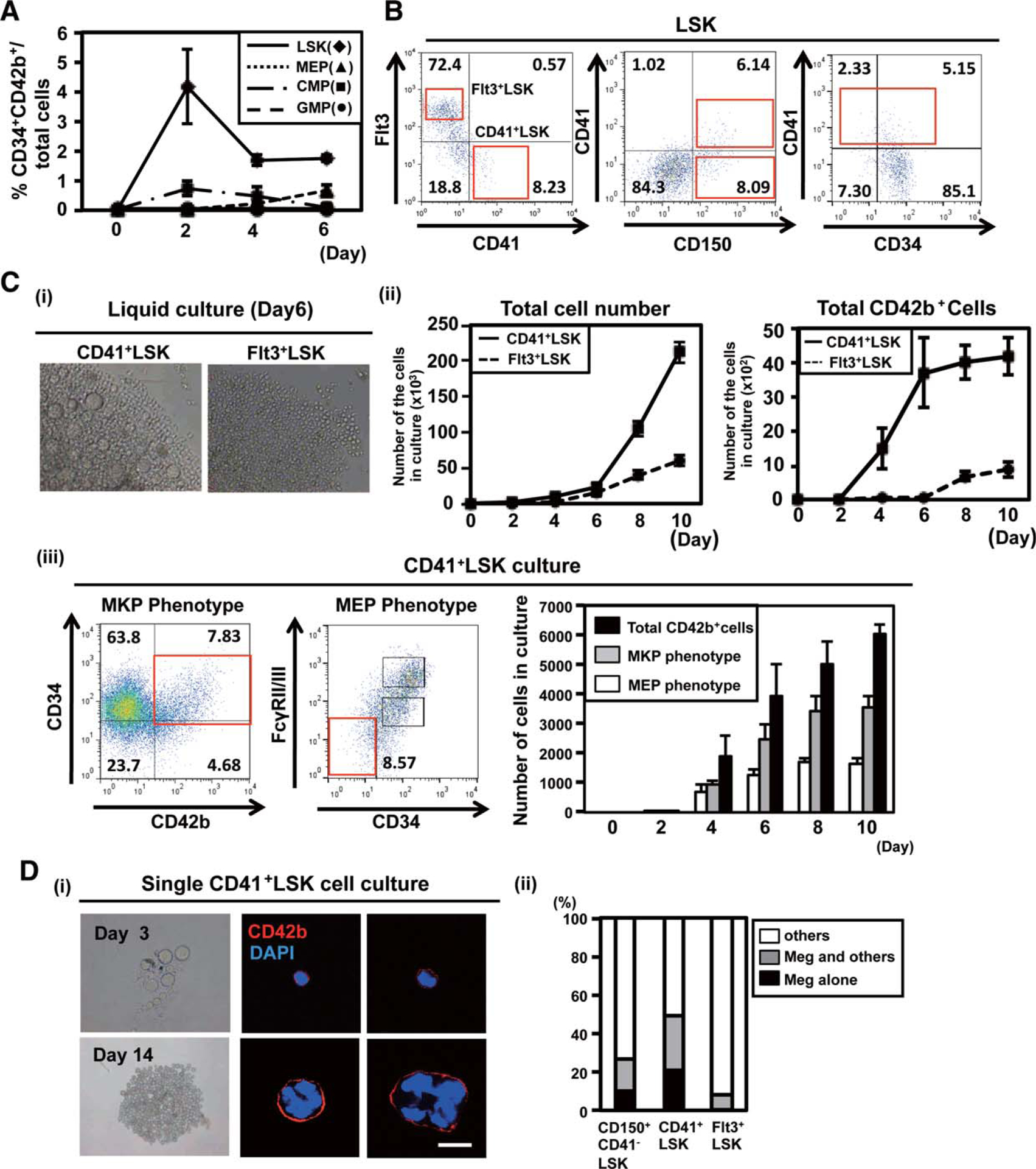
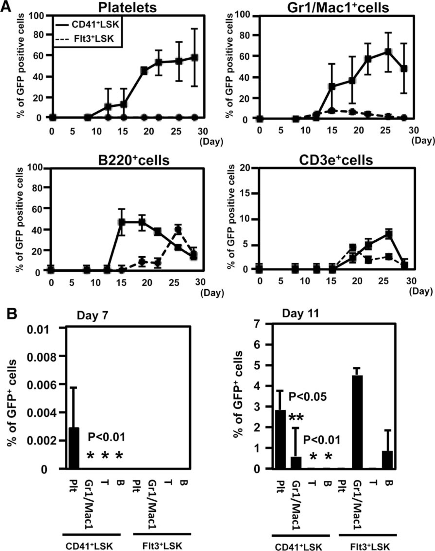
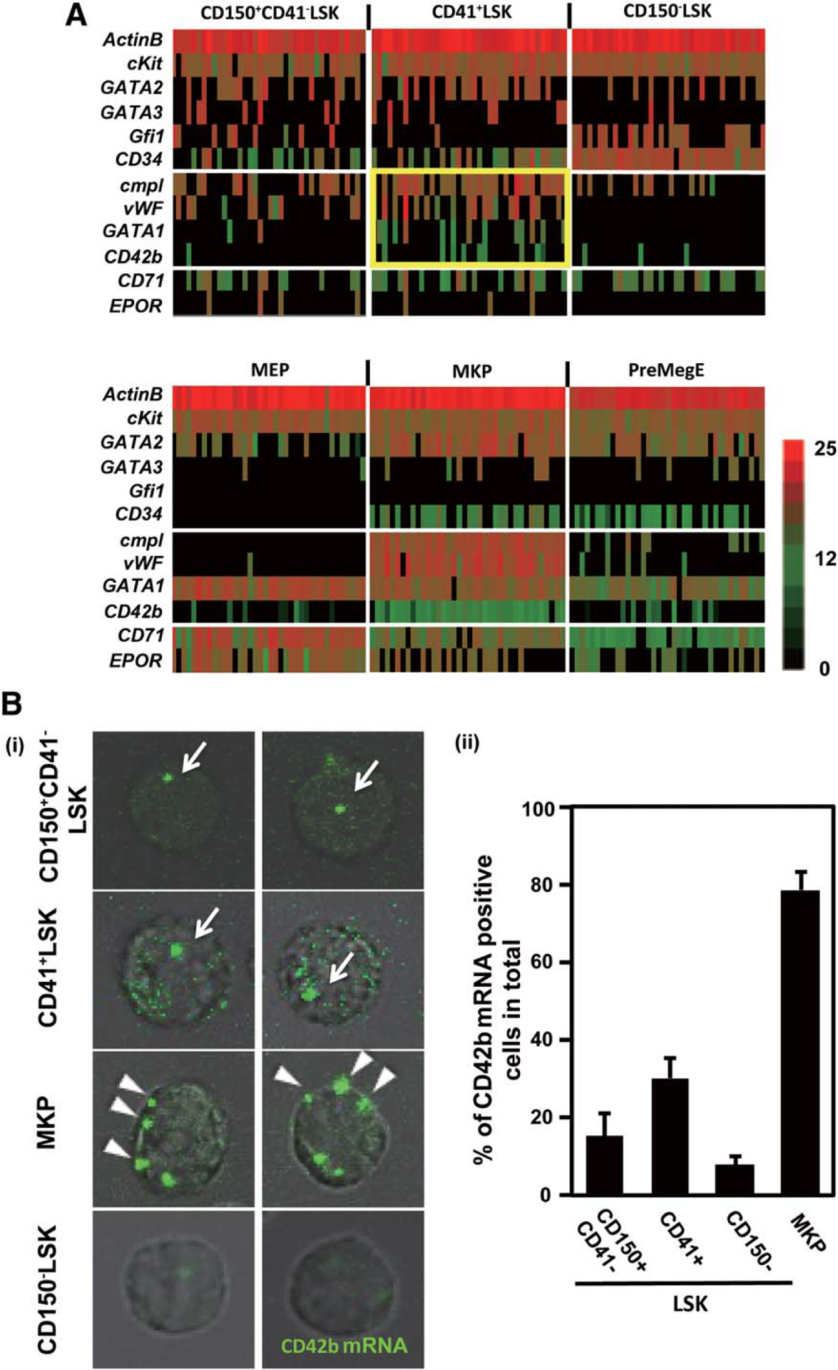
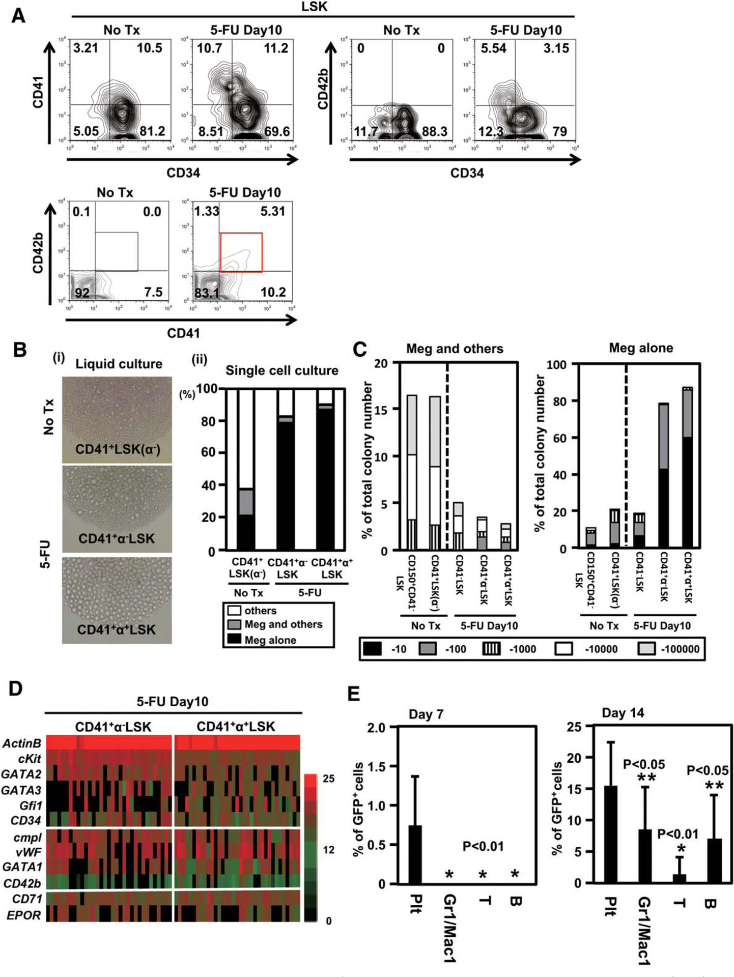
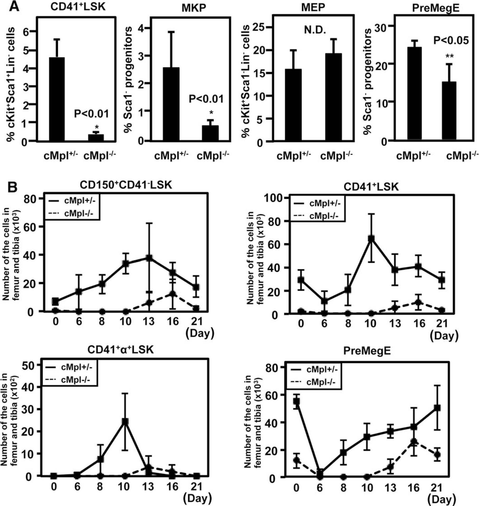
Recent identification of platelet/megakaryocyte-biased hematopoietic stem/repopulating cells requires revision of the intermediate pathway for megakaryopoiesis. Here, we show a unipotent megakaryopoietic pathway bypassing the bipotent megakaryocyte/erythroid progenitors (biEMPs). Cells purified from mouse bone marrow by CD42b (GPIbα) marking were demonstrated to be unipotent megakaryocytic progenitors (MKPs) by culture and transplantation. A subpopulation of freshly isolated CD41(+) cells in the lineage Sca1(+) cKit(+) (LSK) fraction (subCD41(+) LSK) differentiated only into MKP and mature megakaryocytes in culture. Although CD41(+) LSK cells as a whole were capable of differentiating into all myeloid and lymphoid cells in vivo, they produced unipotent MKP, mature megakaryocytes, and platelets in vitro and in vivo much more efficiently than Flt3(+) CD41(-) LSK cells, especially at the early phase after transplantation. In single cell polymerase chain reaction and thrombopoietin (TPO) signaling analyses, the MKP and a fraction of CD41(+) LSK, but not the biEMP, showed the similarities in mRNA expression profile and visible TPO-mediated phosphorylation. On increased demand of platelet production after 5-FU treatment, a part of CD41(+) LSK population expressed CD42b on the surface, and 90% of them showed unipotent megakaryopoietic capacity in single cell culture and predominantly produced platelets in vivo at the early phase after transplantation. These results suggest that the CD41(+) CD42b(+) LSK are straightforward progenies of megakaryocytes/platelet-biased stem/repopulating cells, but not progenies of biEMP. Consequently, we show a unipotent/highly biased megakaryopoietic pathway interconnecting stem/repopulating cells and mature megakaryocytes, the one that may play physiologic roles especially in emergency megakaryopoiesis.

Keywords: Adult hematopoietic stem cells; Hematopoietic progenitors; Megakaryocyte; Thrombopoiesis; Thrombopoietin.


Conflict of interest statement

DISCLOSURE OF POTENTIAL CONFLICTS OF INTEREST

The authors indicate no potential conflicts of interest.

Figures

Figure 1.
Figure 1.
CD34+CD42b+ cells have a restricted capacity of megakaryocyte differentiation in vitro and platelet production in vivo. (A): Identification of CD34+CD42b+ population in bone marrow cells (BMCs). Adult mouse BMCs were stained with antibodies for cKit, Sca1, lineage marker (Lin), CD34, CD16/32 (FccRII/III), CD42b, CD41, CD150, and CD9. Note that only CD34+ fraction of the Lin population expressed CD42b (3rd figure from the left in the upper panels) and that the CD34+CD42b+ population was confined to the Sca1cKit+ population (right in the upper panels), mainly in the common myeloid progenitor (CMP) fraction (the lower panels). A representative result from five independent experiments is shown. (B): (i) The representative morphologies of the colonies derived from indicated cell types in 96-well-plate liquid culture. The number of cells seeded in one well was 500 for LSK/CMP (CD42b-), 2000 for megakaryocyte–erythroid progenitor (MEP)/megakaryocytic progenitor (MKP). Arrowheads indicate mature megakaryocytes. (ii) Frequencies of vWF+ and TER119+ cells in the liquid culture shown in (i). (C): Capacity of CD34+CD42b+ cells (MKP) and MEP to generate platelet in vivo. Sublethally irradiated (4.5 Gy) mice were transplanted with 1 × 10⁴ CD34+CD42b+ cells or MEP from green fluorescent protein transgenic mice. On days 4, 7, 11, and 14 after transplantation, peripheral blood was collected and analyzed for platelet differentiation using CD41+ platelet-sized cells (n 5 4). Abbreviations: CMP, common myeloid progenitor; FSC, forward scatter; GFP, green fluorescent protein; GMP, granulocyte–monocyte progenitor; LSK, lineageSca1+cKit+; MEP, megakaryocyte–erythroid progenitor; MKP, megakaryocytic progenitor; SSC, side scatter.
Figure 2.
Figure 2.
A subpopulation of CD41+LSK is the source of unipotent megakaryopoiesis through megakaryocytic progenitor (MKP). (A):Frequencies of CD34+CD42b+ cells after liquid culture of each of the progenitor cells. Two thousand LineageSca1+cKit+ (LSK), megakaryocyte–erythroid progenitor (MEP), common myeloid progenitor (CMP) depleted with MKP, and granulocyte–monocyte progenitor (GMP) were sorted from mouse bone marrow and cultured in the presence of stem cell factor (SCF), thrombopoietin (TPO), interleukin (IL)26, and IL-11. Cells in culture were stained with antibodies for CD34 and CD42b, and analyzed by flow cytometry. The means of triplicate were shown. (B): Expression pattern of CD150, CD41, CD34, and Flt3 in the LSK population. Note that CD41 and Flt3 are expressed in an almost mutually exclusive manner. (C): (i) A representative morphology of CD41+LSK-and Flt3+LSK-derived colonyforming cells (day 6; 3200, left panel). (ii) Numbers of total CD42b+ cells after culturing 500 CD41+LSK or Flt3+LSK were calculated on flow cytometry, using the fixed numbers of beads (left). (iii) The numbers of total CD42b+, LinSca1cKit+CD34+CD42b+ (MKP phenotype), and Lin Sca1cKit+FcγRII/IIICD34 cells (MEP phenotype) after culturing 500 CD41+LSK. Representative results of day 10 were shown in left panel. The cell numbers were calculated using the same methods (right graph). (D): (i) Typical morphologies of pure megakaryocyte-lineage colonies derived from a single CD41+LSK in 96-well (upper panels, day 3; lower panels, day 14 of culture). The right panels show immunostaining of the cells from the pure megakaryocytes-lineage colonies (red, CD42b; blue, DAPI). Scalebar 5 20 lm. (ii) Preferential differentiation of CD41+LSK into megakaryocytes assessed by single cell culture. CD150+CD41LSK (HSC-enriched population), CD41+LSK, and Flt3+LSK were single cell-sorted into each well of 96-well dishes and cultured in the presence of SCF, TPO, IL-6, and IL-11. Colonies formed in each well were classified into those containing megakaryocytes alone, containing megakaryocytes and others, and without megakaryocytes. The results of three independent experiments are shown. The total number of colonies from each progenitor was 376 in CD41+LSK, 204 in Flt3+LSK, and 446 in CD150+CD41LSK. Abbreviations: CMP, common myeloid progenitor; DAPI, 4’6-diamidino-2-phenylindole; GMP, granulocyte–monocyte progenitor; LSK, lineageSca1+cKit+; MEP, megakaryocyte–erythroid progenitor; MKP, megakaryocytic progenitor; SSC, side scatter.
Figure 3.
Figure 3.
CD41+LSK efficiently differentiate into megakaryocytic progenitor (MKP) and platelets in vivo. (A): (i) Five hundred CD41+LSK or Flt3+LSK from green fluorescent protein (GFP) transgenic mice were sorted and transplanted into lethally irradiated wild type B6 mice. Peripheral blood was analyzed by flow cytometry at the indicated time points after transplantation (n 5 5, each group). The frequency of GFP-positive cells in each lineage is shown. CD41+ Gr1/Mac1+, B220+, and CD3e+ represent platelets, granulocytes/monocytes, B cells, and T cells, respectively. (B):The frequencies of donor-derived cells at the early time point after transplantation were shown (right panel, day 7; left panel, day 11). Abbreviations: GFP, green fluorescent protein; LSK, lineageSca1+cKit+.
Figure 4.
Figure 4.
A single cell gene expression revealed the proximity between a subpopulation of CD41+LSK and MKP. (A):Single cell gene expression. Single cell real-time polymerase chain reaction was applied to analyze gene expression in single cells constituting CD150+CD41LSK, CD41+LSK, CD150LSK, megakaryocyte–erythroid progenitor, MKP, and PreMegE. Expression of 11 genes in 38 representative cells from each population was shown. The levels of transcripts in individual cells quantified by cycle threshold values and shown by colors indicated (red, highest; black, lowest). Note that cMpl, vWF, and CD42b were densely expressed in MKP, and that the closest pattern is observed in CD411LSK (yellow gate). (B): CD42b mRNA imaging at a single cell level. Sorted CD150+CD41LSK, CD41+LSK, MKP, and CD150CD41LSK from wild type bone marrow are fixed on the poly-L-lysine coated glass slides and stained with a CD42b mRNA specific probe. Typical single cell images were shown in (i), green signals, CD42b mRNA. arrow, nuclear-localized CD42b mRNA; arrowhead, cytoplasm-localized CD42b mRNA. The frequencies of CD42b mRNA-positive cells in each progenitor population are shown in (ii). Each graph shows the result of three independent experiments. Abbreviations: LSK, lineageSca1+cKit+; MEP, megakaryocyte–erythroid progenitor; MKP, megakaryocytic progenitor; PreMegE, pre-megakaryocyte/erythrocyte progenitors.
Figure 5.
Figure 5.
Thrombopoietin (TPO)/cMpl signaling in single cell in subCD41+LSK and megakaryocytic progenitor (MKP). (A): (i) Sorted CD41+LSK, MKP, and megakaryocyte–erythroid progenitor (MEP) from bone marrow were stimulated with TPO or EPO on a glass slide and stained with anti-phospho-STAT5 (left), AKT (middle), and JAK2 (right) antibody. Representative pictures from each progenitor are shown (green; p-STAT5/P-AKT/p-JAK2). Scale bar 5 50 lm. (B): Frequencies of phospho-STAT5, AKT, and JAK2-positive cells in each progenitor population. Each graph shows the result from three independent experiments. Abbreviations: DAPI, 4ʹ 6-diamidino-2-phenylindole; EPO, erythropoetin; LSK, lineageSca1+cKit+; MEP, megakaryocyte–erythroid progenitor; MKP, megakaryocytic progenitor; TPO, thrombopoietin.
Figure 6.
Figure 6.
Emergency megakaryopoiesis is driven at the CD41+LSK stage. (A): Expression of CD42b in lineageSca1+cKit+ (LSK) compartment during the recovery phase after 5-FU treatment. Expression of CD42b in LSK was analyzed 10 days after a single 5-FU (150 mg/kg) treatment. A representative result of CD34, CD41, and CD42b staining from five independent experiments are shown. (B): (i) Sorted 500 CD41+CD42bLSK [CD41+LSK (α)] from untreated mice and CD41+CD42b LSK cells (CD41+αLSK) and CD41+CD42b+LSK (CD41+α+LSK) from 5-FU-treated mice were cultured in 96-well plates to assess their capacity for megakaryopoiesis. The pictures show representative morphologies of differentiated cells. (No Tx, no treatment). (ii) Preferential megakaryocyte differentiation from CD41+α+LSK and CD41+αLSK after 5-FU treatment, compared with CD41+LSK (α) at steady-state condition, assessed by single cell culture. Indicated progenitor cells were single cell-sorted into each well of 96-well dishes and cultured in the presence of stem cell factor (SCF), thrombopoietin (TPO), interleukin (IL)26, and IL-11. Colonies formed in each well were classified into those containing megakaryocytes alone, containing megakaryocytes and others, and without megakaryocytes. Three separated experiments were combined. (C): Proliferative capacity of indicated progenitors in single cell culture. Clone-sorted cells were cultured in 96 wells in the presence of TPO, SCF, IL-6, and IL-11. The frequency of cell proliferation was assessed on day 10. The numbers of megakaryocyte-containing (Meg1others, left) and megakaryocytes (Meg Alone, right) colonies were classified into five categories. (D): The results of single cell PCR using the samples from CD41+αLSK and CD41+α+LSK from 5-FU treated mice. Expression of 11 genes in 38 representative cells from each population is shown. The levels of transcripts in individual cells are quantified by cycle threshold values and shown by indicated colors (red, highest; black, lowest). (E): Five hundred CD41+LSK from 5-FU-treated green fluorescent protein (GFP) transgenic mice were sorted and transplanted into lethally irradiated wild type mice. The frequencies of GFP+ cells in peripheral blood at very early time points are shown (n 5 10, each group). Abbreviations: GFP, green fluorescent protein; LSK, lineageSca1+cKit+; Meg, megakaryocyte.
Figure 7.
Figure 7.
Distinct megakaryocyte progenitors at the steady and platelet-demanding state show variable degrees of dependency on thrombopoietin/cMpl signaling. (A): Frequencies of CD41+LSK, megakaryocytic progenitor (MKP), megakaryocyte–erythroid progenitor (MEP), and PreMegE in cMpl-deficient (cMpl−/−) and heterozygous (cMpl+/) mice. The means of quadlicates are shown. (B): The total number of each progenitor cell population in cMpl/ and cMpl+/ mice at several time points after 5-FU treatment (n 5 4, each group). Abbreviations: LSK, lineageSca1+cKit+; MEP, megakaryocyte–erythroid progenitor; MKP, megakaryocytic progenitor; PreMegE, pre-megakaryocyte/erythrocyte progenitors.

References

    1. Akashi K, Traver D, Miyamoto T et al. A clonogenic common myeloid progenitor that gives rise to all myeloid lineages. Nature 2000;404:193–197. - PubMed
    1. Pronk CJ, Rossi DJ, Mansson R et al. Elucidation of the phenotypic, functional, and molecular topography of a myeloerythroid progenitor cell hierarchy. Cell Stem Cell 2007; 1:428–442. - PubMed
    1. Cornejo MG, Mabialah V, Sykes SM et al. Crosstalk between NOTCH and AKT signaling during murine megakaryocyte lineage specification. Blood 2012;118:1264–1273. - PMC - PubMed
    1. Mercher T, Cornejo MG, Sears C et al. Notch signaling specifies megakaryocyte development from hematopoietic stem cells. Cell Stem Cell 2008;3:314–326. - PMC - PubMed
    1. Sanchez M, Weissman IL, Pallavicini M et al. Differential amplification of murine bipotent megakaryocytic/erythroid progenitor and precursor cells during recovery from acute and chronic erythroid stress. STEM CELLS 2006;24:337–348. - PubMed

Publication types