. 2015 Sep 1;523(13):1913-24.
doi: 10.1002/cne.23770. Epub 2015 Jul 1.

Organization of TNIK in dendritic spines

Affiliations

Organization of TNIK in dendritic spines

Alain C Burette et al. J Comp Neurol. .

Abstract

Tumor necrosis factor receptor-associated factor 2 (TRAF2)- and noncatalytic region of tyrosine kinase (NCK)-interacting kinase (TNIK) has been identified as an interactor in the psychiatric risk factor, Disrupted in Schizophrenia 1 (DISC1). As a step toward deciphering its function in the brain, we performed high-resolution light and electron microscopic immunocytochemistry. We demonstrate here that TNIK is expressed in neurons throughout the adult mouse brain. In striatum and cerebral cortex, TNIK concentrates in dendritic spines, especially in the vicinity of the lateral edge of the synapse. Thus, TNIK is highly enriched at a microdomain critical for glutamatergic signaling.

Keywords: PSD; RRID:AB_11213019; RRID:AB_1858225; RRID:AB_94396; RRID:nif-0000-30467; TNIK; cerebral cortex; dendritic spine; striatum RRID:AB_11212843.


Conflict of interest statement

Conflict of interest statement

BB, QW, MDE, ML, GF, QL, NT are/were employees and/or shareholders of Pfizer, Inc. The other authors verify that they have no known or potential conflict of interest including any financial, personal, or other relationships with other people or organizations within 3 years of beginning the submitted work that could inappropriately influence, or be perceived to influence, this work.

Figures
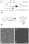
Figure 1
Figure 1. Preparation of the knockout mice
A: Gene targeting of TNIK locus and RT-PCR of whole brain mRNA from targeted mice. Genomic locus illustrating the region of exons 5–7. Targeting vector indicated showing homology arms, LoxP sites flanking exon 7 (▶), and neomycin (G418) resistance cassette used for selection. Neor cassette is flanked by Frt sites (●). After introduction into the germline, exon 7 was removed by Cre recombinase-mediated deletion. B: RT-PCR of whole brain RNA from animals of the indicated genotypes. PCR primers located in exons 6 and 8 indicated by arrows. Deletion of exon 7 results in a novel transcript in animals carrying the Δ7 allele that contains early termination codons as indicated. Identity of PCR products was verified by sequencing (data not shown). C: Immunofluorescence staining for TNIK in neocortex from WT (A) and TNIKΔ7/TNIKΔ7 (B) mouse brain. Scale bar = 50 μm in C and D.
Figure 2
Figure 2. Immunoperoxidase staining for TNIK in the mouse brain
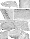
A: Parasagittal section of whole mouse brain. Staining is conspicuous in gray matter, largely sparing white matter. Higher magnification views show patterns of staining in specific regions of brain, including B: Olfactory bulb; C: Piriform cortex; D: Neocortex; E: Hippocampus; F: Striatum; G: Thalamus; H: Hypothalamus; I: Cerebellum. Scale bar = 2 mm in A; 500 μm in B, 200 μm in C; 250 μm in D, E, F; 500 μm in G; 250 μm in H; 500 μm in I.
Figure 3
Figure 3. Immunofluorescence labeling for TNIK in neocortex
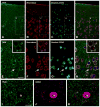
A–D: Sections were counterstained with Hoechst 33342 to visualize nuclei, and with NeuroTrace 640-660 to visualize neuronal somata. Most NeuroTrace-identified neurons are also immunopositive for TNIK, though to varying degrees. E–H: Enlargement of boxed area from upper panel; white arrow points to a small neuron immunonegative for TNIK (extended-focus confocal images). Insets: enlarged single-focus images from the same field; note that many somatic TNIK “blobs” in E correspond to Nissl bodies as defined by Neurotrace (F); some examples are outlined with white circles. I–K; Double labeling for TNIK and GABA; a GABAergic soma (asterisk) is immunonegative for TNIK. Scale bar = 30 μm in A–D; 20 μm in E–H; 10 μm in I–K.
Figure 4
Figure 4. Immunofluorescence labeling for TNIK in striatum
A–H: Sections were counterstained with Hoechst 33342 to visualize nuclei, and NeuroTrace 640-660 to visualize neuronal somata. Most neurons in the field were immunopositive for TNIK. Boxed area in upper panel is shown at higher magnification in middle panel (E–H). The asterisk marks a neuron immunonegative for TNIK. I–K: Double labeling for TNIK and ChAT; white arrows point to ChAT-positive neurons that are immunonegative for TNIK. Scale bar = 30 μm in A–D; 10 μm in E–H; 25 μm in I–K.
Figure 5
Figure 5. Colocalization of TNIK with VGLUT1
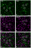
Double labeling in mouse neocortex (A–C) and striatum (D–F) shows that many TNIK puncta (green) are closely apposed to, or partially overlap with, the vesicular glutamate transporter VGLUT1 (magenta), suggesting that TNIK concentrates at synapses. Scale bar = 4 μm in A–F.
Figure 6
Figure 6. Ultrastructural analysis using postembedding immunogold electron microscopy reveals association of TNIK with the postsynaptic density (PSD)
A: an immunopositive spine in cerebral cortex; arrows point to two particles lying close to the edge of the PSD. B: micrograph shows a thin dendritic shaft in cortex receiving two asymmetric synapses; both are immunopositive for TNIK. The lower arrow points to a gold particle lying in the middle of the synapse, whereas the upper arrow points to a particle lying at the edge of the PSD. C: view of a large immunopositive spine in striatum; two gold particles (arrows) lie close to the postsynaptic membrane. D: histograms show the distribution of immunolabeling in the axodendritic axis (20 nm bins, cerebral cortex: N=135 synaptic particles, striatum: N=133 particles). Diagram at bottom illustrates measurement technique; positive values correspond to particles lying inside the postsynaptic plasma membrane. The pattern of labeling was consistent for cortex (left) and striatum (right), peaking just inside the postsynaptic membrane. E: histograms show distribution of label tangentially along the synapse, normalized such that 0 corresponds to the center of the PSD, and 1.0 to its edge (see diagram at bottom; cortex: N=120 synaptic particles, striatum N=116 particles). Labeling in both cortex (left) and striatum (right) concentrated in the vicinity of the edge of the PSD. An additional pool of TNIK lay at the center of the PSD in striatum, but not in cortex. X-axis for each histogram has been normalized so that the largest bin corresponds to 1.0 units, and the origin corresponds to zero. Scale bar = 200 nm in A, B, C

References

    1. Armstrong DM, Saper CB, Levey AI, Wainer BH, Terry RD. Distribution of cholinergic neurons in rat brain: demonstrated by the immunocytochemical localization of choline acetyltransferase. J Comp Neurol. 1983;216(1):53–68. - PubMed
    1. Ayalew M, Le-Niculescu H, Levey DF, Jain N, Changala B, Patel SD, Winiger E, Breier A, Shekhar A, Amdur R, Koller D, Nurnberger JI, Corvin A, Geyer M, Tsuang MT, Salomon D, Schork NJ, Fanous AH, O’Donovan MC, Niculescu AB. Convergent functional genomics of schizophrenia: from comprehensive understanding to genetic risk prediction. Molecular psychiatry. 2012;17(9):887–905. - PMC - PubMed
    1. Benson DL, Tanaka H. N-cadherin redistribution during synaptogenesis in hippocampal neurons. The Journal of neuroscience: the official journal of the Society for Neuroscience. 1998;18(17):6892–6904. - PMC - PubMed
    1. Bernard V, Bolam JP. Subcellular and subsynaptic distribution of the NR1 subunit of the NMDA receptor in the neostriatum and globus pallidus of the rat: co-localization at synapses with the GluR2/3 subunit of the AMPA receptor. Eur J Neurosci. 1998;10(12):3721–3736. - PubMed
    1. Brigidi GS, Bamji SX. Cadherin-catenin adhesion complexes at the synapse. Current opinion in neurobiology. 2011;21(2):208–214. - PubMed

Publication types

MeSH terms

Substances