. 2015 Mar 10;10(9):1477-1486.
doi: 10.1016/j.celrep.2015.02.023. Epub 2015 Mar 5.

N-Cadherin Induction by ECM Stiffness and FAK Overrides the Spreading Requirement for Proliferation of Vascular Smooth Muscle Cells

Affiliations

N-Cadherin Induction by ECM Stiffness and FAK Overrides the Spreading Requirement for Proliferation of Vascular Smooth Muscle Cells

Keeley L Mui et al. Cell Rep. .

Abstract

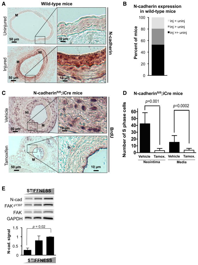
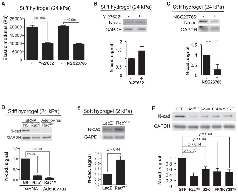
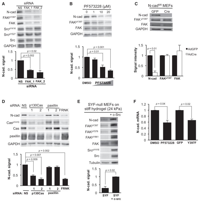
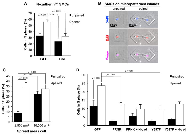
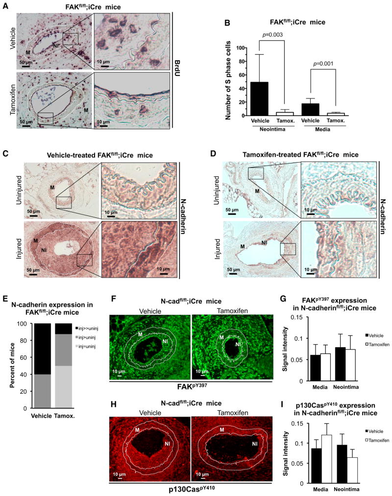
In contrast to the accepted pro-proliferative effect of cell-matrix adhesion, the proliferative effect of cadherin-mediated cell-cell adhesion remains unresolved. Here, we studied the effect of N-cadherin on cell proliferation in the vasculature. We show that N-cadherin is induced in smooth muscle cells (SMCs) in response to vascular injury, an in vivo model of tissue stiffening and proliferation. Complementary experiments performed with deformable substrata demonstrated that stiffness-mediated activation of a focal adhesion kinase (FAK)-p130Cas-Rac signaling pathway induces N-cadherin. Additionally, by culturing paired and unpaired SMCs on microfabricated adhesive islands of different areas, we found that N-cadherin relaxes the spreading requirement for SMC proliferation. In vivo SMC deletion of N-cadherin strongly reduced injury-induced cycling. Finally, SMC-specific deletion of FAK inhibited proliferation after vascular injury, and this was accompanied by reduced induction of N-cadherin. Thus, a stiffness- and FAK-dependent induction of N-cadherin connects cell-matrix to cell-cell adhesion and regulates the degree of cell spreading needed for cycling.


Figures

Figure 1
Figure 1. N-Cadherin Is Essential for In Vivo SMC Proliferation
(A) Uninjured and injured femoral artery sections from wild-type mice (n = 9) were immunohistochemically stained for N-cadherin. The media (M) lies between the dashed white lines, which mark the internal and external elastic laminae. The neointima (NI) seen in injured arteries is the region between internal elastic lamina and lumen. Images on the right are magnifications of the inset black boxes in the left-hand images. (B) N-cadherin expression in the injured and uninjured femoral arteries of each mouse was scored by blinded grouping (inj, injured; uninj, uninjured). (C) In vivo SMC proliferation was examined by in vivo BrdU labeling in injured femoral arteries from vehicle-treated (n = 8) and tamoxifen-treated (n = 7) N-cadherinfl/fl;iCre mice. (D) Results in (C) were quantified by manual counting and plotted as mean + SD. (E) Asynchronous SMCs were cultured on FN-coated PA hydrogels (2, 11, and 24 kPa) for 24 hr in 10% FBS and analyzed by western blot. The bar graph shows N-cadherin levels normalized to control and plots results as mean + SE; n = 3. See also Figure S1.
Figure 2
Figure 2. N-Cadherin Is Regulated by Rac
(A) Serum-starved MEFs were suspended in 10% FBS, seeded on stiff FN-coated hydrogels (24 kPa), and treated with 10 μM of the Rho kinase inhibitor Y-27632 or 150 μM of the Rac inhibitor NSC23766 for 20 hr. The elastic moduli of individual cells were determined by atomic force microscopy; n = 3. (B and C) Serum-starved MEFs treated with (B) 10 μM Y-27632 or (C) 150 μM NSC2376 on stiff FN-coated PA hydrogels (24 kPa) for 20–24 hr in 10% FBS were analyzed by western blot; n = 3–4. (D) Serum-starved MEFs transfected with 200 nM non-specific (NS) or Rac siRNA, or infected with dominant-negative RacN17 adenovirus were cultured on stiff FN-coated PA hydrogels in 10% FBS for 9 hr. Cells were lysed and analyzed by western blot; n = 3. (E) Serum-starved MEFs were infected with adenoviral vectors expressing LacZ or RacV12, and cultured on soft FN-coated PA hydrogels (2 kPa) with 10% FBS for 9 hr and analyzed by western blot; n = 4. (F) MEFs in 10% FBS were infected with adenovirus expressing GFP, RacN17, β2-chimaerin (β2-ch), FRNK, or FAKY397F (Y397F) for 24 hr, lysed, and examined by western blot; n = 3. See also Figure S2.
Figure 3
Figure 3. N-Cadherin Is Induced by FAK-Src-Cas Signaling
(A) MEFs were transfected with 200 nM of two distinct FAK siRNAs or a nonspecific (NS) siRNA. The cells were then serum starved for 48 hr, incubated in 10% FBS for 20 hr, and analyzed by western blot; n = 3. (B) Serum-starved MEFs were treated with different concentrations of the small-molecule FAK inhibitor PF573228 for 20 hr in 10% FBS and analyzed by western blot; n = 4. (C) N-cadherinfl/fl MEFs in 10% FBS were infected overnight with 1,000 moi of adenovirus expressing GFP or Cre and analyzed by western blot; n = 3. (D) Serum-starved MEFs were either transfected with 200 nM of two distinct siRNAs against p130Cas or paxillin, or a non-specific (NS) siRNA, or infected with 1,000 moi adenoviral FRNK. Cells were then incubated with 10% FBS for 24 hr, lysed, and examined by western blot; n = 3. (E) Serum-starved SYF null MEFs cultured on stiff FN-coated PA hydrogels (24 kPa) were incubated with 10% FBS for 9 hr, lysed, and examined by western blot; n = 3. The bar graphs under (A)–(E) show N-cadherin levels normalized to control and are plotted as mean + SE. (F) MEFs in 10% FBS were treated with 20 μM PF573228 for 20 hr or infected with FAKY397F adenovirus for 24 hr. The relative expression of N-cadherin mRNA was examined by quantitative RT-PCR. Results are shown as mean + SE; n = 3. See also Figure S3.
Figure 4
Figure 4. N-Cadherin Determines the Spreading Requirement for Proliferation
(A) Serum-starved N-cadherinfl/fl SMCs infected with adenoviral GFP or Cre were cultured on unpaired or paired micropatterned adhesive islands (4,900 μm2), stimulated with 10% FBS for 48 hr, and analyzed for S-phase entry by EdU incorporation. (B) Representative images of EdU-positive SMCs on 10,000 μm2 unpaired or paired micropatterned adhesive islands. Cells were immunofluorescently co-stained for DAPI and EdU. (C) SMCs were cultured on different-sized micropatterned adhesive islands (2,500 and 10,000 μm2) with 10% FBS for 48 hr and analyzed for S-phase entry by EdU incorporation. (D) Serum-starved SMCs were infected with adenoviral GFP, FRNK or FAKY397F (Y397F) without or with adenoviral N-cadherin, seeded on micropatterned adhesive islands (2,500 μm2) with 10% FBS for 48 hr, and analyzed for S-phase entry. All S-phase results are plotted as mean + SE; n = 3. See also Figure S4.
Figure 5
Figure 5. N-Cadherin Is an Essential Effector of FAK-Regulated SMC Cycling In Vivo
(A) Injured femoral arteries from vehicle-treated (n = 7) and tamoxifen-treated (n = 8) FAKfl/fl;iCre mice were stained for incorporated BrdU. Images on the right are magnifications of the inset black boxes in the left-hand images. (B) The number of S-phase nuclei in the neointima (NI) and media (M) of vehicle-treated (n = 7) and tamoxifen-treated (n = 8) mice was determined by manual counting and plotted as mean + SD. (C and D) Femoral artery sections from (C) vehicle- and (D) tamoxifen-treated FAKfl/fl;iCre mice were stained for N-cadherin. (E) Blinded grouping of the vehicle (n = 5) and tamoxifen (n = 8) data in (C) and (D). Statistical significance was determined by chi-square test; p < 000.1. (F and H) Injured femoral arteries from vehicle- and tamoxifen-treated N-cadherinfl/fl;iCre mice were stained for (F) FAKpY397 and (H) p130CaspY410. The media (M) lies between the dashed white lines, which mark the internal and external elastic laminae. The neointima (NI) is the region between the internal elastic lamina and lumen. (G) Quantification of results from (F); vehicle n = 4, tamoxifen n = 5. (I) Quantification of results from (H); vehicle n = 5, tamoxifen n = 5. Results in (G) and (I) are plotted as mean + SE. See also Figure S5.

Similar articles

Cited by

References

    1. Assoian RK, Klein EA. Growth control by intracellular tension and extracellular stiffness. Trends Cell Biol. 2008;7:24–27. - PMC - PubMed
    1. Bae YH, Mui KL, Hsu BY, Liu S-L, Cretu A, Razinia Z, Xu T, Puré E, Assoian RK. A FAK-Cas-Rac-lamellipodin signaling module transduces extracellular matrix stiffness into mechanosensitive cell cycling. Sci Signal. 2014;7:ra57. - PMC - PubMed
    1. Baumeister U, Funke R, Ebnet K, Vorschmitt H, Koch S, Vestweber D. Association of Csk to VE-cadherin and inhibition of cell proliferation. EMBO J. 2005;24:1686–1695. - PMC - PubMed
    1. Braga VM, Betson M, Li X, Lamarche-Vane N. Activation of the small GTPase Rac is sufficient to disrupt cadherin-dependent cell-cell adhesion in normal human keratinocytes. Mol Biol Cell. 2000;11:3703–3721. - PMC - PubMed
    1. Cary LA, Han DC, Polte TR, Hanks SK, Guan JL. Identification of p130Cas as a mediator of focal adhesion kinase-promoted cell migration. J Cell Biol. 1998;140:211–221. - PMC - PubMed