. 2015 Mar 10:5:8959.
doi: 10.1038/srep08959.

Tuning IL-2 signaling by ADP-ribosylation of CD25

Affiliations

Tuning IL-2 signaling by ADP-ribosylation of CD25

Sophie Teege et al. Sci Rep. .

Abstract

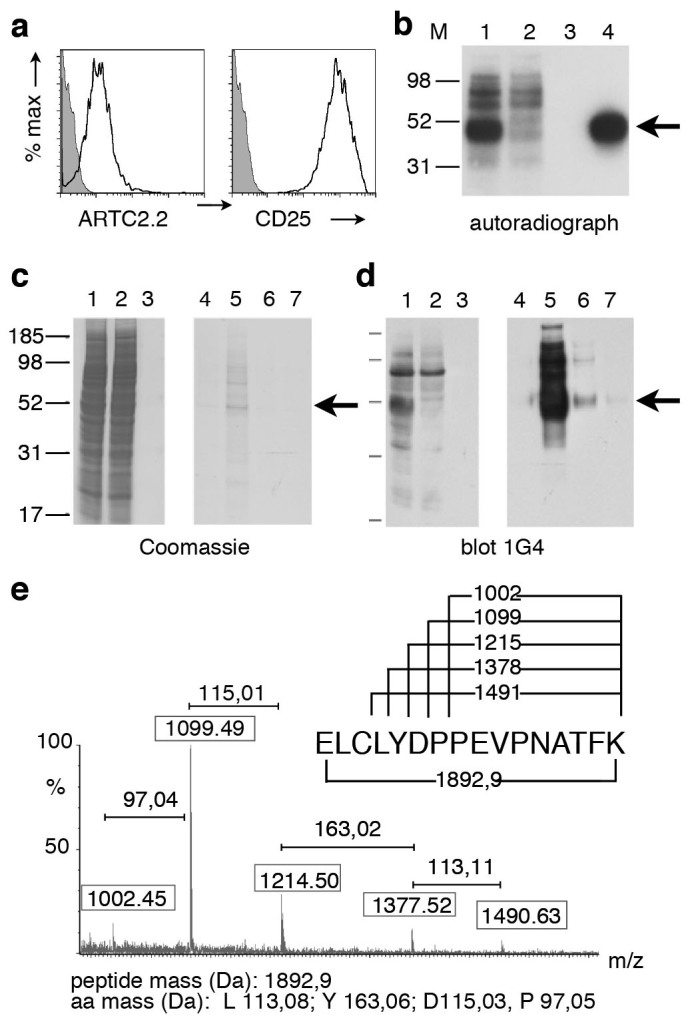
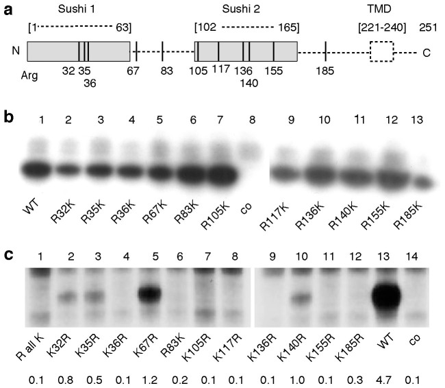
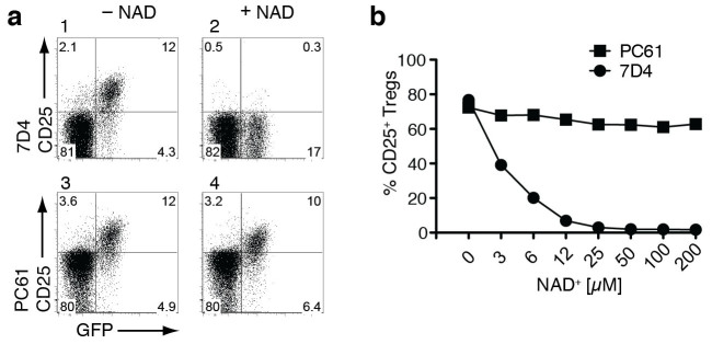
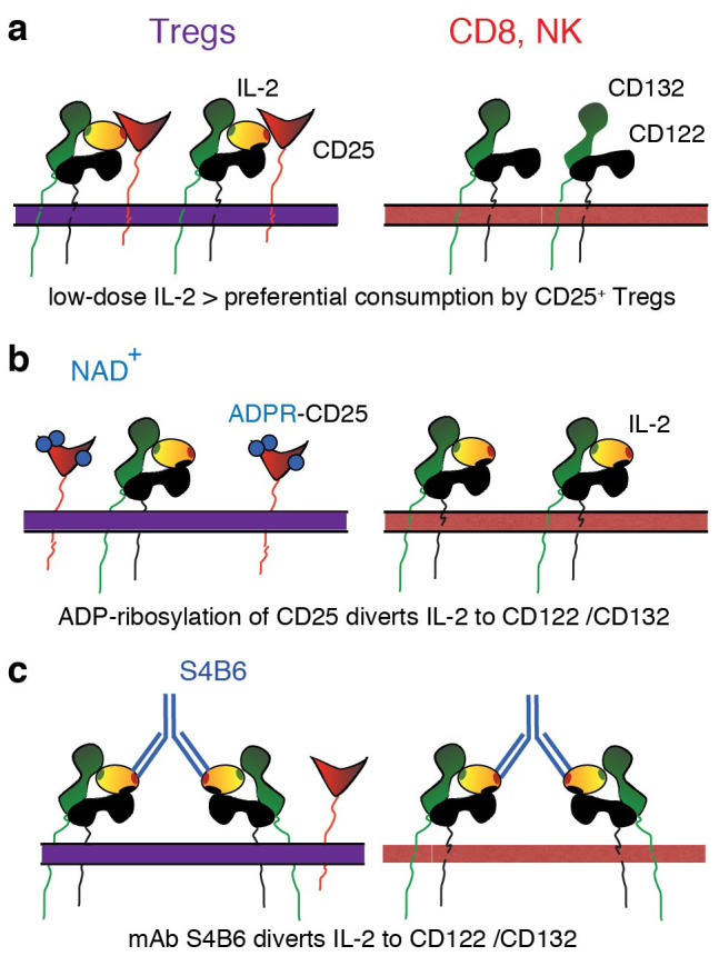
Control of immunologic tolerance and homeostasis rely on Foxp3(+)CD4(+)CD25(+) regulatory T cells (Tregs) that constitutively express the high affinity receptor for Interleukin-2, CD25. Tregs proliferate in response to injections of IL-2/anti-IL-2 antibody complexes or low doses of IL-2. However, little is known about endogenous mechanisms that regulate the sensitivity of CD25 to signaling by IL-2. Here we demonstrate that CD25 is ADP-ribosylated at Arg35 in the IL-2 binding site by ecto-ADP-ribosyltransferase ARTC2.2, a toxin-related GPI-anchored ecto-enzyme. ADP-ribosylation inhibits binding of IL-2 by CD25, IL-2- induced phosphorylation of STAT5, and IL-2-dependent cell proliferation. Our study elucidates an as-yet-unrecognized mechanism to tune IL-2 signaling. This newly found mechanism might thwart Tregs at sites of inflammation and thereby permit a more potent response of activated effector T cells.


Conflict of interest statement

F.K.-N. and F.H. receive royalties from sales of antibodies developed in the lab via MediGate GmbH, a 100% subsidiary of the University Medical Center, Hamburg.

Figures

Figure 1
Figure 1. Immunoprecipitation and mass spectrometry analyses identify CD25 as a major ADP-ribosylation target on YAC-1 cells.
(a) YAC-1 cells were stained with fluorochrome-conjugated mAbs specific for ARTC2.2 and CD25 and analyzed by flow cytometry. (b) YAC-1 cells were incubated with 32P-NAD+. Cell lysates were subjected to immunoprecipitation, proteins were size fractionated by SDS-PAGE, and incorporated radioactivity was detected by autoradiography. Lanes 1, 2: cell lysates before and after precipitation of CD25; lane 3: control precipitation with protein G, lane 4: precipitation with anti-CD25 (PC61). (c–e) YAC-1 cells were incubated with etheno-NAD+. Etheno-ADP-ribosylated proteins were purified from the cell lysate using an affinity column with etheno-adenosine specific mAb 1G4. Bound proteins were eluted in four fractions with etheno-adenosine. Proteins in the cell lysate (lane 1), column flow through (lane 2), wash (lane 3), and eluates (lanes 4–7) were size fractionated by SDS-PAGE. Total protein was visualized by Coomassie staining (c), etheno-ADP-ribosylated proteins were visualized in a parallel immunoblot analysis with mAb 1G4 (d). The 50 kD band in lane 5 (panel c) was excised from the gel and subjected to trypsin digestion and nanospray mass spectrometry. MALDI-TOF spectrum of a fragmented tryptic peptide of 1892,9 kD (e). Boxed numbers indicate the masses of individual peptide peaks, numbers above brackets indicate the mass differences between adjacent peptide peaks. The amino acid sequence of the N-terminal peptide of CD25 is shown on top with numbers corresponding to the masses of the respective peptide fragments. Results are representative of three independent experiments.
Figure 2
Figure 2. Identification of of R32, R35, R67, and R140 as the ADP-ribosylation sites on CD25.
(a) Schematic diagram of the 11 arginine residues in the extracellular domains of human CD25. Numbers correspond to the position within the amino acid sequence of the native protein, i.e. after removal of the signal peptide. Dashed lines indicate residues not visible in the 3D-structure of the IL-2 receptor complexed with IL-2 (2erj). TMD: transmembrane domain. (b, c) HEK cells were co-transfected with expression vectors for ARTC2.2 and either a non-ADP-ribosylated control antigen (co), wild type CD25 (WT), or CD25 variants. Analyzed mutants of CD25 include single R > K mutants (B), a synthetic construct in which all arginine residues in the extracellular domain were replaced by lysine (RallK), and variants of RallK carrying individual K > R back mutations (c). 48 hours post transfection, cells were incubated with 32P-NAD+. Radiolabeled proteins were detected by SDS-PAGE autoradiography. Results are representative of four independent experiments. Numbers indicate the relative labeling intensity of individual bands (mean from four repeats).
Figure 3
Figure 3. ADP-ribosylation inhibits binding of anti-CD25 mAb 7D4 but not of mAb PC61.
Splenocytes from DEREG mice were incubated in the absence or presence of the indicated concentrations of NAD+ before staining with fluorochrome-conjugated mAbs directed against CD4 and CD25 (mAb PC61 or mAb 7D4). Gating was performed on CD4+ cells. (a) Representative dot plots of cells incubated in the absence or presence of 12 μM NAD+. (b) Percentages of CD4+GFP+ cells staining with anti-CD25 mAbs plotted as a function of the concentration of added NAD+. Results are representative of two independent experiments.
Figure 4
Figure 4. ADP-ribosylation inhibits binding of IL-2 and IL-2-dependent proliferation of ARTC2.2-transfected CTLL-2 lymphoma cells.
(a) Untransfected parental and ARTC2.2-transfected CTLL-2 cells were incubated with etheno-NAD+ and stained with fluorochrome-conjugated mAbs specific for CD25, ARTC2.2, and etheno-adenosine and analyzed by flow cytometry. (b) CTLL-2ARTC2.2 cells were incubated with 32P-NAD+ and cell lysates were subjected to immunoprecipitation with anti-CD25 and SDS-PAGE autoradiography as in Fig. 1b. Lanes 1, 2: lysates before and after precipitation of CD25, lane 3: control precipitation with protein G, lane 4: precipitation with anti-CD25. (c) CTLL-2 and CTLL-2ARTC2.2 cells were pre-incubated in the absence or presence of NAD+ or unlabeled IL-2. Cells were then incubated with biotinylated IL-2 before addition of fluorochrome-conjugated streptavidin and analysis by flow cytometry. (d) CTLL-2 and CTLL-2ARTC2.2 cells were seeded in a 24 well culture plate (3 × 10⁴/well) and incubated for four days in medium containing the indicated concentrations of IL-2. Medium lacking or containing NAD+ was added every 12 hours. Cell numbers were assessed by flow cytometry with the aid of Trucount beads (BD). Results are representative of two independent experiments.
Figure 5
Figure 5. ADP-ribosylation inhibits IL-2-induced phosphorylation of STAT5 by CTLL-2ARTC2.2 cells and by ARTC2+/+ Tregs.
(a) CTLL-2 and CTLL-2ARTC2.2 cells were pre-incubated in the absence or presence of NAD+ before stimulation with IL-2 for 15 min. Cells were fixed and stained with fluorochrome-conjugated antibodies against phosphorylated STAT5 (pSTAT5). Control cells were incubated without NAD+ or IL-2. (b) CTLL-2 and CTLL-2ARTC2.2 cells were pre-incubated with the ARTC2.2-blocking nanobody s+16a (nb) before addition of NAD+ and stimulation with IL-2. (c) Tregs were FACS-sorted from wildtype DEREG mice and incubated in the absence or presence of NAD+ before stimulation with IL-2 and staining for pSTAT5. Results are representative of two independent experiments.
Figure 6
Figure 6. Model for the tuning of IL-2 signaling by ADP-ribosylation of CD25.
(a) Tregs constitutively express high levels of CD25 while CD8+ T cells and NK cells express only the β and γ chains of the IL-2 receptor (CD132, CD122). In a non-inflammatory environment, Tregs consume IL-2 due to the higher affinity of CD25 compared to CD122/CD132, thereby depriving neighboring CD8 cells and NK cells of this cytokine. (b) In an inflammatory environment, i.e. following the release of NAD+ from damaged cells, ADP-ribosylation of CD25 (blue circles) diverts IL-2 from the high affinity receptor to the low affinity β and γ chains, allowing efficient expansion of CD8 cells and NK cells. (c) Systemic injection of IL-2 in complex with antibodies that prevent binding of IL-2 to CD25 similarly results in preferential expansion of CD8 cells and NK cells.

Similar articles

Cited by

References

    1. Sakaguchi S. Naturally arising Foxp3-expressing CD25+CD4+ regulatory T cells in immunological tolerance to self and non-self. Nat Immunol 6, 345–352 (2005). - PubMed
    1. Malek T. R. & Castro I. Interleukin-2 receptor signaling: at the interface between tolerance and immunity. Immunity 33, 153–165 (2010). - PMC - PubMed
    1. Bluestone J. A. The yin and yang of interleukin-2-mediated immunotherapy. N Engl J Med 365, 2129–2131 (2011). - PubMed
    1. Boyman O. & Sprent J. The role of interleukin-2 during homeostasis and activation of the immune system. Nat Rev Immunol 12, 180–190 (2012). - PubMed
    1. Matko J. et al. GPI-microdomains (membrane rafts) and signaling of the multi-chain interleukin-2 receptor in human lymphoma/leukemia T cell lines. Eur J Biochem 269, 1199–1208 (2002). - PubMed

Publication types

MeSH terms

Substances