

Solid-phase synthesis, characterization, and cellular activities of collagen-model nanodiamond-peptide conjugates
- PMID: 25753561
- PMCID: PMC4507405
- DOI: 10.1002/bip.22636
Solid-phase synthesis, characterization, and cellular activities of collagen-model nanodiamond-peptide conjugates
Abstract
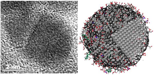
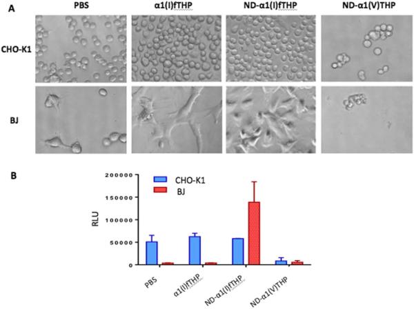
Nanodiamonds (NDs) have received considerable attention as potential drug delivery vehicles. NDs are small (∼5 nm diameter), can be surface modified in a controllable fashion with a variety of functional groups, and have little observed toxicity in vitro and in vivo. However, most biomedical applications of NDs utilize surface adsorption of biomolecules, as opposed to covalent attachment. Covalent modification provides reliable and reproducible ND-biomolecule ratios, and alleviates concerns over biomolecule desorption prior to delivery. The present study has outlined methods for the efficient solid-phase conjugation of ND to peptides and characterization of ND-peptide conjugates. Utilizing collagen-derived peptides, the ND was found to support or even enhance the cell adhesion and viability activities of the conjugated sequence. Thus, NDs can be incorporated into peptides and proteins in a selective manner, where the presence of the ND could potentially enhance the in vivo activities of the biomolecule it is attached to.
Keywords: cell adhesion; collagen; drug delivery; nanoparticle; triple-helical peptide; wound healing.
© 2015 Wiley Periodicals, Inc.
Figures
Comment in
-
Call for submissions.Biopolymers. 2015 May;104(3):v. doi: 10.1002/bip.22673. Biopolymers. 2015. PMID: 25960026 No abstract available.
References
-
- Mochalin VN, Shenderova O, Ho D, Gogotsi Y. Nat Nanotech. 2012;7:11–23. - PubMed
-
- Schrand AM, Ciftan Hens SA, Shenderova OA. Crit Rev Solid State Mat Sci. 2009;34:18–74.
-
- Yu S-J, Kang M-W, Chang H-C, Chan K-M, Yu Y-C. J Am Chem Soc. 2005;127:17604–17605. - PubMed
-
- Schrand AM, Huang H, Carlson C, Schlager JJ, Osawa E, Hussain SM, Dai L. J Phys Chem B. 2007;111:2–7. - PubMed
-
- Lien Z-Y, Hsu T-C, Liu K-K, Liao W-S, Hwang K-C, Chao J-I. Biomaterials. 2012;33:6172–6185. - PubMed
Publication types
MeSH terms
Substances
Grants and funding
LinkOut - more resources
Full Text Sources
Other Literature Sources
