. 2015 Jul;19(7):1720-8.
doi: 10.1111/jcmm.12548. Epub 2015 Mar 6.

Ultrastructure damage of oviduct telocytes in rat model of acute salpingitis

Affiliations

Ultrastructure damage of oviduct telocytes in rat model of acute salpingitis

Jian Yang et al. J Cell Mol Med. 2015 Jul.

Abstract

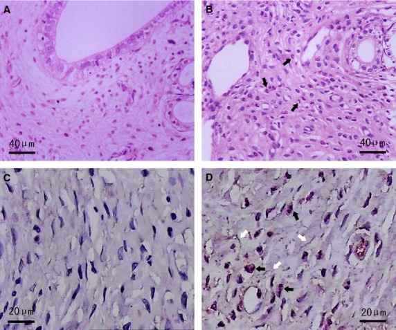
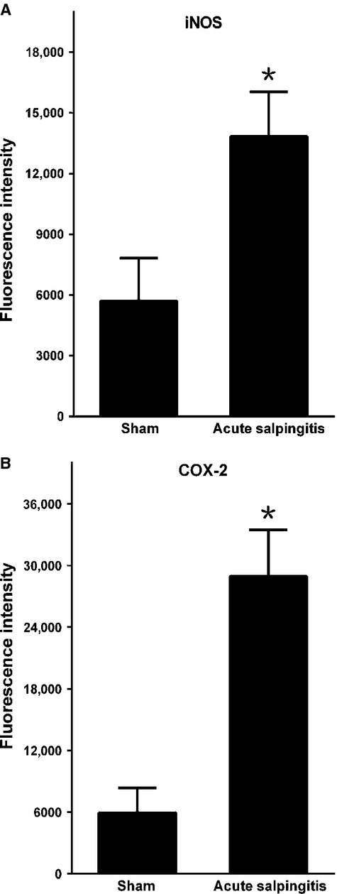
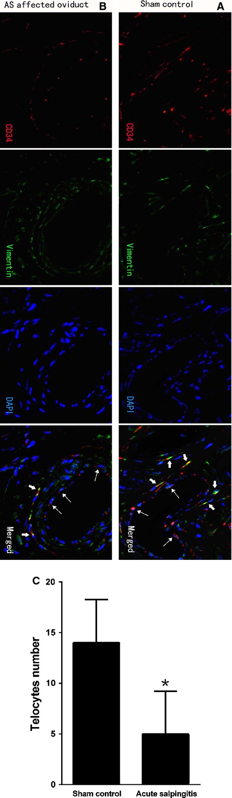
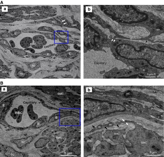
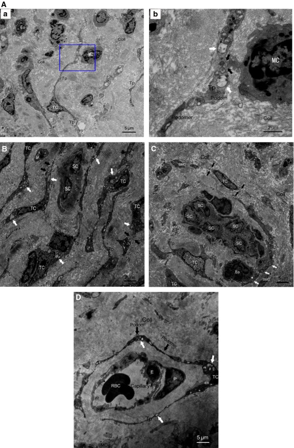
Acute salpingitis (AS) is an inflammatory disease which causes severe damage to a subset of classically described cells lining in oviduct wall and contributes to interstitial fibrosis and fertility problems. Telocytes (TCs), a newly discovered peculiar type of stromal cells, have been identified in many organs, including oviduct, with proposed multiple potential bio-functions. However, with recent increasing reports regarding TCs alterations in disease-affected tissues, there is still lack of evidence about TCs involvement in AS-affected oviduct tissues and potential pathophysiological roles. We presently identified normal TCs by their characteristic ultrastructural features and immunophenotype. However, in AS-affected oviduct tissues, TCs displayed multiple ultrastructural damage both in cellular body and prolongations, with obvious loss of TCs and development of tissue fibrosis. Furthermore, TCs lose their interstitial 3-D network connected by homocellular or heterocellular junctions between TCs and adjacent cells. And especially, TCs connected to the activated immunocytes (mononuclear cells, eosinophils) and affected local immune state (repression or activation). Meanwhile, massive neutrophils infiltration and overproduced Inducible Nitric Oxide Synthase (iNOS), COX-2, suggested mechanism of inflammatory-induced TCs damage. Consequently, TCs damage might contribute to AS-induced structural and reproductive functional abnormalities of oviduct, probably via: (i) substances, energy and functional insufficiency, presumably, e.g. TC-specific genetic material profiles, ion channels, cytoskeletal elements, Tps dynamics, etc., (ii) impaired TCs-mediated multicellular signalling, such as homeostasis/angiogenesis, tissue repair/regeneration, neurotransmission, (iii) derangement of 3-D network and impaired mechanical support for TCs-mediated multicellular signals within the stromal compartment, consequently induced interstitial fibrosis, (iv) involvement in local inflammatory process/ immunoregulation and possibly immune-mediated early pregnancy failure.

Keywords: acute salpingitis; fibrosis; immunoregulation; inflammatory factors; oviduct; rat model; stromal cells; telocytes; tubal ectopic pregnancy; tubal factor infertility.


Figures

Figure 1
Figure 1
Haematoxylin and eosin and CD177 IHC staining in AS-affected and -unaffected oviduct tissues. (A) Normal oviduct tissues from the sham group displayed no obvious changes (haematoxylin and eosin staining). (B) Representative microphotographs of acute inflammation in AS-affected oviduct tissues, including SMCs and capillaries swelling, inflammatory congestion and exudation, interstitial oedema and infiltration of neutrophils (black arrows) (haematoxylin and eosin staining). (C) Totally negative CD177 immunostaining indicating no obvious infiltration of inflammatory cells in sham group. (D) Extensive infiltration of neutrophils (black arrows) as indicated by strong positive CD177 immunostaining, together with interstitial fibrosis (white arrows) in AS-affected oviduct tissues.
Figure 2
Figure 2
Inflammatory factors in AS-affected oviduct tissues were significantly higher than that in sham control. (A) iNOS, (B) COX-2. *P < 0.05 versus sham control; error bars = SD.
Figure 3
Figure 3
TCs immunodiagnostics by double-labelled immunofluorescence. Dotted arrows indicated CD34-positive vascular endothelial cells. Negative c-kit staining was not shown here; scale bar = 20 μm. (A) CD34 (red) in moniliform cells overlying vimentin (green) cells with DAPI counterstaining (blue) in sham control (solid arrows), indicated the existence of perivascular TCs with special immunophenotype of CD34/vimentin double-positive. (B and C) CD34/vimentin double-positive cells with specific TCs morphology and well-defined nuclei was significantly less densely stained, reduced, sparse or completely absent (solid arrows) in AS-affected oviduct tissues. A statistically significant decrease in the mean number of TCs occurred (P = 0.000). *P < 0.05 versus sham control; error bars = SD.
Figure 4
Figure 4
Normal TCs and Tps distributed in perivascular space or SMCs bundles. RBC: red blood cells; E: vascular endothelial cells. (A) Perivascular TCs. (a) A number of TCs, by their extremely long/thin Tps, surrounded and formed an almost complete circle around capillaries. Tps was composed of podoms and podomers. TCs established heterocellular contact with SMCs (white arrows). Mitochondria (m), rough endoplasmic reticulum (rER) and secretory granules can be observed. (b) Higher magnification of the boxed area; TCs frequency formed homocellular junctions (black arrow) and heterocellular junctions (white arrows) with E. (B) Perivascular TCs. (a) TCs surrounded capillaries and scattered among SMCs. b higher magnification of the boxed area. (b) Abundant mitochondria (m) and microvesicles (black arrows) contained in podom. Tps established heterocellular contacts with SMCs (white arrows).
Figure 5
Figure 5
TCs and Tps damage in AS-affected oviduct tissues, accompanied by excessive amount of collagen fibres (Coll) and tissue fibrosis. (A) Intercellular connection between damaged TCs and activated mononuclear cells (MC). (a) Degenerated Tps established closed contact to activated MC which contained dense secretory granules, together with granulocyte infiltration, mainly eosinophils (Eo) and neutrophils (PMN). b higher magnification of the boxed area; (b) synapse (black arrows) between activated MC and degenerated Tps, which contained lots of swollen mitochondria (m) and vacuoles (white arrows), thus indicating degeneration, functional insufficiency of TCs and involvement of TCs in local immunoregulation. (B) Degeneration, discontinue or dissolution of TCs and Tps (black arrows), with cytoplasmic vacuolization (white arrows), accompanied by nearly normal scattered putative stem cells (SCs). Intercellular contacts between TCs and SCs was getting wider or disappeared (black asterisks). (C) Disrupted TC-SC niches which composed of a group of damaged Tps and putative SCs in myosalpinx, with heterocellular contacts getting wider or disappeared between Tps and SCs (black asterisks), Tps and activated Eosinophils (Eo) (white asterisk) with dense secretory granules respectively. Degeneration, discontinue or dissolution of TCs and Tps (black arrows), with swollen mitochondria (m), cytoplasmic vacuolization (white arrows) in Tps, swollen and dissolution of SMCs can be observed. (D) Severely damaged perivascular TCs and Tps, with swollen mitochondria (m), rough endoplasmic reticulum (rER) dilatation and cytoplasmic vacuolization (white arrows), together with damaged endothelial cell (E) and pericytes (P).

Similar articles

Cited by

References

    1. Nowicki S, Ram P, Pham T, et al. Pelvic inflammatory disease isolates of Neisseria gonorrhoeae are distinguished by C1q-dependent virulence for newborn rats and by the sac-4 region. Infect Immun. 1997;65:2094–9. - PMC - PubMed
    1. Pearlman MD, McNeeley SG, Frank TS, et al. Antiendotoxin antibody is protective against tubal damage in an Escherichia coli rabbit salpingitis model. Am J Obstet Gynecol. 1994;171:1588–93. - PubMed
    1. Popescu LM, Faussone-Pellegrini MS. TELOCYTES - a case of serendipity: the winding way from Interstitial Cells of Cajal (ICC), via Interstitial Cajal-Like Cells (ICLC) to TELOCYTES. J Cell Mol Med. 2010;14:729–40. - PMC - PubMed
    1. Suciu L, Popescu LM, Gherghiceanu M, et al. Telocytes in human term placenta: morphology and phenotype. Cells Tissues Organs. 2010;192:325–39. - PubMed
    1. Gherghiceanu M, Popescu LM. Cardiomyocyte precursors and telocytes in epicardial stem cell niche: electron microscope images. J Cell Mol Med. 2010;14:871–7. - PMC - PubMed

Publication types

Substances