Review
. 2015 Apr 6;212(4):435-45.
doi: 10.1084/jem.20150295. Epub 2015 Mar 9.

The interaction of anticancer therapies with tumor-associated macrophages

Affiliations
Review

The interaction of anticancer therapies with tumor-associated macrophages

Alberto Mantovani et al. J Exp Med. .

Abstract

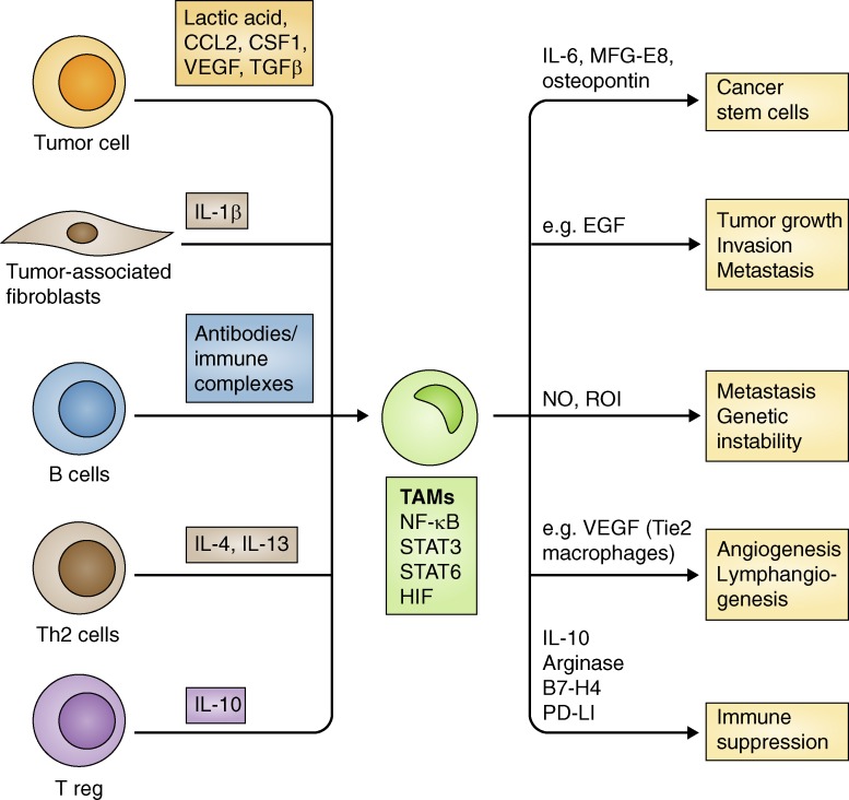
Macrophages are essential components of the inflammatory microenvironment of tumors. Conventional treatment modalities (chemotherapy and radiotherapy), targeted drugs, antiangiogenic agents, and immunotherapy, including checkpoint blockade, all profoundly influence or depend on the function of tumor-associated macrophages (TAMs). Chemotherapy and radiotherapy can have dual influences on TAMs in that a misdirected macrophage-orchestrated tissue repair response can result in chemoresistance, but in other circumstances, TAMs are essential for effective therapy. A better understanding of the interaction of anticancer therapies with innate immunity, and TAMs in particular, may pave the way to better patient selection and innovative combinations of conventional approaches with immunotherapy.


Figures

Figure 1.
Figure 1.
A snapshot of monocyte and macrophage diversity. Two main phenotypically distinct subsets can be identified in the blood: inflammatory monocytes (CCR2+Ly6C+ in mice; CCR2+CD14+CD16 in humans) and patrolling monocytes (CX3CR1+ in mice; CX3CR1+CD14+/−CD16+ in humans). In tissues, macrophages in different organs have different morphological and functional features (e.g., peritoneal macrophages, alveolar macrophages, and liver Kupffer cells). Upon activation with specific signal, macrophages initiate functional programs that are dictated by transcription factors (in rectangles). Two main functional polarizations can be distinguished: classical or M1 and alternative or M2. Other signals, including immune complexes in conjunction with LPS or IL-1, and immune-suppressive cytokines, including IL-10 and TGFβ, also turn on macrophages along an M2-like polarization.
Figure 2.
Figure 2.
Schematic representation of cells and mediators influencing the function of TAMs. On the left side, different cells belonging to the immunological network, as well as tumor cells and tumor-associated fibroblasts, all may influence the functional conditioning of TAMs by producing specific soluble mediators such cytokines, chemokines, and growth factors. For instance, Th2 cytokines (IL-4/IL-13) and other cytokines such as IL-10 and TGFβ, metabolic products derived by tumor cells (lactic acid) and immune complexes, drive TAM polarization into tumor-promoting macrophages. On the right side are listed the major protumor functions of TAMs. For instance, by producing survival factors (IL-6 and MFG-E8) and osteopontin, TAMs protect CSCs form the toxic effect of chemotherapy or directly stimulate tumor cell proliferation via epidermal growth factor (EGF). TAM production of nitric oxide (NO) and reactive oxygen species (ROI) induces genetic instability. TAMs switch on neoangiogenesis by secreting VEGF and suppress immune responses because they express inhibitory molecules (PD-L1 and B7-4) and produce immunosuppressive cytokines/mediators (IL-10 and arginase). In the rectangle, selected transcription factors that orchestrate TAM function are highlighted.
Figure 3.
Figure 3.
Dual role of macrophages in the response to selected therapeutic approaches. Macrophages can either limit (−) the antineoplastic efficacy of selected chemotherapeutic agents or contribute (+) to therapy responses. The general context, including tumor immunogenicity, tissue of origin, and microbial conditioning, defines the set point of balance. The left side of the table lists examples of myelomonocytic cells limiting the efficacy of anticancer therapies. The right side of the table lists examples in which myelomonocytic cells contribute to the efficacy of therapy.

References

    1. Affara N.I., Ruffell B., Medler T.R., Gunderson A.J., Johansson M., Bornstein S., Bergsland E., Steinhoff M., Li Y., Gong Q., et al. . 2014. B cells regulate macrophage phenotype and response to chemotherapy in squamous carcinomas. Cancer Cell. 25:809–821 10.1016/j.ccr.2014.04.026 - DOI - PMC - PubMed
    1. Aharinejad S., Paulus P., Sioud M., Hofmann M., Zins K., Schäfer R., Stanley E.R., and Abraham D.. 2004. Colony-stimulating factor-1 blockade by antisense oligonucleotides and small interfering RNAs suppresses growth of human mammary tumor xenografts in mice. Cancer Res. 64:5378–5384 10.1158/0008-5472.CAN-04-0961 - DOI - PubMed
    1. Alizadeh D., Trad M., Hanke N.T., Larmonier C.B., Janikashvili N., Bonnotte B., Katsanis E., and Larmonier N.. 2014. Doxorubicin eliminates myeloid-derived suppressor cells and enhances the efficacy of adoptive T-cell transfer in breast cancer. Cancer Res. 74:104–118 10.1158/0008-5472.CAN-13-1545 - DOI - PMC - PubMed
    1. Bain C.C., Bravo-Blas A., Scott C.L., Gomez Perdiguero E., Geissmann F., Henri S., Malissen B., Osborne L.C., Artis D., and Mowat A.M.. 2014. Constant replenishment from circulating monocytes maintains the macrophage pool in the intestine of adult mice. Nat. Immunol. 15:929–937 10.1038/ni.2967 - DOI - PMC - PubMed
    1. Beatty G.L., Chiorean E.G., Fishman M.P., Saboury B., Teitelbaum U.R., Sun W., Huhn R.D., Song W., Li D., Sharp L.L., et al. . 2011. CD40 agonists alter tumor stroma and show efficacy against pancreatic carcinoma in mice and humans. Science. 331:1612–1616 10.1126/science.1198443 - DOI - PMC - PubMed

Publication types