. 2015 May;59(5):2849-54.
doi: 10.1128/AAC.04895-14. Epub 2015 Mar 9.

Structural characterization of an alternative mode of tigecycline binding to the bacterial ribosome

Affiliations

Structural characterization of an alternative mode of tigecycline binding to the bacterial ribosome

Andreas Schedlbauer et al. Antimicrob Agents Chemother. 2015 May.

Abstract

Although both tetracycline and tigecycline inhibit protein synthesis by sterically hindering the binding of tRNA to the ribosomal A site, tigecycline shows increased efficacy in both in vitro and in vivo activity assays and escapes the most common resistance mechanisms associated with the tetracycline class of antibiotics. These differences in activities are attributed to the tert-butyl-glycylamido side chain found in tigecycline. Our structural analysis by X-ray crystallography shows that tigecycline binds the bacterial 30S ribosomal subunit with its tail in an extended conformation and makes extensive interactions with the 16S rRNA nucleotide C1054. These interactions restrict the mobility of C1054 and contribute to the antimicrobial activity of tigecycline, including its resistance to the ribosomal protection proteins.


Figures

FIG 1
FIG 1
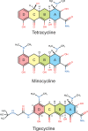
Chemical structures of tetracycline, minocycline, and tigecycline, drawn schematically with their common backbone ring structures (A, B, C, D) colored distinctly. Carbon atom assignments for the 4-ring backbone are indicated on tigecycline.
FIG 2
FIG 2
The environment of the tigecycline-binding site on the 30S ribosomal subunit. TIG binds the 30S ribosomal subunit in a pocket formed by h34 (blue) and h31 (red). The final model with 2Fo-Fc electron density map (1.0σ) surrounding the TIG-binding site is shown from two perspectives in panels A and B. A similar view is shown in Fig. S1B in the supplemental material with the final 2Fo-Fc map rendered at a slightly higher contour level (1.4σ). The relatively weak density for the terminal tert-butyl moiety probably results from the fact that it is connected to the rest of the molecule by two freely rotatable bonds and does not make direct interactions with the binding pocket. TIG is shown in an orientation similar to that of Fig. 1 where the A ring is on the right and the D ring with the tert-butyl-glycylamido side chain is on the left. The two Mg2+ ions are drawn as yellow spheres. The inset in panel A shows the position of the TIG binding pocket on the 30S subunit (viewed from the subunit interface side) with subunit landmarks (pt, platform; h, head; sp, spur). 16S rRNA helices 18, 31, and 34 are colored orange, pink, and blue, respectively. (C) The TIG binding pocket is shown from the same perspective as that in panel B with its putative interactions indicated with dotted lines, including hydrogen bonds (yellow), coordination of the Mg2+ ions (orange), and stacking interactions (gray). These interactions are summarized schematically in panel D. Here the tert-butyl-glycylamido side chain is delineated in light green, and its interactions with C1054 are indicated with cyan and violet arrows. Note that the hydrogen bond between the secondary amine of the side chain and O2 of C1054 is chemically possible but ambiguous in the electron density map. The hydroxyl moiety at position 3 is drawn in a deprotonated form as predicted by nuclear magnetic resonance (NMR) and molecular dynamics (MD) studies (22, 25).
FIG 3
FIG 3
Comparison of TIG's binding mode in the 30S and 70S structures. (A) The 30S-TIG structure (this study) and the 70S-TIG structure (6) have been superimposed using 16S rRNA residues surrounding the TIG binding pocket as the guide. TIG as seen in the 30S structure is colored green, and TIG as seen in the 70S structure is red. h18, h34, and h31 of the 30S-TIG complex are in orange, blue, and red, respectively, and those from the 70S-TIG complex are in shades of gray. Although for the most part the two structures are superimposable, there are notable differences in the placement of h18 (shifts closer to the A site by ∼5 Å in the 70S structure), the position of the base in C1054 (it shifts by 2.5 Å, favoring a parallel shifted π stacking interaction with the heteroaromatic D ring on the 30S structure) and the conformation of the tert-butyl-glycylamido side chain (extended versus bent in the 30S and 70S structures, respectively). (B) To illustrate that the extended side chain conformation of TIG is permissible when the binding pocket is configured as seen in the 70S structure (specifically the shifting of h18), TIG has been aligned to the 70S structure (6) and rendered as a van der Waals surface. This shows that the side chain of TIG in the extended conformation does not significantly clash with h18. (C) Likewise, if the newer tetracycline derivative eravacycline (Erv) (9) is modeled with its side chain extended similar to TIG, it would also not clash with h18 as seen in the 70S structure.

Similar articles

Cited by

References

    1. Wilson DN. 2014. Ribosome-targeting antibiotics and mechanisms of bacterial resistance. Nat Rev Microbiol 12:35–48. doi:10.1038/nrmicro3155. - DOI - PubMed
    1. Wilson DN. 2009. The A-Z of bacterial translation inhibitors. Crit Rev Biochem Mol Biol 44:393–433. doi:10.3109/10409230903307311. - DOI - PubMed
    1. Nguyen F, Starosta AL, Arenz S, Sohmen D, Donhofer A, Wilson DN. 2014. Tetracycline antibiotics and resistance mechanisms. Biol Chem 395:559–575. doi:10.1515/hsz-2013-0292. - DOI - PubMed
    1. Pioletti M, Schlunzen F, Harms J, Zarivach R, Gluhmann M, Avila H, Bashan A, Bartels H, Auerbach T, Jacobi C, Hartsch T, Yonath A, Franceschi F. 2001. Crystal structures of complexes of the small ribosomal subunit with tetracycline, edeine and IF3. EMBO J 20:1829–1839. doi:10.1093/emboj/20.8.1829. - DOI - PMC - PubMed
    1. Brodersen DE, Clemons WM Jr, Carter AP, Morgan-Warren RJ, Wimberly BT, Ramakrishnan V. 2000. The structural basis for the action of the antibiotics tetracycline, pactamycin, and hygromycin B on the 30S ribosomal subunit. Cell 103:1143–1154. doi:10.1016/S0092-8674(00)00216-6. - DOI - PubMed

Publication types

MeSH terms

LinkOut - more resources